Review
. 2016 Jan;24(1):5-17.
doi: 10.1007/s10577-015-9506-4.

Centrosomes are multifunctional regulators of genome stability

Affiliations
Review

Centrosomes are multifunctional regulators of genome stability

Dorothy A Lerit et al. Chromosome Res. 2016 Jan.

Abstract

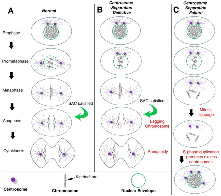
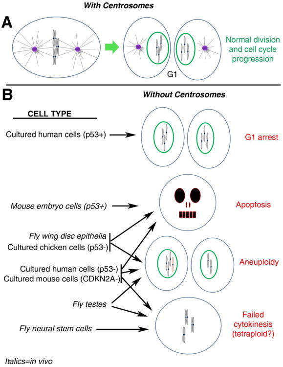
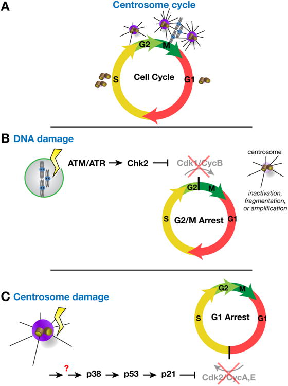
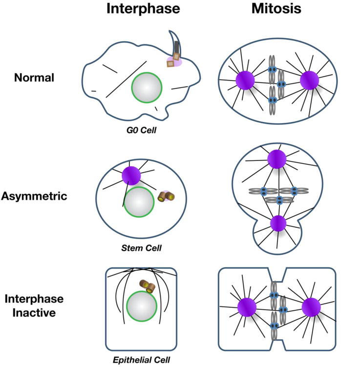
The maintenance of genome stability is critical for proper cell function, and loss of this stability contributes to many human diseases and developmental disorders. Therefore, cells have evolved partially redundant mechanisms to monitor and protect the genome. One subcellular organelle implicated in the maintenance of genome stability is the centrosome, best known as the primary microtubule organizing center of most animal cells. Centrosomes serve many different roles throughout the cell cycle, and many of those roles, including mitotic spindle assembly, nucleation of the interphase microtubule array, DNA damage response, and efficient cell cycle progression, have been proposed to help maintain genome stability. As a result, the centrosome is itself a highly regulated entity. Here, we review evidence concerning the significance of the centrosome in promoting genome integrity. Recent advances permitting acute and persistent centrosome removal suggest we still have much to learn regarding the specific function and actual importance of centrosomes in different contexts, as well as how cells may compensate for centrosome dysfunction to maintain the integrity of the genome. Although many animal cells survive and proliferate in the absence of centrosomes, they do so aberrantly. Based on these and other studies, we conclude that centrosomes serve as critical, multifunctional organelles that promote genome stability.

Keywords: Acentrosomal; Aneuploidy; Asymmetric division; Cell cycle; Centrosome; Centrosome separation; Chromosomal instability; DNA damage; Genome stability; Interphase; Mitosis; PCM; p53.


Figures

Figure 1
Figure 1. Centrosomes promote efficient bipolar spindle assembly
(A) In most cell types, the pair of centrosomes (purple) is closely associated at the entry into mitosis (Prophase). During prophase and prometaphase, centrosomes separate from one another and take up residence on opposite sides of the nucleus. In prometaphase, the nuclear envelope (green) breaks down, allowing MTs (black lines) emanating from the centrosomes to enter the nuclear space, find the kinetochores, and ultimately establish amphitelic attachments (i.e. the kinetochores of sister chromatids are individually attached to MTs from opposing spindle poles). The chromosomes will then congress to the metaphase plate. Once all kinetochores are attached to MTs, the SAC is satisfied, providing the “go anaphase” signal. During anaphase, sister chromatids are segregated towards opposite poles of the spindle. Finally, during telophase, the cytokinetic furrow ingresses, dividing the cell into two daughter cells with identical chromosome complements (abscission and daughter cell formation not depicted). (B) When there are moderate early defects in centrosome separation, the centrosomes may remain closely apposed as the cell enters prometaphase. This is believed to favor incorrect attachments of MTs from both poles to the kinetochore of the same chromatid. The red MT indicates the inappropriate attachment to the affected chromosome (pink). Even in the presence of these merotelic attachments, the SAC can be satisfied, allowing anaphase onset. The opposing forces of MTs from opposite poles on the same kinetochore can result in a lagging chromosome, which, in some cases, will mis-segregate into the wrong daughter cell, resulting in aneuploidy. (C) When centrosomes fail to separate, MTs may not attach to some kinetochores, leaving the SAC unsatisfied. Eventually, mitotic slippage may allow for mitotic exit into G1 without chromosome segregation or cytokinesis, resulting in a tetraploid cell with both centrosomes. In the subsequent S-phase, both centrosomes duplicate, resulting in extra centrosomes.
Figure 2
Figure 2. Differential effects of centrosome loss among model systems
(A) In normal cells with centrosomes, a metaphase cell progresses through cytokinesis (not shown) to produce 2 daughter cells in G1, each with a normal chromosome complement and a single centrosome. To help visualize the consequences of chromosome segregation errors (e.g., aneuploidy), G1 chromosomes are depicted as condensed in this figure, though they would actually decondense upon mitotic exit. (B) The outcomes of mitosis in different cell types depleted of centrosomes are shown. p53 status is indicated for some relevant cell types (if not indicated, p53 is normal). Italics denote cells from in vivo studies. See text for more thorough descriptions of these studies. Not represented is the potential connection between mitotic error and accumulation of DNA damage; as observed in fly wing disc epithelia and cultured chicken cells. Note that lack of a connection to a specific outcome for a particular cell type does not necessarily indicate the outcome does not occur for that cell type. Rather it may simply have not been examined or reported in the relevant studies.
Figure 3
Figure 3. Centrosomes as effectors and regulators of cell cycle progression
(A) The centrosome cycle is entrained with the cell cycle. Similar to the chromosomes, centrosomes duplicate in S-phase. Centrosome maturation, the recruitment of PCM and concordant increase in MTOC activity, occurs during G2 and into M-phase to support the elaboration of the bipolar mitotic spindle. Centrosome activity attenuates in late mitosis such that each centrosome that segregates to a daughter cell (G1) is associated with a basal level of PCM. (B) Activation of the DNA damage response pathway leads to centrosome dysfunction (e.g., inactivation in Drosophila embryos and fragmentation or amplification in mammalian cells) and triggers a block to mitotic entry. (C) Centrosome dysfunction can itself alter cell cycle progression. In some cells, a centrosome integrity checkpoint impedes interphase progression by triggering a G1 arrest.
Figure 4
Figure 4. Strategies for regulating centrosome activity
(A) MT nucleation is differentially regulated in various cell types. In many normal mammalian cells, quiescent (G0) cells form primary cilia during interphase when the centriole docks at the plasma membrane. Cilia are reabsorbed by the cell prior to mitotic onset, when centrosomes function as MTOCs and contribute to spindle formation. Variations on interphase centrosome activity control are observed in Drosophila asymmetrically dividing stem cells, which display asymmetric interphase centrosome activity (one off, one on). During mitosis, both centrosomes are active in stem cells. In contrast, Drosophila and C. elegans epithelial cells deregulate interphase centrosome activity and MTs are nucleated from the apical cortex. Centrosomes mature, or become active, upon mitotic onset. Centrioles (brown barrels) denote inactive centrosomes.

References

    1. Abraham RT. Cell cycle checkpoint signaling through the ATM and ATR kinases. Genes Dev. 2001;15:2177–2196. - PubMed
    1. Arquint C, Gabryjonczyk AM, Nigg EA. Centrosomes as signalling centres. Philos Trans R Soc Lond B Biol Sci. 2014;369 - PMC - PubMed
    1. Basto R, Lau J, Vinogradova T, Gardiol A, Woods CG, Khodjakov A, Raff JW. Flies without centrioles. Cell. 2006;125:1375–1386. - PubMed
    1. Baumbach J, Novak ZA, Raff JW, Wainman A. Dissecting the function and assembly of acentriolar microtubule organizing centers in Drosophila cells in vivo. PLoS Genet. 2015;11:e1005261. - PMC - PubMed
    1. Bazzi H, Anderson KV. Acentriolar mitosis activates a p53-dependent apoptosis pathway in the mouse embryo. Proc Natl Acad Sci U S A. 2014;111:E1491–1500. - PMC - PubMed

Publication types

LinkOut - more resources