. 2015 Dec 9;35(49):16077-93.
doi: 10.1523/JNEUROSCI.1890-15.2015.

Re-Opening the Critical Window for Estrogen Therapy

Affiliations

Re-Opening the Critical Window for Estrogen Therapy

Linda A Bean et al. J Neurosci. .

Abstract

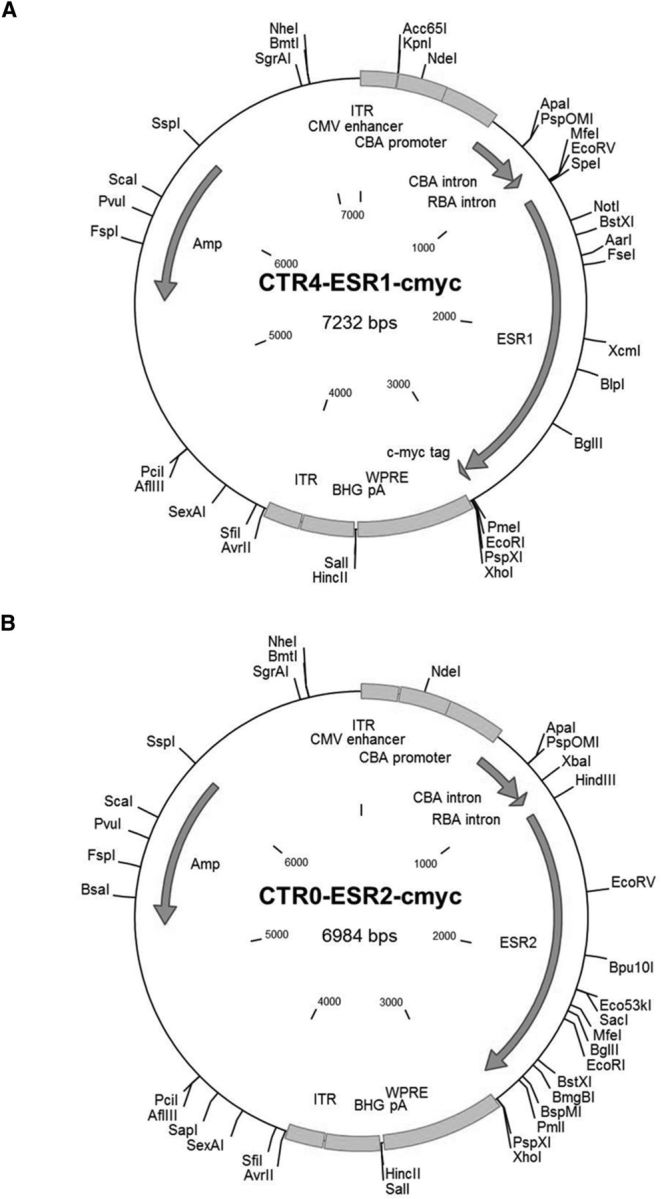
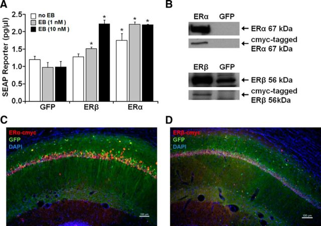
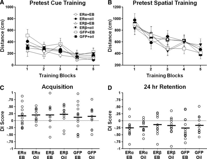
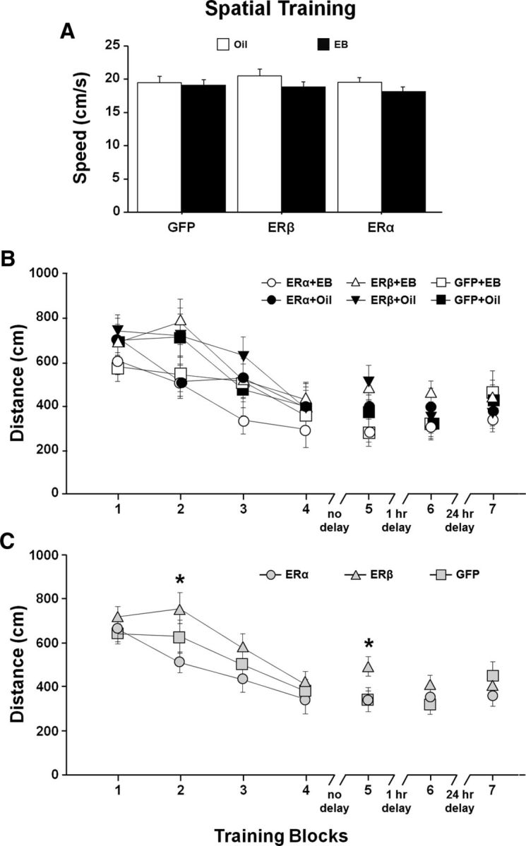
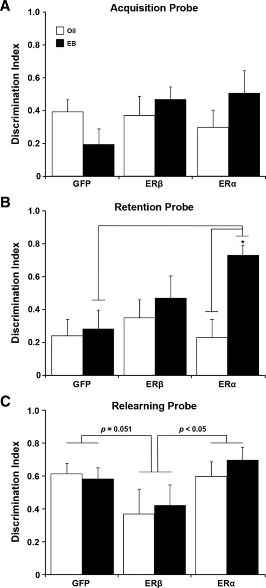
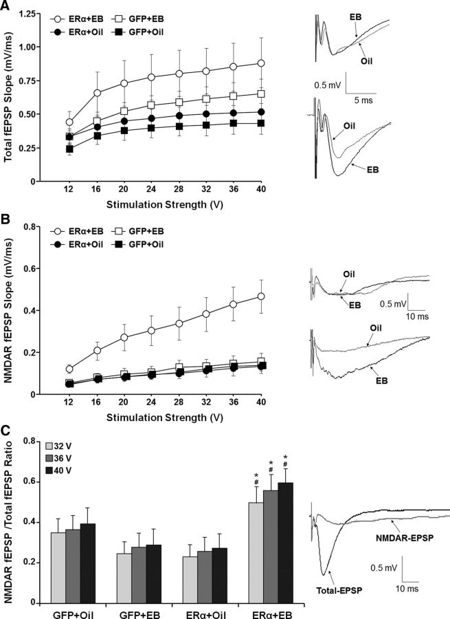
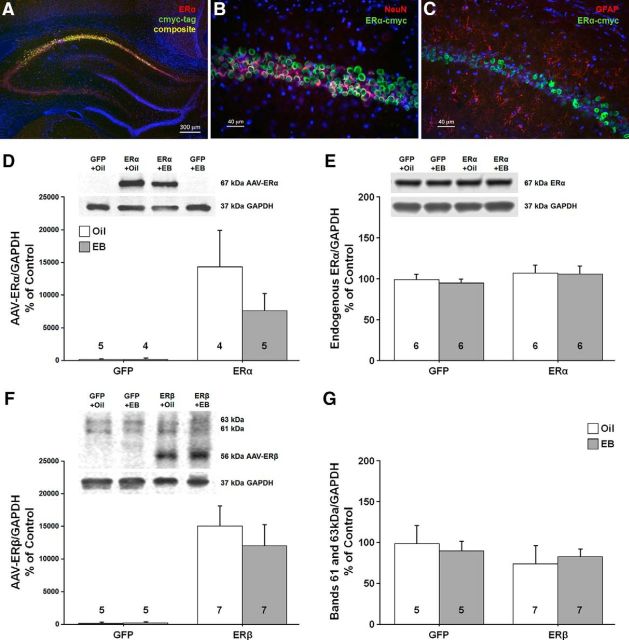
A decline in estradiol (E2)-mediated cognitive benefits denotes a critical window for the therapeutic effects of E2, but the mechanism for closing of the critical window is unknown. We hypothesized that upregulating the expression of estrogen receptor α (ERα) or estrogen receptor β (ERβ) in the hippocampus of aged animals would restore the therapeutic potential of E2 treatments and rejuvenate E2-induced hippocampal plasticity. Female rats (15 months) were ovariectomized, and, 14 weeks later, adeno-associated viral vectors were used to express ERα, ERβ, or green fluorescent protein (GFP) in the CA1 region of the dorsal hippocampus. Animals were subsequently treated for 5 weeks with cyclic injections of 17β-estradiol-3-benzoate (EB, 10 μg) or oil vehicle. Spatial memory was examined 48 h after EB/oil treatment. EB treatment in the GFP (GFP + EB) and ERβ (ERβ + EB) groups failed to improve episodic spatial memory relative to oil-treated animals, indicating closing of the critical window. Expression of ERβ failed to improve cognition and was associated with a modest learning impairment. Cognitive benefits were specific to animals expressing ERα that received EB treatment (ERα + EB), such that memory was improved relative to ERα + oil and GFP + EB. Similarly, ERα + EB animals exhibited enhanced NMDAR-mediated synaptic transmission compared with the ERα + oil and GFP + EB groups. This is the first demonstration that the window for E2-mediated benefits on cognition and hippocampal E2 responsiveness can be reinstated by increased expression of ERα.

Significance statement: Estradiol is neuroprotective, promotes synaptic plasticity in the hippocampus, and protects against cognitive decline associated with aging and neurodegenerative diseases. However, animal models and clinical studies indicate a critical window for the therapeutic treatment such that the beneficial effects are lost with advanced age and/or with extended hormone deprivation. We used gene therapy to upregulate expression of the estrogen receptors ERα and ERβ and demonstrate that the window for estradiol's beneficial effects on memory and hippocampal synaptic function can be reinstated by enhancing the expression of ERα. Our findings suggest that the activity of ERα controls the therapeutic window by regulating synaptic plasticity mechanisms involved in memory.

Keywords: ERα and ERβ; NMDA receptor; aging; estrogen; hippocampus; learning and memory.


Figures

Figure 1.
Figure 1.
Experimental timeline. Female Fischer 344 rats were received at 14 months of age and lavaged to examine the estrous cycle before removal of the ovaries at 15 months. Five to 6 weeks after OVX, the animals were behaviorally tested in the spatial water maze and assigned to ER or EB/oil treatment groups. Eight weeks later, 14 weeks after OVX, stereotaxic surgery was used to deliver viral vectors encoding ERα, ERβ, or GFP bilaterally into the dorsal hippocampi. The animals were allowed 1 week to recover and were then given cyclic injections of EB or oil for 5 weeks. Final behavioral assessment in the water maze was at 20 months of age and initiated 48 h after the seventh cycle of EB/oil treatment. Cyclic EB/oil treatments continued (1–7 additional cycles) and rats were sacrificed (Sac) 48 h after a final EB/oil treatment for electrophysiology, histology, and Western blot analysis.
Figure 2.
Figure 2.
Plasmid maps. ESR1 cDNA for ERα expression (A) and ESR2 cDNA for ERβ expression (B) were cloned into vectors containing a CBA promoter with a CMV enhancer element. Plasmids were packaged into an AAV1 capsid before stereotaxic injections into the dorsal hippocampus of aged OVX female rats.
Figure 3.
Figure 3.
Analysis of functionality and in vivo expression of viral vectors. A, Plasmids expressing cmyc-tagged ERα, cmyc-tagged ERβ, or GFP were cotransfected with an ERE-SEAP reporter plasmid in HEK293T cells with the addition of 1 or 10 nm EB treatment or vehicle. SEAP reporter levels were measured 24 h after application of treatments and compared with control groups. These results confirm functionality of the encoded ERs in vitro. Asterisks indicate a significant (p < 0.05) increase in SEAP expression relative to control. B, Western analysis of HEK293T cells treated with AAV-ERα, AAV-ERβ, or the GFP control virus. Top, For cells treated with AAV-ERα, antibodies for ERα (Ab17) and cmyc-tag (A21281) detected the 67 kDa full-length protein for ERα. Bottom, For cells treated with AAV-ERβ, antibodies for ERβ (H-150) and cmyc-tag (A21281) detected a band at ∼56 kDa. Note that HEK293T cells treated with the GFP virus exhibit a band at ∼56 kDa, which is increased in cultures treated with AAV-ERβ-cmyc. C, D, Immunofluorescent staining for the cmyc-tag, verified hippocampal expression of ERα and ERβ 4 weeks after injection of ER-encoding vectors. AAV-GFP was mixed with the ER virus in a 1:4 ratio to visualize distribution of expression vectors.
Figure 4.
Figure 4.
Water maze Pretest indicates no initial difference in performance of the treatment groups. Rats were pseudorandomly assigned to ER and EB/oil treatment groups. For this and all subsequent figures, circle = ERα, triangle = ERβ, square = GFP, open = EB, and filled = oil. Five to 6 weeks after OVX and before virus injections, animals were tested on the cue task (A) and spatial discrimination task (B). The symbols indicate the mean escape distance (±SEM) for each training block. Individual DI scores and mean (bar) for the acquisition (C) and 24 h retention probe (D) trials. No group differences were observed. Across all groups, animals exhibited acquisition of a spatial discrimination with impaired retention 24 h later.
Figure 5.
Figure 5.
Spatial discrimination performance after differential expression of ER and EB/oil treatment. A, Mean swim speed (+ SEM) across training Blocks 1–5 for EB (filled bars)- and oil (open bars)-treated animals. Symbols indicate the mean (±SEM). B, Escape path length to the submerged platform for the ER and treatment groups. C, Mean (±SEM) escape path length collapsed across EB/oil treatments (gray) for each ER group, illustrating the shortest and longest escape distances in ERα and ERβ groups, respectively. The asterisks indicate trial blocks during acquisition training with significant (p < 0.05) differences between ERβ and other groups.
Figure 6.
Figure 6.
Expression of AAV-ERα in conjunction with EB treatment improved retention of spatial information for platform location. AC, Bars represent the mean (±SEM) DI score calculated from performance on the probe trials for rats injected with AAV vectors encoding ERα, ERβ, or GFP and subsequently treated with EB (filled bars) or oil (open bars). A, Acquisition probe trial directly followed Block 4 of the spatial discrimination task. B, Retention probe trial followed a 1 h delay between Blocks 5 and 6. Superior performance was seen in EB-treated rats injected with the AAV expressing ERα. Asterisk indicates significant difference (p < 0.05). C, The probe trial immediately followed a refresher block the day after spatial training. The p-values above the bars indicate for differences between the ERα and GFP groups and animals with viral-mediated expression of ERβ.
Figure 7.
Figure 7.
Expression of ERα, ERβ, or GFP and EB/oil treatments did not influence sensory-motor abilities examined as average swim speed (A) or distance (B) to find the visible platform across the training blocks on a cue discrimination task administered after the completion of the probe trial on day 2.
Figure 8.
Figure 8.
Expression of ERα in conjunction with EB treatment enhanced the NMDAR-synaptic response in the hippocampus of aged female rats. Input–output curves are illustrated for the mean slope of the total-fEPSP (A) and NMDAR-fEPSP (B). EB treatment was associated with an increase in the total-fEPSP response, which was particularly evident for animals that received AAV-ERα. After collection of the total-fEPSP, the NMDAR component of the synaptic response was isolated. In contrast to the total-fEPSP, the EB-mediated increase in NMDAR-fEPSP was specific to animals that received AAV-ERα. The insets in A and B show representative traces of synaptic responses from animals injected with AAV-GFP (top) and AAV-ERα (bottom) and treated with EB (black line) or oil (gray line). C, The ratio of the NMDAR-fEPSP/total-fEPSP for the highest stimulation intensities (32–40 V) was increased in the ERα + EB group. Asterisks indicate significant (p > 0.05) treatment effect, with greater NMDAR-fEPSP/total-fEPSP ratio in the ERα + EB group relative to the ERα + oil group. Pound signs indicate a significant ER group effect with a greater NMDAR-fEPSP/total-fEPSP ratio in ERα + EB group relative to GFP + EB group. The inset shows the total-fEPSP and isolation of NMDAR-fEPSP from the same slice.
Figure 9.
Figure 9.
Western blots and histology were used to confirm increased ER expression in neurons of the dorsal hippocampus. Immunofluorescent chemistry (AC) showing expression of ERα within the CA1 region of the hippocampus. A, Merged image shows expression of AAV-ERα tagged with cmyc (green), and ERα (red). B, C, Cmyc (green) tagged to ERα was expressed mainly in neurons immunostained with a neural marker (NeuN, red; B), but not in astrocytes immunostained with the glial marker, glial fibrillary acidic protein (GFAP, red; C). Images AC were counterstained with the nuclear marker DAPI (blue). D, E, Western blots using an antibody selective against the human ERα (D) or rat ERα (E) confirmed a band at ∼67 kDa. Animals injected with virus carrying ERα exhibited increased expression of the human ERα and no difference was observed for endogenous ERα. F, G, An antibody against ERβ confirmed that the ERβ vector increased the expression of human ERβ (∼56 kDa; F) in the absence of a change in the 63 and 61 kDa bands (G). For these and subsequent Western blots, the numbers in or above the bars indicate the number of samples used in the analysis.
Figure 10.
Figure 10.
Western blots to assess the possible growth of dendritic spines in the CA1 region of the hippocampus for animals expressing ERα or GFP. The bars represent the means (±SEM) for the expression of the synaptic proteins PSD95 (A) and synaptophysin (B).
Figure 11.
Figure 11.
Western blots of glutamate receptor expression in the CA1 region of the hippocampus for animals expressing ERα or GFP. The bars represent the means (±SEM) for the expression of the NR2B subunit (A), the NR2A subunit (B), and the GluR1 subunit (C).
Figure 12.
Figure 12.
Western blots to assess possible changes in phosphorylation and kinase activity in the CA1 region of the hippocampus for animals expressing ERα or GFP. The bars represent the means (±SEM) for the phosphorylation of NR2B at S1303 (A), GluR1 at S831 (B), and ERK1/2 (C).

References

    1. Adams MM, Oung T, Morrison JH, Gore AC. Length of postovariectomy interval and age, but not estrogen replacement, regulate N-methyl-D-aspartate receptor mRNA levels in the hippocampus of female rats. Exp Neurol. 2001a;170:345–356. doi: 10.1006/exnr.2001.7716. - DOI - PubMed
    1. Adams MM, Shah RA, Janssen WG, Morrison JH. Different modes of hippocampal plasticity in response to estrogen in young and aged female rats. Proc Natl Acad Sci U S A. 2001b;98:8071–8076. doi: 10.1073/pnas.141215898. - DOI - PMC - PubMed
    1. Adams MM, Fink SE, Shah RA, Janssen WG, Hayashi S, Milner TA, McEwen BS, Morrison JH. Estrogen and aging affect the subcellular distribution of estrogen receptor-alpha in the hippocampus of female rats. J Neurosci. 2002;22:3608–3614. - PMC - PubMed
    1. Adams MM, Fink SE, Janssen WG, Shah RA, Morrison JH. Estrogen modulates synaptic N-methyl-D-aspartate receptor subunit distribution in the aged hippocampus. J Comp Neurol. 2004;474:419–426. doi: 10.1002/cne.20148. - DOI - PubMed
    1. Aenlle KK, Foster TC. Aging alters the expression of genes for neuroprotection and synaptic function following acute estradiol treatment. Hippocampus. 2010;20:1047–1060. doi: 10.1002/hipo.20703. - DOI - PMC - PubMed

Publication types

MeSH terms

LinkOut - more resources