. 2016 Feb 15:127:123-134.
doi: 10.1016/j.neuroimage.2015.11.060. Epub 2015 Dec 4.

Network mechanisms of intentional learning

Affiliations

Network mechanisms of intentional learning

Adam Hampshire et al. Neuroimage. .

Abstract

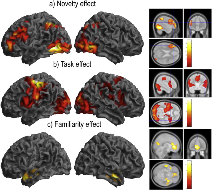
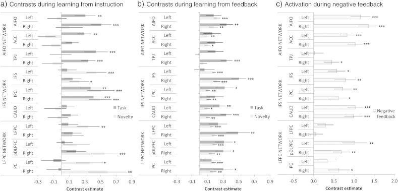
The ability to learn new tasks rapidly is a prominent characteristic of human behaviour. This ability relies on flexible cognitive systems that adapt in order to encode temporary programs for processing non-automated tasks. Previous functional imaging studies have revealed distinct roles for the lateral frontal cortices (LFCs) and the ventral striatum in intentional learning processes. However, the human LFCs are complex; they house multiple distinct sub-regions, each of which co-activates with a different functional network. It remains unclear how these LFC networks differ in their functions and how they coordinate with each other, and the ventral striatum, to support intentional learning. Here, we apply a suite of fMRI connectivity methods to determine how LFC networks activate and interact at different stages of two novel tasks, in which arbitrary stimulus-response rules are learnt either from explicit instruction or by trial-and-error. We report that the networks activate en masse and in synchrony when novel rules are being learnt from instruction. However, these networks are not homogeneous in their functions; instead, the directed connectivities between them vary asymmetrically across the learning timecourse and they disengage from the task sequentially along a rostro-caudal axis. Furthermore, when negative feedback indicates the need to switch to alternative stimulus-response rules, there is additional input to the LFC networks from the ventral striatum. These results support the hypotheses that LFC networks interact as a hierarchical system during intentional learning and that signals from the ventral striatum have a driving influence on this system when the internal program for processing the task is updated.

Keywords: Caudate; Dynamic causal modelling; Frontal cortex; Functional connectivity; Learning.


Figures

Fig. S1
Fig. S1
Mean number of incorrect responses made at each of the five stages of learning by trial and error. Error bars report the standard error of the mean.
Fig. S2
Fig. S2
Supplemental whole-brain analyses of data from study 1 (all rendered at p < 0.05 FWE cluster corrected for the whole brain volume). Voxelwise analysis using a novelty contrast during learning by instruction (Stages 1–6 weighted as 3 2 1 − 1 − 2 − 3) rendered significant effects across a set of brain regions including the left IFS and the IPC bilaterally (S2a). A Task contrast (Stages 1–6 all weighted as 1) rendered activation across a set of brain regions including the AIFO and ACC (S2b). The negative effect of Novelty rendered significant voxels within brain regions that approximately conform to the default mode network (Buckner et al., 2008) (S2c). There were no significant voxels within the striatum for any of the above contrasts even at the uncorrected threshold of p < 0.05 one-tailed.
Fig. S3
Fig. S3
Supplemental whole-brain analyses of data from Study 2 (all rendered at p < 0.05 FWE cluster corrected for the whole brain volume). Voxelwise analysis using a Novelty contrast during learning from feedback (Stages 1–5 weighted as 2 1 0 − 1 − 2) rendered significant activation across the frontoparietal cortices and within the striatum (S3a). A similar pattern of activation was evident for the Task contrast (Stages 1–5 all weighted as 1) (S3b). A set of regions within the medial orbitofrontal cortices and temporal lobes were more active when rules were familiar (S3c). Negative feedback generated strong activation within the frontoparietal cortices and within the striatum (S3d). Notably, peak coordinates within the caudate varied between the contrasts (S3e), with mid/ventral regions including the nucleus accumbens, putamen and ventral caudate activated by negative feedback (rendered in red on the sagittal images) but not Novelty (rendered in blue on the sagittal images). Comparing directly between these contrasts (inset on the right) showed that the feedback-familiarity difference was significant at the whole brain corrected threshold, whereas the reverse contrast was not significant even at the uncorrected threshold of p < 0.05. The peak striatum coordinates for this contrast of contrasts (left x = − 12 y = 5 z = 4; right x = 15 y = 8 z = 4) were within 5 mm of the centres of the caudate ROIs (left x = − 12 y = 10 z = 4; right x = 12 y = 12 z = 4).
Fig. 1
Fig. 1
Paradigms that are used to probe LFC function often treat learning effects as nuisance variables. This can lead to overly static interpretations of LFC function. For example, one prominent hypothesis states that a sub-region of the right inferior frontal gyrus (pIFG) is involved in the effortful cancellation of dominant motor responses. The Stop Signal Task is designed to probe motor inhibition processes and shows significant activation within this region. However, the pIFG is most active when the task is initially being learnt. Other LFC sub-regions, including the anterior insula inferior frontal operculum (AIFO), inferior frontal sulcus (IFS) and posterior dorsolateral prefrontal cortex (pDLPFC), show similar learning effects. Behavioural performance measures correlate with changes in functional connectivity between these LFC sub-regions. These results (Erika-Florence et al., 2014) indicate that distributed LFC networks work in a coordinated manner to support novel tasks. As a task becomes automated, the involvement of these networks diminishes.
Fig. 2
Fig. 2
a) & b) In a previous study (Parkin et al., 2015) we applied spatial ICA to decompose the LFCs into functionally distinct sub-regions in a data-driven manner. The timecourses of these sub-regions were used in seed analyses to characterise their cortically and sub-cortically distributed functional networks. One network (red) included the AIFO. Seed analyses identified the anterior cingulate cortex/pre-supplementary motor area (ACC) and temporal parietal junction (TPJ) bilaterally. A second network (green) included the IFS. Seed analysis identified the caudate nucleus bilaterally and a region extending from the superior occipital lobe into the inferior parietal cortex (IPC). A third network (blue) included the lateral frontopolar cortices (LFPC). Seed analyses identified the pDLPFC and superior parietal cortices bilaterally (PC). c) 10 mm radius spherical regions of interest were defined based on peak coordinates from the ICA and seed analyses. These ROIs were formed into a two-tier network model, with the lower tier consisting of intra-network connections and the upper tier consisting of connections between different LFC sub-regions.
Fig. 3
Fig. 3
a) In Study 1 participants learnt novel discrimination rules from explicit instruction. Initially, a slide was presented with a discrimination rule. Subsequently, a sequence of coloured shapes was presented for 3 min and the participant was required to respond with the relevant button press as quickly and accurately as possible. After 3 min a new rule slide was displayed followed by another sequence of coloured shapes. b) In Study 2, there were no rule slides. Instead, feedback indicating whether the previous response was correct or incorrect was presented randomly after 50% of trials. Therefore, participants were required to derive the discrimination rules by a process of trial-and-error. c) The two studies used the same stimulus sets. d) In Study 1, response times followed a non-monotonic decrease when stimulus–response rules were being learnt from instruction. Specifically, there was a rapid decrease in RT from stages one to three, followed by a small increase in RT then a more gradual decrease. e) In Study 2, a similar non-monotonic decrease was also evident in RTs when stimulus-response rules were being learnt by exploration with feedback. (***p < 0.001, **p < 0.01, *p < 0.05 two tailed significance).
Fig. 4
Fig. 4
a) Placement of the LFC network regions of interest. b) The IFS and LFPC networks showed smooth downwards curves in task-related activation as the stimulus–response rules transitioned from novel to familiar. By contrast, the AIFO network still showed significant task-related activation at the sixth and final stage of the learning process. c) All three networks showed decreases in task-related activation as the rules transitioned from novel to familiar during learning by exploration with feedback. The LFPC showed the sharpest decline, with a large early peak, whereas the AIFO showed the slowest decline, with significant activation through the third stage. The networks also showed significant activation during negative feedback (left bar).
Fig. 5
Fig. 5
a) Task and Novelty effects for each region of interest during learning from instruction. Notably, the caudate ROIs showed no significant effects of task or novelty whereas the rest of the IFS network was significantly activated by either one or both contrasts. b) During learning from feedback, there were significant effects of task relative to fixation and of rule novelty throughout all three networks. In contrast to learning by instruction, these effects were evident within the caudate (highlighted) and the AIFO network ROIs. c) Negative feedback generated heightened activation within all three networks including the caudate ROIs. (***p < 0.001, **p < 0.01, *p < 0.05 2-tailed significance. Error bars report the standard error of the mean).
Fig. 6
Fig. 6
a) In Study 1, analysis of ROI phase synchrony showed an increase in network functional connectivity when processing novel rules. b) This effect was also evident when examining phase synchrony across all voxels within the brain. (Error bars report the standard error of the mean). c) In contrast to learning from instruction, there was only an increase in network phase synchrony at the first stage during learning by exploration with feedback. Unexpectedly, there was also a decrease in network phase synchrony during the reception of negative feedback. d) In Study 2, the phase synchrony effects were uncoupled from the activation magnitude effects during learning by exploration with feedback. (Error bars report the standard error of the mean).
Fig. 7
Fig. 7
a) Psychophysiological interaction of the IFS ROI timecourse with the rule novelty contrast showed greater functional connectivity with the IPC but not the caudate ROIs during learning by instruction. 7b) Psychophysiological interaction of the IFS ROI negative feedback event timecourses showed a significant increase in function connectivity with the caudate but not the IPC ROIs during learning by exploration with feedback. (Error bars report the standard error of the mean).
Fig. 8
Fig. 8
a) In Study 1, Bayesian model selection with fixed effects analysis favoured a dynamic causal model in which rule novelty modulated top-down connections from the LFPC to the IFS and from the IFS to the AIFO. b) In Study 2, Bayesian model selection with fixed effects analysis favoured the family of models in which negative feedback was a driving input. Model 1, in which the driving input was via the caudate ROI provided the best explanation of the data.

References

    1. Badre D. Cognitive control, hierarchy, and the rostro-caudal organization of the frontal lobes. Trends Cogn. Sci. 2008;12:193–200. - PubMed
    1. Badre D., Hoffman J., Cooney J.W., D'Esposito M. Hierarchical cognitive control deficits following damage to the human frontal lobe. Nat. Neurosci. 2009;12:515–522. - PMC - PubMed
    1. Beckmann C.F., Smith S.M. Probabilistic independent component analysis for functional magnetic resonance imaging. IEEE Trans. Med. Imaging. 2004;23:137–152. - PubMed
    1. Boettiger C.A., D'Esposito M. Frontal networks for learning and executing arbitrary stimulus–response associations. J. Neurosci. 2005;25:2723–2732. - PMC - PubMed
    1. Brett M., Anton J., Valabregue R., Poline J. 8th International Conference on Functional Mapping of the Human Brain, Sendai, Japan. 2002. Region of interest analysis using an SPM toolbox [abstract]

Publication types