. 2015 Dec 11:5:17943.
doi: 10.1038/srep17943.

A novel, native-format bispecific antibody triggering T-cell killing of B-cells is robustly active in mouse tumor models and cynomolgus monkeys

Affiliations

A novel, native-format bispecific antibody triggering T-cell killing of B-cells is robustly active in mouse tumor models and cynomolgus monkeys

Eric J Smith et al. Sci Rep. .

Abstract

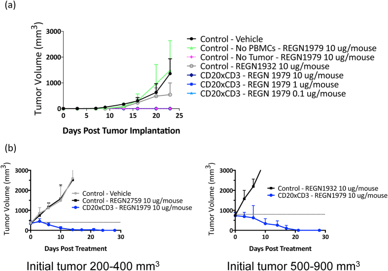
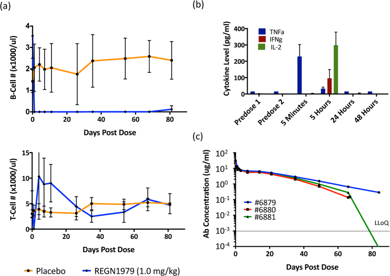
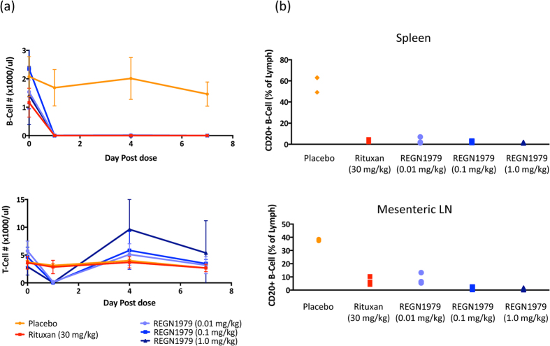
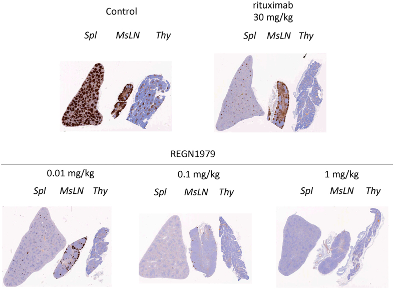
Bispecific antibodies, while showing great therapeutic potential, pose formidable challenges with respect to their assembly, stability, immunogenicity, and pharmacodynamics. Here we describe a novel class of bispecific antibodies with native human immunoglobulin format. The design exploits differences in the affinities of the immunoglobulin isotypes for Protein A, allowing efficient large-scale purification. Using this format, we generated a bispecific antibody, REGN1979, targeting the B cell marker, CD20, and the CD3 component of the T cell receptor, which triggers redirected killing of B cells. In mice, this antibody prevented growth of B cell tumors and also caused regression of large established tumors. In cynomolgus monkeys, low doses of REGN1979 caused prolonged depletion of B cells in peripheral blood with a serum half-life of approximately 14 days. Further, the antibody induced a deeper depletion of B cells in lymphoid organs than rituximab. This format has broad applicability for development of clinical bispecific antibodies.


Conflict of interest statement

There is potential competing interest. All authors are employees of Regeneron Phamaceuticals.

Figures

Figure 1
Figure 1. Description of bispecific antibody format.
(a) Alignment of the sequences of the CH3 domain of human IgG1 and IgG3. (b) Localization of the Fc* substitution within the crystal structure of human IgG1. Residues that are conserved between IgG1 and IgG3 are shown in yellow; H435 and Y436 are shown in magenta; other non-conserved residues are shown in green. (c) Schematic diagram of bispecific antibody format. Heavy chain variable regions of different specificity are in red and cyan; common light chain variable regions are in orange.
Figure 2
Figure 2. Isolation of bispecific antibody by selective Protein A affinity chromatograpy.
(a) Chromatogram illustrating the separation of REGN2280 bispecific from the binding (FcFc) and non-binding (Fc*Fc*) homodimers, using a recombinant Protein A resin. The dotted trace shows pH, and the solid trace shows the absorbance at 280 nm of load, wash and gradient elution steps. (b,c) Flow-cytometric analysis of CD20xCD3 bispecific antibodies REGN2280 and REGN1979 binding to CD20+/CD3− Raji (b) and CD3+/CD20− Jurkat (c) cells. Bound antibody is proportional to mean fluorescence intensity (MFI). A non-specific hIgG1 isotype control antibody showed no binding to either cell line.
Figure 3
Figure 3. Redirected T cell-mediated cytotoxicity of bispecific antibodies in vitro.
(a,b) Effects of treatment of CD20 positive Raji cells with CD20 x CD3 bispecific antibodies and pre-activated human (a) or cynomolgus (b) effector lymphocytes (E:T-ratio 10:1). Cell viability was measured by Calcein release assay after 2 hours of incubation. (c) Specific lysis of CD20+ target cells upon treatment with 10 ug/ml of CD20 x CD3 bispecific antibodies and effector lymphocytes (E:T ratio 4:1). B16F10.9 cells expressing exogenous CD20 are labeled with Violet cell tracker (upper left quadrants), while control B16F10.9 cells are labeled with CFDA-SE (lower right quadrants). Both REGN2280 and REGN1979 selectively deplete cells expressing CD20. (d) Effects of treatment of CD20-positive B16F10.9 cells with CD20 x CD3 bispecific antibodies and effector lymphocytes (E:T ratio 4:1). Cell viability was measured by FACS after 48 hours incubation. (e) Dose-dependent effect of the CD20 x CD3 bispecific antibodies on the expression of CD69 on CD2+ lymphocytes in the presence of B16F10.9 expressing CD20 cells after incubation for 48 hours. The surface expression of CD69 on effector cells was detected by flow cytometry using an antigen-specific PE conjugated antibody.
Figure 4
Figure 4. Effects of REGN1979 on tumor growth in in vivo mouse tumor models.
(a) Raji tumor cells were co-implanted with human PBMCs subcutaneously into NSG mice and treated with the indicated amount of REGN1979 or a control antibody via intraperitoneal injection twice a week starting at day 0 for the length of the study. No tumor growth was observed at all dose levels of REGN1979 tested. (b) Raji tumor cells were co-implanted with hPBMCs and tumors were allowed to develop until they reached the indicated initial volume (either 200–400 mm3 or 500–900 mm3). Treatment was then initiated with REGN1979 or a control antibody. These were dosed twice weekly for the duration of the study. Animals dosed with REGN1979 showed complete tumor regression in both initial tumor sizes.
Figure 5
Figure 5. Effects of REGN1979 on circulating B cells and on cytokine levels in cynomolgus monkeys.
(a) Cynomolgus monkeys were injected with a single dose of REGN1979 (1 mg/kg), and levels of peripheral B and T cells were examined by flow cytometry. Treatment with REGN1979 resulted in complete depletion of measurable circulating B cells for at least 60 days post dose, and a transient loss of circulating T cells. (b) Cytokine levels were measured at the indicated time post dose. Compared to pre-dose levels, treatment with REGN1979 resulted in transient increase in all measured cytokines, which returned to base line levels by T = 24 hours. (c) Levels of REGN1979 in circulation are shown for the three animals in the cohort (I06879, I06880, I06881). LLoQ represents the lower limit of quantification for the assay.
Figure 6
Figure 6. Depth of killing of B cells in peripheral lymphoid tissues of cynomolgus monkeys treated with REGN1979.
Cynomolgus monkeys were dosed with the indicated amounts of REGN1979 or rituximab, and levels of circulating B and T cells were analyzed by FACS (a). Seven days post dose the animals were sacrificed, and levels of B cells in the peripheral lymphoid tissues were examined by tissue FACS as described in Methods (b).
Figure 7
Figure 7. Depth of killing of B cells in peripheral lymphoid tissues of cynomolgus monkeys treated with REGN1979, immunohistochemical analysis.
Seven days post dose with the indicated amounts of REGN1979 or rituximab, the animals were sacrificed, and the indicated peripheral lymphoid tissues were stained with anti-CD20 antibodies. Similar results were obtained with antibodies against the B cell markers, CD19 and CD79a (data not shown). Spl, spleen; MsLN, mesenteric lymph nodes; Thy, thymus.

References

    1. Spiess C., Zhai Q. & Carter P. J. Alternative molecular formats and therapeutic applications for bispecific antibodies. Mol. Immunol. 67, 95–106 (2015). - PubMed
    1. Ridgway J. B., Presta L. G. & Carter P. ‘Knobs-into-holes’ engineering of antibody CH3 domains for heavy chain heterodimerization. Protein Eng. 9, 617–21 (1996). - PubMed
    1. Gunasekaran K. et al. Enhancing antibody Fc heterodimer formation through electrostatic steering effects: applications to bispecific molecules and monovalent IgG. J. Biol. Chem. 285, 19637–46 (2010). - PMC - PubMed
    1. Lindhofer H., Mocikat R., Steipe B. & Thierfelder S. Preferential species-restricted heavy/light chain pairing in rat/mouse quadromas. Implications for a single-step purification of bispecific antibodies. J. Immunol. 155, 219–25 (1995). - PubMed
    1. Staerz U. D. & Bevan M. J. Hybrid hybridoma producing a bispecific monoclonal antibody that can focus effector T-cell activity. Proc. Natl. Acad. Sci. USA 83, 1453–7 (1986). - PMC - PubMed

MeSH terms