. 2015 Dec 11;10(12):e0144509.
doi: 10.1371/journal.pone.0144509. eCollection 2015.

Free Radical-Scavenging, Anti-Inflammatory/Anti-Fibrotic and Hepatoprotective Actions of Taurine and Silymarin against CCl4 Induced Rat Liver Damage

Affiliations

Free Radical-Scavenging, Anti-Inflammatory/Anti-Fibrotic and Hepatoprotective Actions of Taurine and Silymarin against CCl4 Induced Rat Liver Damage

Ashraf M Abdel-Moneim et al. PLoS One. .

Abstract

The present study aims to investigate the hepatoprotective effect of taurine (TAU) alone or in combination with silymarin (SIL) on CCl4-induced liver damage. Twenty five male rats were randomized into 5 groups: normal control (vehicle treated), toxin control (CCl4 treated), CCl4+TAU, CCl4+SIL and CCl4+TAU+SIL. CCl4 provoked significant increases in the levels of hepatic TBARS, NO and NOS compared to control group, but the levels of endogenous antioxidants such as SOD, GPx, GR, GST and GSH were significantly decreased. Serum pro-inflammatory and fibrogenic cytokines including TNF-α, TGF-β1, IL-6, leptin and resistin were increased while the anti-inflammatory (adiponectin) cytokine was decreased in all treated rats. Our results also showed that CCl4 induced an increase in liver injury parameters like serum ALT, AST, ALP, GGT and bilirubin. In addition, a significant increase in liver tissue hydroxyproline (a major component of collagen) was detected in rats exposed to CCl4. Moreover, the concentrations of serum TG, TC, HDL-C, LDL-C, VLDL-C and FFA were significantly increased by CCl4. Both TAU and SIL (i.e., antioxidants) post-treatments were effectively able to relieve most of the above mentioned imbalances. However, the combination therapy was more effective than single applications in reducing TBARS levels, NO production, hydroxyproline content in fibrotic liver and the activity of serum GGT. Combined treatment (but not TAU- or SIL-alone) was also able to effectively prevent CCl4-induced decrease in adiponectin serum levels. Of note, the combined post-treatment with TAU+SIL (but not monotherapy) normalized serum FFA in CCl4-treated rats. The biochemical results were confirmed by histological and ultrastructural changes as compared to CCl4-poisoned rats. Therefore, on the basis of our work, TAU may be used in combination with SIL as an additional adjunct therapy to cure liver diseases such as fibrosis, cirrhosis and viral hepatitis.


Conflict of interest statement

Competing Interests: The authors have declared that no competing interests exist.

Figures

Fig 1
Fig 1. Schematic diagram of in vivo experimental protocol.
Fig 2
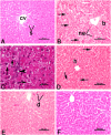
Fig 2. Photomicrographs of rat liver (H&E stain).
(A) Control liver showing normal hepatocytes arranged in cords, obvious sinusoids (s), and central vein (cv). (B, C) Hepatic injury induced by CCl4. Note hepatic cells with ballooning degeneration (b), focal necrotic cell death (ne), and diffuse fatty changes (arrows) (in B). Microvesicular steatosis (i.e., accumulation of small fat droplets) in hepatocyte cytosol (arrows), and inflammatory infiltrates (arrowhead) are evident in liver tissue (in C). (D) CCl4+TAU group showing an improvement of cellular structure and uniform sinusoidal arrays (compared to B). Pathological fatty deposition (arrows) and also normal centrilobular hepatocytes (h) with well-defined cell borders, dense cytoplasm and central nuclei are visible. (E) CCl4+SIL group with less severe liver injury. Focal hepatocellular degeneration (d) is observed. (F) CCl4+TAU+SIL group indicating no pathologic lesions.
Fig 3
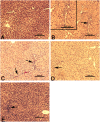
Fig 3. Photomicrographs of rat liver (Masson trichrome stain).
(A) Normal liver of control group. (B) CCl4 group with notable signs of portal fibrosis. The upper left inset depicts a higher magnification view. (C) CCl4+TAU and (D) CCl4+SIL, groups showing less extensive fibrotic areas. (E) CCl4+TAU+SIL group showing an obvious reduction in fibrosis (compared to C and D). Arrows point to fibrotic foci stained by trichrome staining (bluish color).
Fig 4
Fig 4. Electron micrographs of rat hepatocytes.
(A) Control hepatocytes depicting normal architecture. Note part of nucleus (N), rough endoplasmic reticulum (RER), mitochondria (M), primary lysosomes (Ly), glycogen granules (Gly), bile canaliculus (bc), and blood sinusoid (s). (B-D) CCl4 group showing severe hepatocyte damage. Observe nucleus (N) with increased heterochromatin patches, disruption of RER membranes, proliferated smooth endoplasmic reticulum (SER), mitochondria (M) with broken cristae, large lipid droplets (L), and glycogen loss (in B). Severe reduction of RER, large autophagic vesicles and lysosomes are seen (in C). Nucleus (N) with irregular nuclear envelope and dense clumped chromatin, scanty RER membranes, and progressive mitochondrial (M) swelling are also discernible (in D). (E) CCl4+TAU group demonstrating RER fragments (arrows), mitochondria (M) with obvious cristolysis, normal glycogen rosettes (Gly), nucleus (N), and few lipid droplets (L). (F) CCl4+SIL group showing nucleus with normal chromatin content, an increase in RER cisternae (arrows) compared to CCl4 group, and electron-dense mitochondria (M). (G) CCl4+TAU+SIL group illustrating numerous RER profiles, normal-looking mitochondria (M), nucleus (N), and small lipid droplets (L).

References

    1. Fujii T, Fuchs BC, Yamada S, Lauwers GY, Kulu Y, Goodwin JM, et al. Mouse model of carbon tetrachloride induced liver fibrosis: Histopathological changes and expression of CD133 and epidermal growth factor. BMC Gastroenterol. 2010; 10:79 10.1186/1471-230X-10-79 - DOI - PMC - PubMed
    1. Hamdy N, El-Demerdash E. New therapeutic aspect for carvedilol: Antifibrotic effects of carvedilol in chronic carbon tetrachloride-induced liver damage. Toxicol Appl Pharmacol. 2012; 261(3):292–299. 10.1016/j.taap.2012.04.012 - DOI - PubMed
    1. Abdelaziz DHA, Ali SA. The protective effect of Phoenix dactylifera L. seeds against CCl4-induced hepatotoxicity in rats. J Ethnopharmacol. 2014; 155(1):736–743. 10.1016/j.jep.2014.06.026 - DOI - PubMed
    1. Ranawat L, Bhatt J, Patel J. Hepatoprotective activity of ethanolic extracts of bark of Zanthoxylum armatum DC in CCl4 induced hepatic damage in rats. J Ethnopharmacol. 2010; 127(3):777–780. 10.1016/j.jep.2009.10.019 - DOI - PubMed
    1. Huo HZ, Wang B, Liang YK, Bao YY, Gu Y. Hepatoprotective and antioxidant effects of Licorice extract against CCl4-induced oxidative damage in rats. Int J Mol Sci. 2011; 12(10):6529–6543. 10.3390/ijms12106529 - DOI - PMC - PubMed

Publication types

MeSH terms