Review
. 2016 Feb;131(2):159-184.
doi: 10.1007/s00401-015-1511-3. Epub 2015 Dec 10.

Neurotropic virus infections as the cause of immediate and delayed neuropathology

Affiliations
Review

Neurotropic virus infections as the cause of immediate and delayed neuropathology

Martin Ludlow et al. Acta Neuropathol. 2016 Feb.

Abstract

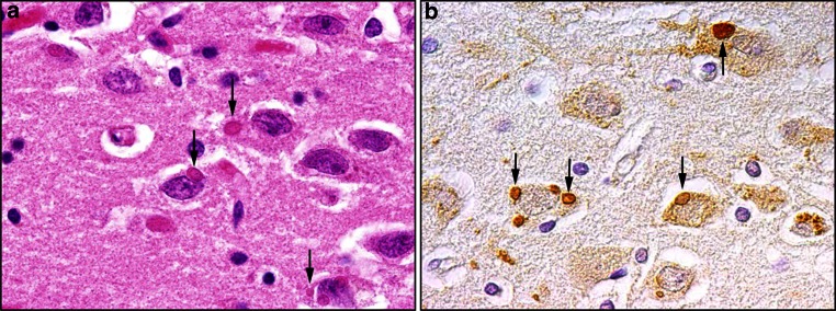
A wide range of viruses from different virus families in different geographical areas, may cause immediate or delayed neuropathological changes and neurological manifestations in humans and animals. Infection by neurotropic viruses as well as the resulting immune response can irreversibly disrupt the complex structural and functional architecture of the central nervous system, frequently leaving the patient or affected animal with a poor or fatal prognosis. Mechanisms that govern neuropathogenesis and immunopathogenesis of viral infections are highlighted, using examples of well-studied virus infections that are associated with these alterations in different populations throughout the world. A better understanding of the molecular, epidemiological and biological characteristics of these infections and in particular of mechanisms that underlie their clinical manifestations may be expected to provide tools for the development of more effective intervention strategies and treatment regimens.

Keywords: Alphavirus; Bornavirus; Bunyavirus; Central nervous system; Flavivirus; Herpesvirus; Influenza virus; Neuroinfectiology; Neuropathology; Paramyxovirus; Picornavirus; Rhabdovirus; Virus infection.


Figures

Fig. 1
Fig. 1
Routes of virus spread into the central nervous system. a Infection of peripheral nerves. (i) Virus spread from mucosal epithelium (ME) to sensory and autonomic neurons (SN) following infection of axon termini. Retrograde axonal transport results in virus spread to the spinal cord (SC). (ii) Virus infection of motor neurons (MN) at neuromuscular junctions in smooth muscle (SM) results in retrograde axonal transport to the spinal cord and the brain. b Blood–brain barrier (BBB). Virus-infected lymphocytes (green) (1) in blood vessels (BV) ‘roll’ along the endothelium (2), attach to the endothelial cells (3) and transverse the endothelial cell layer (EC) (4) and the glia limitans (GL). Virus spread to neurons (5) is assumed to occur following contacts with uninfected neurons. Alternatively, direct virus infection of endothelial cells may occur with subsequent spread into the brain parenchyma resulting in neuronal infection. c Infection of olfactory neurons. Virus present in the mucosa (M) of the upper respiratory tract can directly infect olfactory sensory neurons (OSN) present in olfactory epithelium (OE). Anterograde axonal transport leads to spread of virus within axonal bundles passing through the cribriform plate (CP) into the olfactory bulb (OB). Trans-synaptic spread to mitral cells (MC) results in virus spread along the olfactory tract (OT) to other brain regions. d Meningeal blood–cerebrospinal fluid (CSF) barrier. Virus-infected leukocytes in meningeal blood vessels present within the sub-arachnoid space between the pia and arachnoid roll, attach to the endothelium and transverse endothelial cells into the CSF. Direct infection of endothelial cells may also lead to virus spread into the CSF. e Blood–cerebrospinal fluid barrier. Virus-infected leukocytes or cell-free virus present within blood vessels of the choroid plexus (CHP) transverse the endothelium as described previously in b, d. This can lead to infection of epithelial cells and apical release of virus or spread of virus-infected leukocytes across the CHP epithelium into the CSF. Figure was composed using ©Motifolio.com Biomedical PowerPoint Toolkit Suite
Fig. 2
Fig. 2
Immunohistochemcial detection of the X-protein of novel Bornavirus (variegated squirrel Borna virus-1; VSBV-1) in human brain sections (a) and demonstration of Borna disease virus 1 (BoDV-1) X-protein in the hippocampus of a horse which had neurological signs of BoDV-1 infection (b). Viral X-protein is present in the nuclei, cytoplasm and cellular processes of infected neurons
Fig. 3
Fig. 3
Geographical distribution of selected flaviviruses capable of causing neurotropic infections in humans. a Cases of tick-borne encephalitis (demarcated within red-shaded zone) are distributed from central and eastern Europe across a wide band of Eurasia. b Distribution of cases of West Nile fever in the European region and Mediterranean basin in 2015 and previous seasons. c Gradient map showing cumulative cases of St. Louis encephalitis virus neuroinvasive disease in the USA from 1964 to 2010. d Cases of Japanese encephalitis virus (JEV) (red-shaded area) are diagnosed in many parts of South East Asia. a Adapted from [73]; b adapted from European Center for Disease Control and Prevention (ECDC) map of reported cases of West Nile fever, transmission season 2015 and previous transmission seasons; c adapted from Centers for Disease Control and Prevention (CDC) map; d adapted from CDC map of geographical distribution of Japanese encephalitis virus
Fig. 4
Fig. 4
Neuropathology in the spinal cord of a 3-year-old dog (Bernese mountain dog) infected with tick-borne encephalitis virus (TBE). a Hematoxylin and eosin-stained section showing severe lympho-histiocytic myelitis with perivascular predominantly lymphocytic cuffing (arrowheads). b Intralesional immunolabelling of TBE antigen (arrows), detected using a rabbit polyclonal antibody
Fig. 5
Fig. 5
Magnetic resonance imaging and microscopic images of typical herpes simplex virus (HSV) encephalitis pathology in humans. Magnetic resonance imaging (MRI) of an acute HSV encephalitis case. a Swelling of the left temporal lobe (Fluid attenuation inversion recovery, FLAIR). b Cortical cytotoxic edema typical for this neurotropic virus at the time of symptom onset (diffusion-weighted image, reduced apparent diffusion coefficient values not shown). c Parenchymal atrophy with residual gliosis 2 months later (FLAIR). Microscopic imaging of autopsy material of the basal ganglia (d, f) and the frontal lobe (e) of a patient with fulminant HSV encephalitis. d Immunohistochemically stained sections showing CD45-positive perivascular infiltrates. e Hematoxylin and eosin staining (H&E) showing additional intravascular thrombosis. f Several parenchymal hemorrhages in an H&E-stained brain section. ac Images courtesy of Dr. Peter Raab (Department of Neuroradiology, Hannover Medical School, Hannover, Germany); df Images courtesy of Prof. Christian Hartmann (Department of Neuropathology, Hannover Medical School, Hannover, Germany)
Fig. 6
Fig. 6
Central nervous system complications of influenza virus infections in humans. Detection of influenza virus antigen in neurons in the olfactory bulb of an H3N2 virus-infected immunocompromised child. a Hematoxylin and eosin-stained tissue section showing a lack of cellular infiltrates. b Immunohistochemcial detection of influenza virus antigen in neurons using an anti-nucleoprotein monoclonal antibody
Fig. 7
Fig. 7
Morbillivirus infection of the CNS. Immunocytochemical detection of measles virus (MV) antigen using anti-nucleoprotein monoclonal antibody (a) or SSPE serum (b, c) in human brain sections from subacute sclerosing panencephalitis (SSPE) patients. d, e Hematoxylin and eosin-stained brain tissue sections from canine distemper virus (CDV)-infected dogs. gi Immunohistochemical detection of CDV-infected cells in dog brain tissue sections. a MV antigen (green) is present in neurons and associated processes in the occipital lobe of an SSPE case. b MV antigen (green) is restricted to oligodendrocytes and is not present in GFAP-positive astrocytes (red) in SSPE white matter. c MV-positive oligodendrocytes in close proximity to CD68-positive macrophages and GFAP-positive astrocytes (blue) in white matter from an SSPE case. d Cerebrum of a dog with CDV-induced polioencephalitis with perivascular lymphocytic cuffs and diffuse gliosis. e Acute leukoencephalitis with severe demyelination in the medulla of a CDV-infected dog. f Chronic leukoencephalitis with severe demyelination (arrows) and perivascular lymphocytic cuffs is evident in the cerebellum of a CDV-infected dog. g Cerebrum of the same dog shown in panel d with CDV antigen evident in neurons and neuronal processes. h Immunolabelling of CDV antigen in astrocytes and gitter cells in the medulla of the same dog shown in panel e. i Cerebellum of the same dog shown in panel f with CDV antigen present in astrocytes and gitter cells. ac Images courtesy of Dr. Stephen McQuaid (Belfast Health and Social Care Trust, Northern Ireland)
Fig. 8
Fig. 8
Measles virus (MV) spread in the CNS. a Schematic diagrams illustrating the trans-synaptic spread of MV. (i) Neurons are initially infected by MV via an uncharacterized route of infection. Following virus entry MV is able to spread to connected uninfected neurons. (ii) Viral ribonucleoprotein (RNP) complexes consisting of viral genome encapsidated by nucleoprotein with associated phosphoprotein and polymerase are transported together with the fusion (F) and hemagluttinin (H) glycoproteins along axons to the synaptic termini. Axonal transport of viral RNPs is known to occur via both anterograde (AG) or retrograde (RG) axonal transport. (iii) Aggregation of viral RNPs and viral glycoproteins at the pre-synaptic terminal. (iv) A number of questions relating to the mechanism and consequences of MV spread in the CNS remain to be addressed. (bd) MV transneuronal spread in human brain (b) and animal models (c, d). b Interconnected MV-infected neurons are present in brain sections from an SSPE patient (b), a rMV-infected Ifnarko-CD46Ge mouse (c). d A number of extended neuronal processes (arrows) connect infected neurons in the polymorph cell layer of the hippocampus of a C57/BL/6 mouse infected with the rodent-adapted MVCAM/RB strain. a Plate was composed using ©Motifolio.com Biomedical PowerPoint Toolkit Suite; b image courtesy of Dr. Stephen McQuaid (Belfast Health and Social Care Trust, Northern Ireland); c adapted from [76]; d adapted from [75]
Fig. 9
Fig. 9
Characterisation of fatal rabies encephalitis in the hippocampus of a 46-year-old human who received a lung transplant from a rabies virus-infected donor. a Hematoxylin and eosin staining showing eosinophilic cytoplasmic inclusion bodies (Negri bodies) in the perikaryon of neurons (arrows). b Immunohistochemical labeling using polyclonal goat anti-rabies antisera of rabies viral antigen in the perikaryon of neurons (arrows)

Similar articles

Cited by

References

    1. Abdel-Wahab KS, El Baz LM, El-Tayeb EM, Omar H, Ossman MA, Yasin W. Rift Valley Fever virus infections in Egypt: pathological and virological findings in man. Trans R Soc Trop Med Hyg. 1978;72:392–396. doi: 10.1016/0035-9203(78)90134-7. - DOI - PubMed
    1. Abul-Kasim K, Palm L, Maly P, Sundgren PC. The neuroanatomic localization of Epstein–Barr virus encephalitis may be a predictive factor for its clinical outcome: a case report and review of 100 cases in 28 reports. J Child Neurol. 2009;24:720–726. doi: 10.1177/0883073808327842. - DOI - PubMed
    1. Agamanolis DP, Tan JS, Parker DL. Immunosuppressive measles encephalitis in a patient with a renal transplant. Arch Neurol. 1979;36:686–690. doi: 10.1001/archneur.1979.00500470056011. - DOI - PubMed
    1. Agut H, Bonnafous P, Gautheret-Dejean A. Laboratory and clinical aspects of human herpesvirus 6 infections. Clin Microbiol Rev. 2015;28:313–335. doi: 10.1128/CMR.00122-14. - DOI - PMC - PubMed
    1. Alker A. West Nile virus-associated acute flaccid paralysis. BMJ Case Rep. 2015 - PMC - PubMed

Publication types

MeSH terms