. 2016 Mar 3;127(9):e1-e11.
doi: 10.1182/blood-2015-06-649434. Epub 2015 Dec 10.

Circular RNA enrichment in platelets is a signature of transcriptome degradation

Affiliations

Circular RNA enrichment in platelets is a signature of transcriptome degradation

Abd A Alhasan et al. Blood. .

Abstract

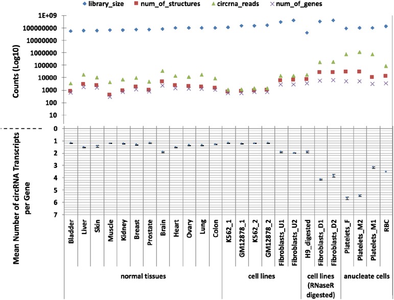
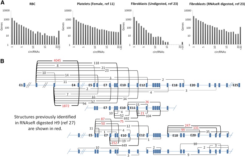
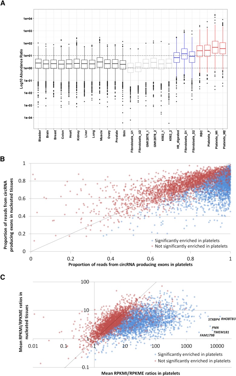
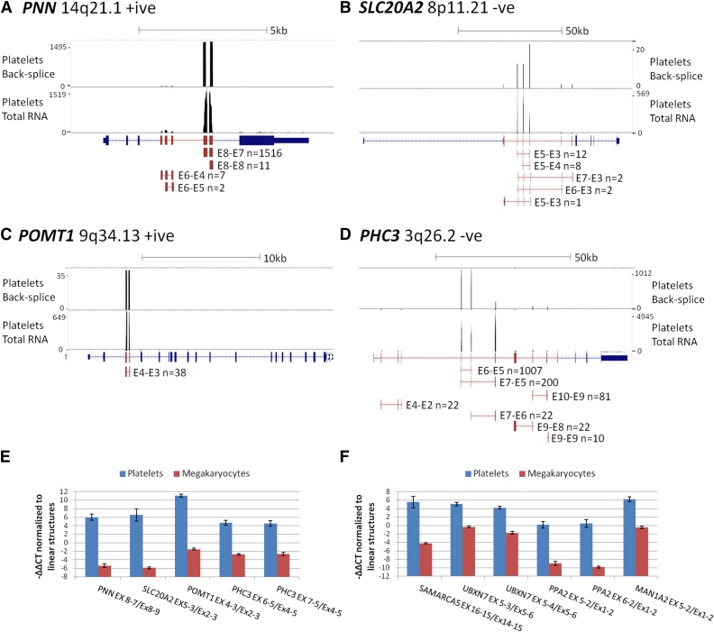
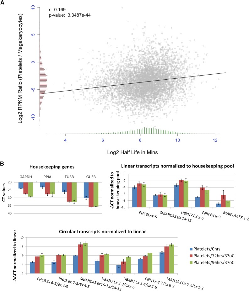
In platelets, splicing and translation occur in the absence of a nucleus. However, the integrity and stability of mRNAs derived from megakaryocyte progenitor cells remain poorly quantified on a transcriptome-wide level. As circular RNAs (circRNAs) are resistant to degradation by exonucleases, their abundance relative to linear RNAs can be used as a surrogate marker for mRNA stability in the absence of transcription. Here we show that circRNAs are enriched in human platelets 17- to 188-fold relative to nucleated tissues and 14- to 26-fold relative to samples digested with RNAse R to selectively remove linear RNA. We compare RNAseq read depths inside and outside circRNAs to provide in silico evidence of transcript circularity, show that exons within circRNAs are enriched on average 12.7 times in platelets relative to nucleated tissues and identify 3162 genes significantly enriched for circRNAs, including some where all RNAseq reads appear to be derived from circular molecules. We also confirm that this is a feature of other anucleate cells through transcriptome sequencing of mature erythrocytes, demonstrate that circRNAs are not enriched in cultured megakaryocytes, and demonstrate that linear RNAs decay more rapidly than circRNAs in platelet preparations. Collectively, these results suggest that circulating platelets have lost >90% of their progenitor mRNAs and that translation in platelets occurs against the backdrop of a highly degraded transcriptome. Finally, we find that transcripts previously classified as products of reverse transcriptase template switching are both enriched in platelets and resistant to decay, countering the recent suggestion that up to 50% of rearranged RNAs are artifacts.


Figures

Figure 1
Figure 1
CircRNAs in human tissues. The number of circRNAs (structure counts), junction reads (back-splice read counts), circRNA producing genes (gene counts), mean number of circRNA transcripts per circRNA producing genes, and RNAseq library sizes (read counts) are shown. Samples are presented as 4 groups: nucleated tissues, nucleated cell lines, nucleated cell lines digested with RNAse R, and anucleated cell types. The number of back-splice reads is significantly higher in anucleate samples (platelets and RBCs) than all others (P = 2.7 × 10⁴, Wilcoxon rank sum test; figures adjusted for library size). All data were processed using PTESFinder, and all were publicly available with the exception of the data from RBCs (see “Materials and methods” and supplemental Table S1 for details of datasets, and supplemental Table S3 for details of structures).
Figure 2
Figure 2
circRNA structures per gene. (A) Histograms showing circRNA numbers per gene established using PTESFinder for RBCs (this study), platelets, fibroblasts, and fibroblasts digested with RNAse R. (B) Schematic diagram showing XPO1 intron/exon organization with read counts and inferred structure of all circRNAs identified within the 3 platelet samples. Inferred structures assume that all internal Refseq exons are present within the structure defined by each back-splice. Read counts for the 11 structures identified previously within RNAse R digested H9 ES cells are highlighted in red.
Figure 3
Figure 3
Confirmation of circRNA abundance and resistance to RNAse R. (A) Schema of qPCR assays using an E5-E2 circRNA as an example. All assays use a common reporter probe and use either an exon downstream of the circRNA to assay linear expression (probe in donor exon) or an exon upstream (probe in acceptor exon). (B) (Left) Expression levels (−∆CT values) of linear (Ex1-2) and circular (Ex5-2 and Ex4-2) MAN1A2 transcripts relative to housekeeping pool. (Right) Expression levels of circRNAs relative to linear RNAs from the same loci normalized to housekeeping pool (−∆∆CT values). (C) Expression of 9 circRNAs relative to linear forms from the same loci, normalized to housekeeping pool (−∆∆CT values). (D) Change in CT values of circRNAs relative to linear forms from the same loci in RNAse R digested RNAs, normalized to mock digested RNAs (−∆∆CT values). Templates, circRNAs, and linear forms assayed are indicated.
Figure 4
Figure 4
Differential read depth defines genes with significant circRNA enrichment in platelets. (A) Box and whisker plots showing the ratio of RPKM from circRNA producing exons (RPKMI) to RPKM from exons that do not produce circRNAs (RPKME) for all genes in each sample. The median and upper and lower quartiles are shown, with outliers as solid circles. (B) The proportion of reads from circRNA producing exons averaged across all nucleated samples (y-axis) and platelets (x-axis). (C) Fold enrichment of reads from circRNA producing exons in platelets relative to nucleated tissues. All genes with an average RPKMI >1 in platelets and expressed in 8 or more nucleated tissues are shown. Blue, genes significantly enriched in platelets; red, genes not significantly enriched in platelets. The data points corresponding to the 5 most enriched genes are indicated. The slope x = y is shown as a dashed line.
Figure 5
Figure 5
circRNA enrichment occurs in platelets but not megakaryocytes. (A-D) Adapted University of California Santa Cruz screen shots for 4 genes with extreme circRNA enrichment in platelets. Each panel shows RNAseq read abundance in a single platelet total RNA sample (F1), together with the position and abundance of back-splice exon junctions in the same sample. The intron-exon structure is shown below, with exons known to contribute to circRNAs shown in red. CircRNA structures present in the sample together with back-splice frequencies are also shown (for PHC3, only structures with ≥10 back-splice reads are shown). Above each panel, the gene names, cytogenetic locations, coding strand, and scale in kilobases are indicated. (E-F) Expression levels of circRNAs relative to linear RNAs from the same loci in platelets and cultured megakaryocytes, normalized to the housekeeping pool (−∆∆CT values).
Figure 6
Figure 6
Degradation of platelet RNA. (A) Correlation of the change in gene expression between megakaryocte polyA+ RNA and platelet polyA+ RNA (y-axis), and mRNA half-life estimates (x-axis). Distributions of both are shown as histograms on each axis. (B) qPCR analysis of differential decay of linear and circRNAs in platelets following incubation at 37°C for 0, 72, and 96 hours. Data from 3 biological replicates are shown. (Top left) Expression levels of housekeeping genes (CT values). (Top right) Expression levels of linear structures from 5 circRNA-producing genes relative to the housekeeping pool (−∆CT values). (Bottom) Expression levels of 7 circRNAs relative to linear transcripts from the same loci, both normalized to the housekeeping pool (−∆∆CT values).

Comment in

References

    1. Deutsch VR, Tomer A. Megakaryocyte development and platelet production. Br J Haematol. 2006;134(5):453–466. - PubMed
    1. Garraud O, Cognasse F. Are Platelets Cells? And if Yes, are They Immune Cells? Front Immunol. 2015;6:70. - PMC - PubMed
    1. Migliaccio AR. Erythroblast enucleation. Haematologica. 2010;95(12):1985–1988. - PMC - PubMed
    1. Harker LA, Roskos LK, Marzec UM, et al. Effects of megakaryocyte growth and development factor on platelet production, platelet life span, and platelet function in healthy human volunteers. Blood. 2000;95(8):2514–2522. - PubMed
    1. Weyrich AS, Schwertz H, Kraiss LW, Zimmerman GA. Protein synthesis by platelets: historical and new perspectives. J Thromb Haemost. 2009;7(2):241–246. - PMC - PubMed

Publication types