. 2017 Feb;43(1):137-143.
doi: 10.1007/s00068-015-0606-9. Epub 2015 Dec 11.

Treatment of tibia avulsion fracture of posterior cruciate ligament with high-strength suture fixation under arthroscopy

Affiliations

Treatment of tibia avulsion fracture of posterior cruciate ligament with high-strength suture fixation under arthroscopy

W Zhu et al. Eur J Trauma Emerg Surg. 2017 Feb.

Abstract

Aim: To evaluate the outcome of arthroscopy treatment using high-strength line in the treatment of tibial avulsion fracture of posterior cruciate ligament.

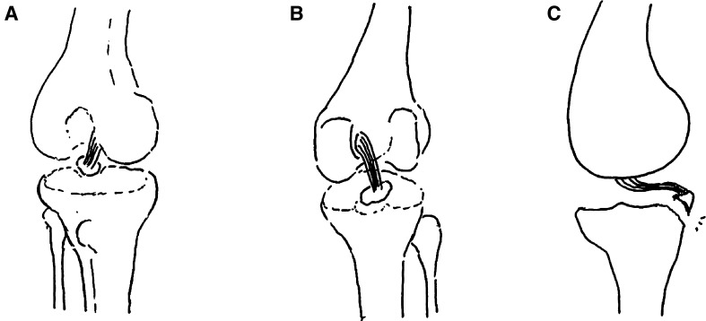
Methods: Both the avulsed bone block and the tibia bone bed were refreshed. The procedure was completed with the assistance of PCL director drill guide. The reduction and fixation using high-strength line were used to fix the avulsed bone by from posterior middle portal. Rehabilitation began early postoperatively.

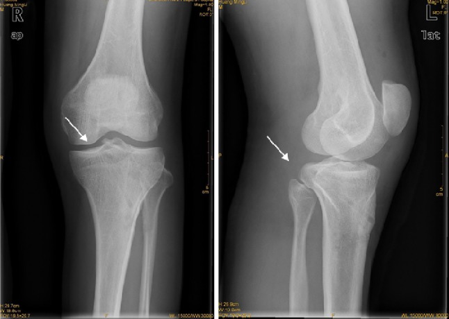
Results: From January 2010 to June 2012, a total of 18 arthroscopically treated cases of PCL tibial avulsion fracture were retrospectively evaluated. Reduction of the avulsion fragment was obtained in all cases. 16 cases were followed up for 7-30 months (average 13.6), and 2 cases were out of follow-up. In the 16 followed patients, flexion and extension were back to normal within 6 weeks, and return to normal walk in 12 weeks. The bone healing was good without any vascular or nerve complications. All the patients regained the preinjury activity level. The mean score (and standard deviation) increased from 38.9 ± 4.9 points to 95.2 ± 3.8 points with the system of Lysholm, from 57.1 ± 10.3 points to 94.3 ± 4.4 points with the system of IKDC. Post-test displacement of KT3000 declined from 3.6 ± 0.39 to 1.1 ± 0.27 mm.

Conclusion: Arthroscopic vertical fixation by high-strength line is a simple, safe, reliable, and micro-invasive treatment to PCL tibial avulsion fracture. It is a kind of real all arthroscopic technique, and good for early postoperative rehabilitation. The total stability of the knee could be gained, and the second operation to remove the internal fixation is avoided.

Keywords: Arthroscopy; Avulsion fracture; High-strength line; Posterior cruciate ligament.


Conflict of interest statement

Compliance with ethical standards Each subject had signed the informed consent before participating in our study. This study was approved by the ethics committee of The First Affiliated Hospital of Shenzhen University and was conducted in conformity with the guidelines outlined in the Declaration of Helsinki statement. Conflict of interest Weimin Zhu, Wei Lu, Jiaming Cui, Liangquan Peng, kan OuYang, Hao Li, Haifeng Liu, Wei You, Daping Wang, and Yanjun Zeng declare that they have no conflict of interest.

Figures

Fig. 1
Fig. 1
Preoperative X-ray and MRI examinations. Preoperative X-ray and MRI examinations indicate the avulsion fracture at the distal insertion of the posterior cruciate ligament (represented by the arrow)
Fig. 2
Fig. 2
Preoperative schematic drawings. Preoperative schematic drawings indicate the avulsion fracture at the distal insertion of the posterior cruciate ligament. a Anteroposterior position schematic drawing. b Posteroanterior position schematic drawing. c Lateral schematic drawing
Fig. 3
Fig. 3
Operative schematic drawings. Operative schematic drawings indicate that the suture passer for rotator cuff suture was used to place high-strength suture around the distal insertion of the posterior cruciate ligament and knotted. a Retrieve the PDS suture from the suture passer with a suture retriever. b Place a high-strength suture around the loop of the PDS suture. c Retrieve the PDS suture with the high-strength suture and make the high-strength suture around the distal insertion of the posterior cruciate ligament. Retrieve the other end of the high-strength suture through the same portal. d, e, Use the knot pusher to knot at the distal insertion of the posterior cruciate ligament
Fig. 4
Fig. 4
Operative schematic drawings. Operative schematic drawings indicate the high-strength suture around the distal insertion of the posterior cruciate ligament passing through the bone tunnels. a The distal insertion of the posterior cruciate ligament with a high-strength suture knot on it. b A Kirschner wire with the diameter of 2.0 mm was drilled from the lateral area of the tubercles of tibia to the inferolateral area of the fragment. The other Kirschner wire with the diameter of 2.0 mm was drilled from the medial area of the tubercles of tibia to the inferomedial area of the fragment. c, d The PDS lines inserted through two lumbar puncture needles were extracted from the two bone tunnel to the front of the tubercles of tibia. e The high-strength suture was tightened and tied at the front of the tubercles of tibia (anteroposterior position schematic drawing). f The high-strength suture was tightened and tied at the front of the tubercles of tibia (posteroanterior position schematic drawing)
Fig. 5
Fig. 5
Preoperative observation under arthroscope. The avulsion at the distal insertion of the posterior cruciate ligament under the arthroscope (Fig. 5 shows the anterior interior approach and Fig. 5 the posterior exterior approach)
Fig. 6
Fig. 6
Preoperative and postoperative observation under arthroscope. The preoperative and postoperative status under the arthroscope (Fig. 6 shows the preoperative status and Fig. 6 the postoperative status)
Fig. 7
Fig. 7
Postoperative X-ray. Postoperative X-ray shows good avulsion fracture reduction at the distal insertion of the posterior cruciate ligament (represented by the arrow)
Fig. 8
Fig. 8
Postoperative three-dimensional reconstructed CT scan. Postoperative three-dimensional reconstructed CT scan shows good fixation and reduction at the distal insertion of the posterior cruciate ligament (represented by the arrow)

References

    1. Frosch K, Proksch N, Preiss A, et al. Treatment of bony avulsions of the posterior cruciate ligament (PCL) by a minimally invasive dorsal approach. Oper Orthop Traumatol. 2012;24(4–5):348–353. doi: 10.1007/s00064-012-0208-1. - DOI - PubMed
    1. Zhang X, Cai G, Xu J. Wang K.A minimally invasive postero-medial approach with suture anchors for isolated tibial avulsion fracture of the posterior cruciate ligament. Knee. 2013;20(2):96–99. doi: 10.1016/j.knee.2012.10.016. - DOI - PubMed
    1. Chen CW, Chen L, Pan ZE, Yang SW. Open reduction and internal fixation via a posterior approach for posterior fractures of tibial plateau. Zhongguo Gu Shang. 2012;25(7):561–565. - PubMed
    1. Lyshom J, Gillquist J. Evaluation of knee ligament surgery results with special emphasis onuse of a scoring scale [J] Am J Sports Med. 1982;10:150–154. doi: 10.1177/036354658201000306. - DOI - PubMed
    1. Andri GT, Klineberg EO, Wahl CJ, Mills WJ. Treatment of posterior cruciate ligament tibial avulsion fractures through a modified open posterior approach: operative technique and 12- to 48-month outcomes. J Orthop Trauma. 2008;22:317–324. doi: 10.1097/BOT.0b013e31817279d1. - DOI - PubMed

LinkOut - more resources