. 2016 Mar;48(3):859-872.
doi: 10.1007/s00726-015-2134-7. Epub 2015 Dec 10.

Co-regulation of mitochondrial respiration by proline dehydrogenase/oxidase and succinate

Affiliations

Co-regulation of mitochondrial respiration by proline dehydrogenase/oxidase and succinate

Chad N Hancock et al. Amino Acids. 2016 Mar.

Erratum in

Abstract

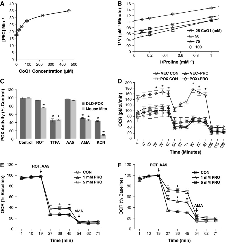
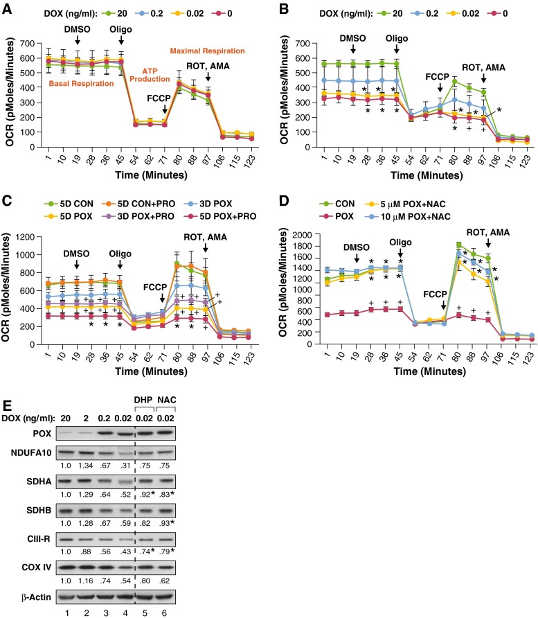
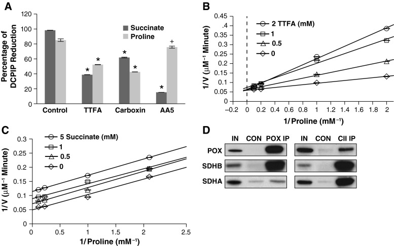
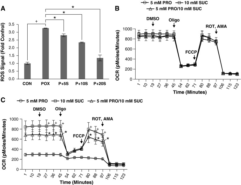
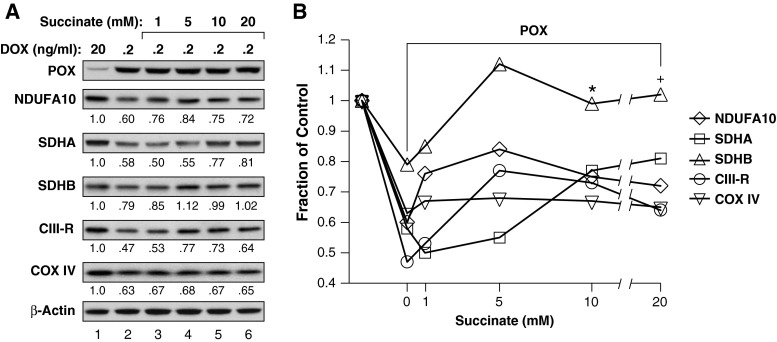
Proline dehydrogenase/oxidase (PRODH/POX) is a mitochondrial protein critical to multiple stress pathways. Because of the roles of PRODH/POX in signaling, and its shared localization to the mitochondrial inner membrane with the electron transport chain (ETC), we investigated whether there was a direct relationship between PRODH/POX and regulation of the ETC. We found that PRODH/POX binds directly to CoQ1 and that CoQ1-dependent PRODH/POX activity required functional Complex III and Complex IV. PRODH/POX supported respiration in living cells during nutrient stress; however, expression of PRODH/POX resulted in an overall decrease in respiratory fitness. Effects on respiratory fitness were inhibited by DHP and NAC, indicating that these effects were mediated by PRODH/POX-dependent reactive oxygen species (ROS) generation. PRODH/POX expression resulted in a dose-dependent down-regulation of Complexes I-IV of the ETC, and this effect was also mitigated by the addition of DHP and NAC. We found that succinate was an uncompetitive inhibitor of PRODH/POX activity, inhibited ROS generation by PRODH/POX, and alleviated PRODH/POX effects on respiratory fitness. The findings demonstrate novel cross-talk between proline and succinate respiration in vivo and provide mechanistic insights into observations from previous animal studies. Our results suggest a potential regulatory loop between PRODH/POX and succinate in regulation of mitochondrial respiration.

Keywords: Energetics; Reactive oxygen species; Redox; Respiration; Signaling.


Figures

Fig. 1
Fig. 1
PRODH/POX supports respiration independent of Complex I and II activity. a POX activity assay showing the increase in POX activity in the presence of increasing CoQ1. DLD-POX cells were grown in 0.2 ng/ml DOX to allow POX expression. Homogenized cell lysate containing 100 μg protein was incubated in assay buffer with 0, 25, 50, 100, 250, and 500 μM of CoQ1 at 37 °C for 20 min. Absorbance at 440 nm was measured to assess the amount of OAB–P5C complex formed. A P5C standard curve was used to calculate the P5C concentration. Data shown represent mean ± SEM. Compared to CoQ1 = 0, all values are significant to p < 0.001. b Double-reciprocal analysis of CoQ1-dependent POX activity. DLD-POX lysate with 100 µg protein was incubated with 1, 2, 5, or 10 mM of proline and 25, 50, 75, or 100 µM of CoQ1 in assay buffer at 37 °C for 20 min. Absorbance at 440 nm was used to measure the amount of OAB–P5Ccomplex formed and a P5C standard curve used to calculate the P5C concentration. c PRODH/POX activity in DLD-POX lysates and mouse mitochondria show similar dependence on ETC function. PRODH/POX-containing lysates (200 µg protein) or mouse mitochondria (50 µg protein) were incubated in assay buffer at 37 °C for 30 min. Sensitivity of PRODH/POX activity to ETC function was measured by the addition of 50 µM ROT, 2 mM TTFA, 5 µM of AA5, 100 µM of AMA, and 1 mM KCN. Data shown represent mean ± SEM. *p < 0.01 compared to its respective control. d PRODH/POX expression is required for proline to support respiration during acute nutrient stress. DLD-VEC control cells (VEC) and DLD-POX cells (POX) were grown in 0.2 ng/ml DOX for 48 h. Cells were incubated in glucose-free media alone (CON) or with the addition of 5 mM proline (+PRO) for 1 h prior to Seahorse XF24 analysis. OCR was measured and a respiratory profile was established by the addition of 2.5 µM DMSO, OLIGO, FCCP, and ROT/AMA at the indicated time points. *p < 0.01 compared to vector control. e Seahorse XF24 oxygen consumption analysis of DLD-POX control cells. Cells were grown in 20 ng/ml DOX to suppress PRODH/POX expression. 1 h prior to analysis, cells were incubated in assay media containing 5 mM glucose and either 0 (CON), 1, or 5 mM proline (PRO). OCR was measured at the indicated timepoints. ROT and AA5 were added at 2.5 uM to inhibit Complex I and Complex II activity, respectively. AMA was added at 2.5 μM to inhibit Complex III. Compared to control, *p < 0.01; + p < 0.02. f Experimental details as described for (e), except in this case, DLD-POX cells were grown in 0.2 ng/ml DOX for 48 h prior to assay. Compared to control, + p < 0.001; *p < 0.01. The following abbreviations apply: AA5 atpenin A5, AMA antimycin A, CoQ1 coenzyme Q1, DMSO dimethyl sulfoxide, DOX doxycycline, FCCP carbonyl cyanide p-trifluoromethoxyphenylhydrazone, KCN potassium cyanide, OAB 2-aminobenzaldehyde, OCR oxygen consumption rate, OLIGO oligomycin, PRODH/POX proline oxidase, ROT rotenone, TTFA 2-thenoyltrifluoroacetone
Fig. 2
Fig. 2
Extended PRODH/POX ROS production decreases total respiratory fitness. Data for each time point represents mean ± SEM (n = 3) for all panels. a DOX concentration does not affect respiration in DLD-VEC control cells. Cells grown for 48 h in the indicated concentration of DOX. Oxygen consumption rate (OCR) was measured and a cellular respiratory profile was established by the addition of 2.5 µM DMSO, OLIGO, FCCP, and ROT/AMA at the indicated timepoints. b Increasing PRODH/POX expression correlates with lower cellular respiration. DLD-POX cells were grown in the indicated amount of DOX and oxygen consumption rate was analyzed as in (a). Values with DOX = 0.02 and 0.0 were compared to DOX = 20; *p ≤ 0.01; + p < 0.02. c Prolonged PRODH/POX expression and addition of proline correlates with greater decreases in respiration. DLD-POX cells were grown in 0.2 ng/ml DOX to allow PRODH/POX expression for 3 days (3D POX) or 5 days (5D POX) alone or in media supplemented with 5 mM proline (3D POX + PRO and 5D POX + PRO). Respiration by these cells was compared to DLD-POX cells that had been grown for 5 days in 20 ng/ml DOX to suppress PRODH/POX expression, alone or in combination with 5 mM proline (5D CON and 5D CON + PRO, respectively). Oxygen consumption rate was measured and a respiratory profile established as described in (a). Values for 3D POX + PRO, 5D POX, and 5D POX + PRO were compared to 5D CON. + p ≤ 0.05; *p < 0.01. d Inhibition of PRODH/POX-mediated ROS decreases effects on respiration. DLD-POX cells were grown for 48 h in either 20 ng/ml DOX (CON), or 0.2 ng/ml DOX alone (POX) or in combination with 5 or 10 mM  of N-acetyl-l-cysteine (NAC). Oxygen consumption rate was measured and a respiratory profile established as described in (a). Compared to control, *p < 0.001; Compared to POX, + p < 0.01. e PRODH/POX expression down-regulated ETC component proteins. DLD-POX cells were grown in the indicated ng/ml concentration of doxycycline (DOX) for 48 h, alone or in combination with 10 mM DHP or NAC. Whole cell lysates were harvested, and protein expression of subunits of Complex I (NDUFA10), Complex II (SDHA and SDHB), Complex III (CIII-R, Reiske Fe-S subunit), and Complex IV (COX IV) were analyzed by Western blotting. β-Actin was used as a protein loading control. The band intensities shown below each panel were quantified using Image Studio, normalized to β-actin control, and expressed as the level relative to untreated control (lane 1 20 ng/ml DOX). Values for DHP and NAC were compared to the mean ± STD of the ratios for 0.2 ng/mL DOX (lane 3 e and lane 2, Fig. 5a) combined with those for 0.02 ng/ml DOX (lane 4 e) Although the DOX concentration for DHP and NAC treatment was 0.02 ng/mL DOX, the values at 0.02 ng/ml DOX were consistently lower than those at 0.2 ng/ml DOX. Thus, the values used represent a higher distribution for the PRODH/POX-mediated effect on ETC proteins. DHP and NAC increased the values of the ETC proteins, i.e., decreased the effect of PRODH/POX. *Value greater than 2 standard deviations of aforementioned distribution, denoting 95 % confidence limits
Fig. 3
Fig. 3
PRODH/POX activity is regulated by succinate and SDH inhibitors and co-localizes with SDH on the mitochondrial inner membrane. a PRODH/POX and SDH share similar sensitivity to SDH inhibitors except AA5. Mouse mitochondria (440 µg protein) were incubated in reaction buffer containing either 10 mM of succinate or proline alone (Control) or in the presence of 1 mM TTFA, 1 mM Carboxin, or 5 µM AA5 at 37 °C for 10 min. Absorbance was measured at 600 nm and results calculated as a percent of unreacted control dye. Data shown represent mean ± SEM (n = 3) of comparisons against their respective controls. *p < 0.001; + p < 0.05. b Double-reciprocal analysis of TTFA-dependent inhibition of PRODH/POX activity. DLD-POX lysate containing 100 µg protein was incubated with 1, 2, 5, or 10 mM of proline and 0, 0.5, 1 and 2 mM of TTFA in assay buffer at 37 °C for 20 min. Absorbance at 440 nm was used to measure the amount of OAB–P5C complex formed and a P5C standard curve used to calculate the P5C concentration. c Double-reciprocal analysis of succinate-dependent inhibition of PRODH/POX activity. DLD-POX lysate containing 100 µg protein was incubated with 1, 2, 5, or 10 mM of proline and 0, 0.5, 1, and 5 mM of succinate in assay buffer at 37 °C for 20 min. Absorbance at 440 nm was used to measure the amount of OAB–P5Ccomplex formed and a P5C standard curve used to calculate the P5C concentration. d Co-immunoprecipitation of PRODH/POX and Complex II. 4 mg of mouse mitochondria was cross-linked with DSP, then solubilized and incubated with either PRODH/POX (POX IP) or Complex II antibody (CII IP). Control samples were incubated with an equal concentration of a non-specific mouse IgG (CON). Samples were incubated with beads coated with anti-mouse IgG overnight. Cross-linker was cleaved and proteins solubilized with SDS-PAGE buffer. Control and IP lysates were immunoblotted versus a 5 % input control (IN) using PRODH/POX, SDHA, and SDHB antibodies. Data is representative of three separate experiments
Fig. 4
Fig. 4
Succinate inhibits PRODH/POX ROS production and effects on respiration. a Succinate inhibits ROS production by PRODH/POX-expressing cells. DLD-POX cells were grown for 48 h in 0.2 ng/ml DOX to allow for PRODH/POX expression alone (POX+) or in media supplemented with 5, 10, or 20 mM succinate (P + 5S, P + 10S, and P + 20S). Control wells contained DLD-POX cells grown in 20 ng/ml to suppress PRODH/POX expression (CON). Cells were treated with DCFDA in DPBS for 20 min and fluorescence measured at 488 absorption/530 emission. Signals were normalized by BCA quantitation of protein levels in each well. Comparisons under brackets, *p < 0.01. b Succinate does not affect respiration in DLD-VEC control cells. Cells grown for 48 h in 0.2 ng/ml of DOX in media supplemented with 5 mM proline (PRO), 10 mM succinate (SUC), or both (PRO/SUC). OCR was measured and a respiratory profile was established by the addition of 2.5 µM DMSO, OLIGO, FCCP, and ROT/AMA at the indicated timepoints. c Succinate inhibits the decrease in respiration induced by PRODH/POX and proline. DLD-POX cells were grown for 48 h in 0.2 ng/ml of DOX to allow PRODH/POX expression. Oxygen consumption rate (OCR) was measured and a respiratory profile was established as described for (b). Comparison of 10 mM succinate to 5 mM PRO, + p < 0.01. Comparison of 5 mM POX to 5 MM POX + 10 mM SUC, *p < 0.05. Data represents mean ± standard error of the mean (n = 3) for all panels
Fig. 5
Fig. 5
Succinate inhibits the effects of PRODH/POX on ETC component proteins. DLD-POX cells were grown in the indicated ng/ml concentration of DOX for 48 h, alone or in combination with increasing concentrations of succinate as suggested by the effects of succinate on ROS (Fig. 4a). a Whole cell lysates were harvested, and protein expression of PRODH/POX and of subunits of Complex I (NDUFA10), Complex II (SDHA, SDHB), Complex III (CIII-R, Reiske Fe-S subunit), and Complex IV (COX IV) was analyzed by Western blotting. The band intensities shown below each panel were quantified using Image Studio, normalized to β-actin control, and expressed as the level relative to untreated control (lane 1 20 ng/ml DOX). b Since succinate decreased PRODH/POX-mediated ROS in a dose-dependent manner (Fig. 4a) and the levels of proteins in a suggested a succinate concentration effect, we constructed a combination plot of protein levels as the fraction of control against succinate concentrations (see text). For the value with treatment by POX (0.2 ng/ml DOX) without succinate, we combined the values from a with those obtained in Fig. 2e. We then compared the values statistically. The value with 10 mM succinate was different from that without succinate, *p = 0.020. At 20 mM succinate, the difference was borderline significant, + p = 0.055

References

    1. Abrahamson JL, Baker LG, Stephenson JT, Wood JM. Proline dehydrogenase from Escherichia coli K12. Properties of the membrane-associated enzyme. Eur J Biochem. 1983;134(1):77–82. doi: 10.1111/j.1432-1033.1983.tb07533.x. - DOI - PubMed
    1. Adachi H, Fujiwara Y, Ishii N. Effects of oxygen on protein carbonyl and aging in Caenorhabditis elegans mutants with long (age-1) and short (mev-1) life spans. J Gerontol A Biol Sci Med Sci. 1998;53(4):B240–B244. doi: 10.1093/gerona/53A.4.B240. - DOI - PubMed
    1. Alvord W (2014) Introduction to sla pdf. http://cran.r-project.org/web/packages/sla/vignettes/introduction-to-sla...
    1. Astuti D, Latif F, Dallol A, Dahia PL, Douglas F, George E, Skoldberg F, Husebye ES, Eng C, Maher ER. Gene mutations in the succinate dehydrogenase subunit SDHB cause susceptibility to familial pheochromocytoma and to familial paraganglioma. Am J Hum Genet. 2001;69(1):49–54. doi: 10.1086/321282. - DOI - PMC - PubMed
    1. N AWaC (2014) sla: two-group striaght line ANCOVA. R package version 0.1. http://CRAN.R-project.org/package=sla

Publication types

LinkOut - more resources