. 2015 Dec 14;10(12):e0144891.
doi: 10.1371/journal.pone.0144891. eCollection 2015.

Characterizing the Human Cone Photoreceptor Mosaic via Dynamic Photopigment Densitometry

Affiliations

Characterizing the Human Cone Photoreceptor Mosaic via Dynamic Photopigment Densitometry

Ramkumar Sabesan et al. PLoS One. .

Abstract

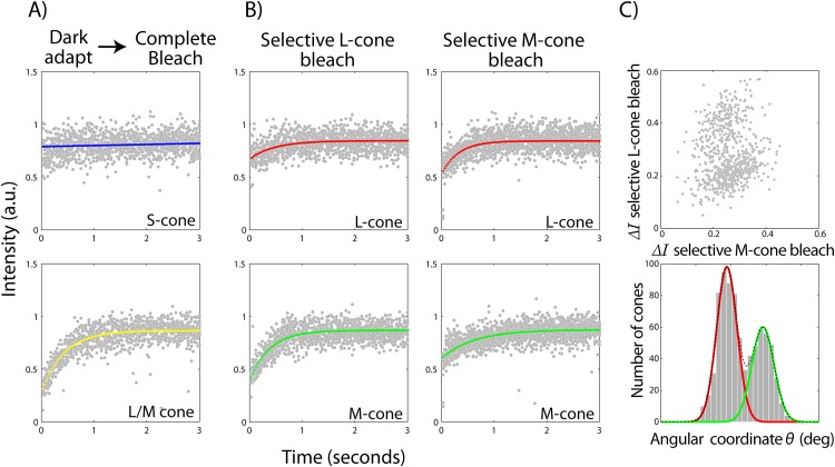
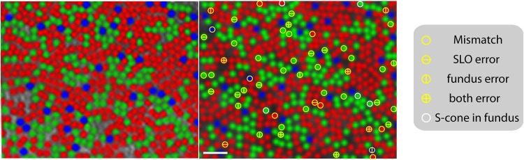
Densitometry is a powerful tool for the biophysical assessment of the retina. Until recently, this was restricted to bulk spatial scales in living humans. The application of adaptive optics (AO) to the conventional fundus camera and scanning laser ophthalmoscope (SLO) has begun to translate these studies to cellular scales. Here, we employ an AOSLO to perform dynamic photopigment densitometry in order to characterize the optical properties and spectral types of the human cone photoreceptor mosaic. Cone-resolved estimates of optical density and photosensitivity agree well with bulk estimates, although show smaller variability than previously reported. Photopigment kinetics of individual cones derived from their selective bleaching allowed efficient mapping of cone sub-types in human retina. Estimated uncertainty in identifying a cone as long vs middle wavelength was less than 5%, and the total time taken per subject ranged from 3-9 hours. Short wavelength cones were delineated in every subject with high fidelity. The lack of a third cone-type was confirmed in a protanopic subject. In one color normal subject, cone assignments showed 91% correspondence against a previously reported cone-typing method from more than a decade ago. Combined with cone-targeted stimulation, this brings us closer in studying the visual percept arising from a specific cone type and its implication for color vision circuitry.


Conflict of interest statement

Competing Interests: Roorda has two patents on technology related to the Adaptive Optics Scanning Laser Ophthalmoscope. These are: USPTO #7,118,216 Method and apparatus for using adaptive optics in a scanning laser ophthalmoscope and USPTO #6,890,076 Method and apparatus for using adaptive optics in a scanning laser ophthalmoscope. These patents are assigned to both the University of Rochester and the University of Houston. The patents are currently licensed to Canon, Inc. Japan. Both Roorda and the company may benefit financially from the publication of this research. This does not alter the authors' adherence to PLOS ONE policies on sharing data and materials.

Figures

Fig 1
Fig 1. Methodology for classifying LMS cones.
Representative examples of cone bleaching curves under a complete bleach from a dark adapted retina (A) and under a selective L and M-cone (B) bleach respectively are shown. Mean intensity timecourses for a given cone are accumulated together across all bleaching cycles. Non-linear least squares fit to the mean intensity of individual cones are shown and colored according to their peak sensitivity as blue, green or red for LMS cones respectively. The change in intensity under selective L- and M-cone bleaches for all L and M cones are plotted in (C) in a scatter plot. Each scatter point represents either an L or M cone. The x-y plot is converted to polar coordinates. The histogram of the angular coordinate is separared into respective L and M cone clusters by fitting a sum of 2 one-dimensional gaussian curves and taking the point of intersection of the component gaussians.
Fig 2
Fig 2. LMS cone mosaics in the retinae of human subjects.
Each column represents an individual subject’s retina at 1.5 deg temporal eccentricity. The last column represents cone assignments for the protanopic subject. S-cones appear as a cluster of weakly reflecting cones under complete bleach starting from a dark adapted retina (top row). L and M cones appear as two distinct clusters of cones on the basis of their relative change in intensity under selective bleaches (middle row). No separation of such clusters are obtained in the protanope. Based on where a cone appears in the S vs L/M and L vs M clustering analysis, it is shaded as ‘blue’, ‘green’ and ‘red’ to represent S, M and L cones respectively (bottom row). Other than S-cones, cone assignments for the protanope are shaded as ‘green’ M-cones on the basis of previous verification of his color deficiency. The scale bar is 2 arc-min.
Fig 3
Fig 3. Comparison of cone classification in the AO fundus camera (left) and SLO (right).
Cone assignments in the former are obtained from Hofer et al. 2005. Mismatched cones in the SLO image are marked as ‘circles’. Cones which have probability less than 0.9 to be reliably identified in the SLO and fundus are marked with ‘horizontal’ & ‘vertical’ lines respectively. The scale bar is 4 arc-min.

References

    1. Williams DR, MacLeod DI, Hayhoe MM. Punctate sensitivity of the blue-sensitive mechanism. Vision Research. 1981;21(9):1357–75. Epub 1981/01/01. . - PubMed
    1. Curcio CA, Allen KA, Sloan KR, Lerea CL, Hurley JB, Klock IB, et al. Distribution and morphology of human cone photoreceptors stained with anti-blue opsin. The Journal of Comparative Neurology. 1991;312(4):610–24. Epub 1991/10/22. 10.1002/cne.903120411 . - DOI - PubMed
    1. Williams DR, MacLeod DI, Hayhoe MM. Foveal tritanopia. Vision Research. 1981;21(9):1341–56. Epub 1981/01/01. . - PubMed
    1. Cicerone CM, Nerger JL. The relative numbers of long-wavelength-sensitive to middle-wavelength-sensitive cones in the human fovea centralis. Vision Research. 1989;29(1):115–28. Epub 1989/01/01. . - PubMed
    1. Carroll J, Neitz J, Neitz M. Estimates of L:M cone ratio from ERG flicker photometry and genetics. Journal of Vision. 2002;2(8):531–42. Epub 2003/04/08. . - PubMed

Publication types