. 2016 Apr;10(4):462-71.
doi: 10.1093/ecco-jcc/jjv223. Epub 2015 Dec 9.

Mucosal Barrier Depletion and Loss of Bacterial Diversity are Primary Abnormalities in Paediatric Ulcerative Colitis

Affiliations

Mucosal Barrier Depletion and Loss of Bacterial Diversity are Primary Abnormalities in Paediatric Ulcerative Colitis

Misagh Alipour et al. J Crohns Colitis. 2016 Apr.

Abstract

Background and aims: Ulcerative colitis [UC] is associated with colonic mucosa barrier defects and bacterial dysbiosis, but these features may simply be the result of inflammation. Therefore, we sought to assess whether these features are inherently abrogated in the terminal ileum [TI] of UC patients, where inflammation is absent.

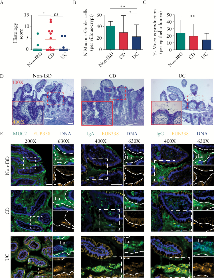
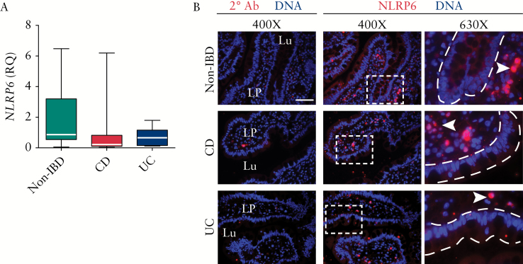
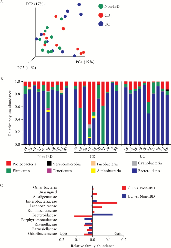
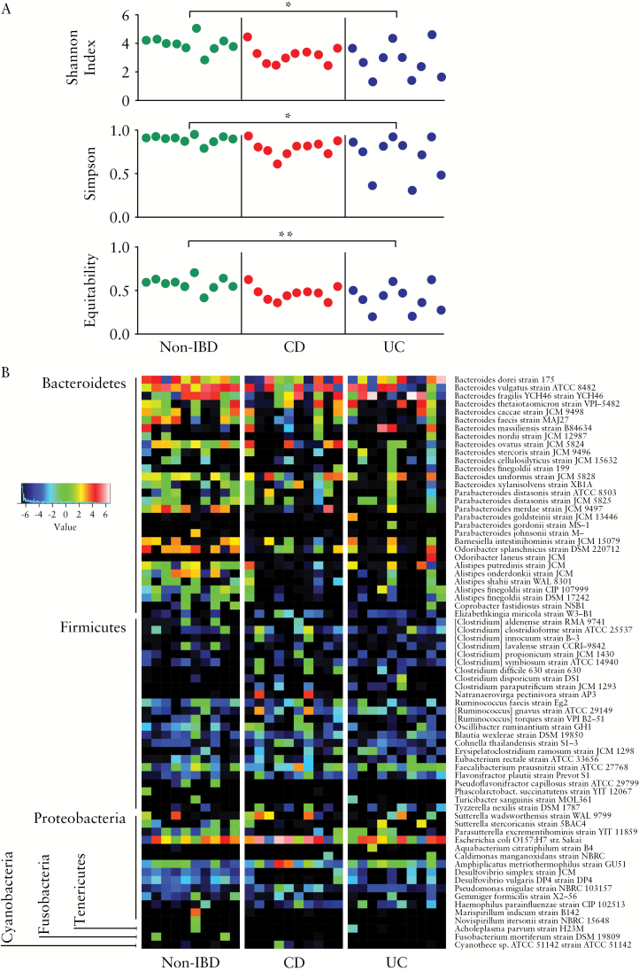
Methods: TI biopsies from paediatric inflammatory bowel disease [IBD] subsets [Crohn's disease [CD; n = 13] and UC [n = 10]], and non-IBD disease controls [n = 12] were histologically graded, and alcian blue/periodic acid-Schiff stained biopsies were quantified. The mucosal barrier was assessed for mucin [MUC2], immunoglobulin [Ig]A, IgG, and total bacteria (fluorescence in-situ hybridisation [FISH probe EUB338]) by immunofluorescence. The regulation of mucin secretion was investigated by NLRP6 gene expression and immunofluorescence. The composition of the active mucosa-associated microbiota was explored by sequencing the 16S rRNA amplicon generated from total RNA.

Results: Despite the absence of ileitis, UC patients displayed ileal barrier depletion illustrated by reductions in mucin-containing goblet cells and mucin production and altered epithelial NLRP6 expression. In both CD patients with ileitis and UC patients with normal histology, bacteria coated with IgA and IgG penetrated the TI mucin layer. Biopsy 16S rRNA sequencing revealed a reduction in α-diversity by three methods [Shannon, Simpson, and Equitability indices] between UC and non-IBD paediatric patients.

Conclusions: These findings suggest an underlying defect in the UC-afflicted intestinal tract even in the absence of inflammation, implicating barrier and microbial changes as primary abnormalities in UC that may play a causative role in disease development.

Keywords: Ulcerative colitis; mucin; mucosal barrier.


Figures

Figure 1.
Figure 1.
Ulcerative colitis [UC] patients demonstrate mucous barrier reduction and an elevated mucosa-bacterial interaction even in the absence of ileitis. [A] Scatterplot representation of histology showed ileitis in Crohn’s disease [CD] and its absence in UC and non-inflammatory bowel disease [IBD] groups. [B] Compared with the non-IBD and CD groups, the mean numbers of mucin-containing goblet cells per villous crypt were significantly reduced in the terminal ileum [TI] of the UC group. [C] Compared with the non-IBD group, mucous production expressed as % of epithelial surface covered with mucin [see Supplementary Figure S1, available as Supplementary data at ECCO-JCC online] in the epithelia-lumen was significantly reduced in the UC group. [D] Representative AB/PAS-stained TI biopsies showing reduced mucin in UC. Bar is 100 µm; inset image is 400X. [E] Immunostaining for mucin [MUC2], IgA, IgG, and bacteria, showing bacteria virtually absent from the mucosal layer in non-IBD patients. In contrast, in CD and UC bacteria were in close proximity to the epithelial lining with an increase in association with IgA and IgG. Lu, lumen; images showing mucin or Ig [top right] and bacteria [bottom right] were cropped digitally using the ZEN software, and the dashed lines were digitally added to mark the epithelial surface; bar is 50 µm. Statistically significant differences between two groups are indicated by an asterisk [p < 0.05] or double asterisk [p < 0.01]; ns, not significant. Note: the epithelial tissue structures visualised [shown in orange] are caused by autofluorescence resulting from fluorescence in-situ hybridisation [FISH]; in contrast, bacteria appear as clear orange clumps.
Figure 2.
Figure 2.
Ileal NLRP6 location is altered in inflammatory bowel disease [IBD]. [A] NLRP6 gene expression in ileal biopsies as measured by quantitative reverse-transcription polymerase chain reaction [qRT-PCR] [relative quantification, RQ]. Data are expressed as median with interquartile range, minimum and maximum. [B] Representative immunofluorescence of biopsies stained for NLRP6 protein showed expression in the lamina propria and proportionately distributed throughout epithelial cells of non-IBD group, with lower and intermittent epithelial distribution in both IBD subsets. Lu, lumen; LP, lamina propria; arrowhead, NLRP6-expressing cell in the LP; bar is 50 µm. The dashed lines were digitally added to mark the epithelial surface.
Figure 3.
Figure 3.
Perturbations in ileal microbial composition in inflammatory bowel disease [IBD]. [A] Principal coordinates analysis [PCoA] of BrayCurtis dissimilarity showed approximately half of Crohn’s disease [CD] and ulcerative colitis [UC] samples had a microbiota composition that was different from most non-IBD patients. [B] Relative abundance at the phylum level for each individual patient [identified by the patient study code number] showing extensive changes in several CD patients. [C] Relative abundance at family level [calculated as change in total fraction compared with non-IBD] showed gains in Enterobacteriaceae in IBD subsets, with Bacteroidaceae loss in CD and gain in UC. No statistically significant difference between the groups was found using BenjaminiHochberg false discovery rate correction.
Figure 4.
Figure 4.
Bacterial α-diversity comparison and heatmap of relative bacterial species abundance. [A] Bacterial α-diversity of individual patients. Statistical comparison between groups, made by one-way analysis of variance with Bonferroni’s multiple comparison post-test, indicated reduced diversity in ulcerative colitis [UC] compared with non-inflammatory bowel disease [IBD]. Asterisk, p < 0.05; double asterisk, p < 0.01. Statistical comparison between non-IBD and CD, as well as CD and UC, did not show significance. [B] Minimum entropy decomposition nodes were identical [100% similarity; coverage 100%] to the reported species/strain. However, in many cases, more than one identical sequence was found in the subject database and therefore the reported taxa represent only taxonomical approximations. Data represented as log2. Patterns of alterations at the species level are observed; however, no statistically significant difference was found between the groups when using Benjamini-Hochberg false discovery rate correction.

References

    1. El Aidy S, Merrifield CA, Derrien M, et al. The gut microbiota elicits a profound metabolic reorientation in the mouse jejunal mucosa during conventionalisation. Gut 2013;62:1306–14. - PubMed
    1. Wlodarska M, Willing BP, Bravo DM, Finlay BB. Phytonutrient diet supplementation promotes beneficial clostridia species and intestinal mucus secretion resulting in protection against enteric infection. Sci Rep 2015;5:9253. - PMC - PubMed
    1. Sanchez de Medina F, Romero-Calvo I, Mascaraque C, Martinez-Augustin O. Intestinal inflammation and mucosal barrier function. Inflamm Bowel Dis 2014;20:2394–404. - PubMed
    1. Johansson ME, Sjovall H, Hansson GC. The gastrointestinal mucus system in health and disease. Nat Rev Gastroenterol Hepatol 2013;10:352–61. - PMC - PubMed
    1. Johansson ME. Mucus layers in inflammatory bowel disease. Inflamm Bowel Dis 2014;20:2124–31. - PubMed

Publication types

Substances