. 2016 Apr;36(4):721-30.
doi: 10.1177/0271678X15608395. Epub 2015 Oct 2.

Stroke neuroprotection revisited: Intra-arterial verapamil is profoundly neuroprotective in experimental acute ischemic stroke

Affiliations

Stroke neuroprotection revisited: Intra-arterial verapamil is profoundly neuroprotective in experimental acute ischemic stroke

Michael E Maniskas et al. J Cereb Blood Flow Metab. 2016 Apr.

Abstract

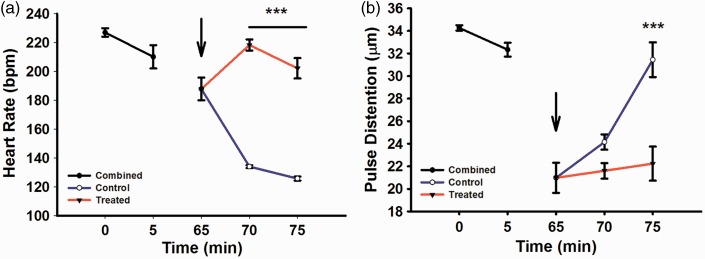
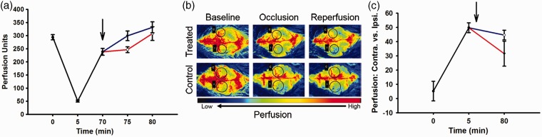
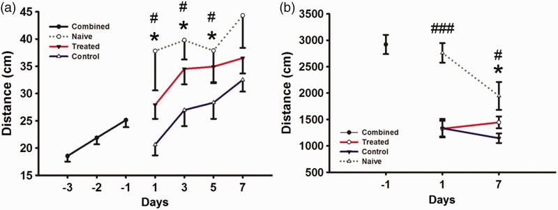
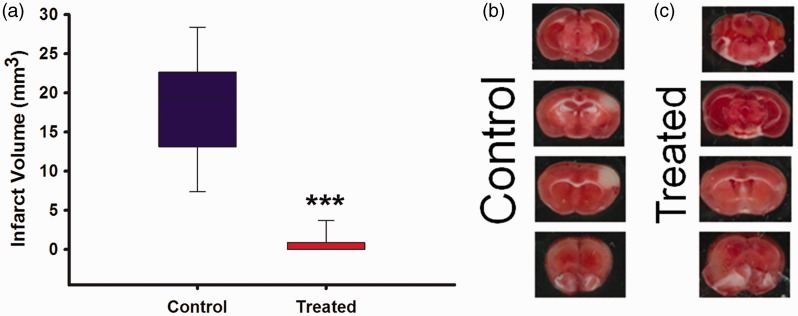
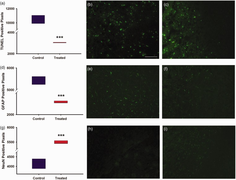
While clinical trials have now solidified the role of thrombectomy in emergent large vessel occlusive stroke, additional therapies are needed to optimize patient outcome. Using our previously described experimental ischemic stroke model for evaluating adjunctive intra-arterial drug therapy after vessel recanalization, we studied the potential neuroprotective effects of verapamil. A calcium channel blocker, verapamil is often infused intra-arterially by neurointerventionalists to treat cerebral vasospasm. Such a direct route of administration allows for both focused targeting of stroke-impacted brain tissue and minimizes potential systemic side effects. Intra-arterial administration of verapamil at a flow rate of 2.5 µl/min and injection volume of 10 µl immediately after middle cerebral artery recanalization in C57/Bl6 mice was shown to be profoundly neuroprotective as compared to intra-arterial vehicle-treated stroke controls. Specifically, we noted a significant (P ≤ 0.05) decrease in infarct volume, astrogliosis, and cellular apoptosis as well as a significant increase in neuronal survival and functional outcome over seven days. Furthermore, intra-arterial administration of verapamil was well tolerated with no hemorrhage, systemic side effects, or increased mortality. Thus, verapamil administered intra-arterially immediately following recanalization in experimental ischemic stroke is both safe and neuroprotective and merits further study as a potential therapeutic adjunct to thrombectomy.

Keywords: Ischemic stroke; cerebral ischemia; intra-arterial; neuroprotection; recanalization; verapamil.


Figures

Figure 1.
Figure 1.
MouseOx plus physiological measurements for combined (black), treated (red) and control (blue) groups. Arrow indicates drug administration after reperfusion/un-occlusion for 5 min. (a) Heart rate in beats per minute (bpm), 0–5 min (baseline measurements), 65 min (reperfusion/un-occlusion and drug administration), 70 and 75 min (reperfusion). (b) Pulse distention measuring vessel diameter (µm), 0–5 min (baseline), 65 min (reperfusion/un-occlusion and drug administration), 70 and 75 min (reperfusion). Combined (N = 30), control (N = 15), treated (N = 15).
Figure 2.
Figure 2.
Perfusion measurements for combined (black), treated (red), and control (blue) groups. Arrow indicates drug administration after reperfusion/un-occlusion for 5 min. (a) Laser Doppler blood perfusion through middle cerebral artery at time points 0 min (baseline measurements), 5 min (occlusion), 70 min (reperfusion/un-occlusion), and 75 and 80 min (reperfusion). Treated (N = 9), control (N = 9). (b) Laser Speckle whole brain perfusion images through contralateral and ipsilateral cortex at time points 0 min (baseline), 5 min (MCA occlusion) and 80 min (reperfusion) for treated (N of 3) and control (N of 3). Black circles indicate regions of interest, ipsilateral (I) and corresponding contralateral (C) for MCA occlusion at aforementioned time points. (c) Laser Speckle graph for whole brain perfusion through contralateral and ipsilateral cortex at time points 0 minutes (baseline measurements), 5 min (MCA occlusion), and 80 (15 min reperfusion/un-occlusion) treated (N = 3), control (N = 3).
Figure 3.
Figure 3.
Behavioral measurements for rotor rod and open field for combined (black), naïve (green dashed), treated (red), and control (blue) groups. (a) Rotor rod forced motor movement test for combined groups to learn task on days (-3, -2, and -1), groups separated following stroke surgery into control, treated, and naïve groups and were tested on days (1, 3, 5, and 7). (b) Open field free roam movement for combined groups on day (-1) and separated into control, treated, and naïve groups (1 and 7). Combined (N = 49), treated (N = 25), control (N = 20), naïve (N = 5). * indicates P ≤ 0.05 for treated versus control. # indicates P ≤ 0.05 for naïve versus control and ### indicates P ≤ 0.001 for naïve versus control and treated.
Figure 4.
Figure 4.
(a) Infarct volume analysis for control versus treated using TTC and cresyl violet stained stroked tissue. Treated (N = 18), control (N = 15); (b) TTC image for control group; (c) TTC image for treated. *** indicates P ≤ 0.001.
Figure 5.
Figure 5.
Graphs and images for immunohistochemistry, magnification at 20X with quantification of positive pixel density. (a) Graph depicting positive pixel for TUNEL stain of control versus treated in infarcted region. (b) Image of control TUNEL stain from infarcted region at magnification 20X. (c) Image of treated TUNEL stain from infarcted region at magnification 20X. (d) Graph depicting positive pixels for GFAP stain of control versus treated in infarcted region. (e) Image of control GFAP stain from infarcted region at magnification 20X. (f) Image of treated GFAP stain from infarcted region at magnification 20X. (g) Graph depicting positive pixels for NeuN stain of control versus treated in infarcted region. (h) Image of control NeuN stain from infarcted region at magnification 20X. (i) Image of control and treated NeuN stain from infarcted region at magnification 20X. Treated (N = 10), control (N = 10). White scale bar, 100 µm. *** indicates P ≤ 0.001.

References

    1. Saver JL, Jahan R, Levy EI, et al. Solitaire flow restoration device versus the Merci Retriever in patients with acute ischaemic stroke (SWIFT): a randomised, parallel-group, non-inferiority trial. Lancet 2012; 380: 1241–1249. - PubMed
    1. Bretz MN, Graves A, West A, et al. Steps against recurrent stroke plus: patient transition program. J Neurosci Nurs 2014; 46: E3–13. quiz E1-2. - PubMed
    1. Go AS, Mozaffarian D, Roger VL, et al. Heart disease and stroke statistics—2014 update: a report from the American Heart Association. Circulation 2014; 129: e28–e292. - PMC - PubMed
    1. Thacker C. Stroke falls to No. 5 cause of death in U.S. American Heart Association Media Alert. 12/30/2014 ed2014.
    1. Broderick JP, Palesch YY, Demchuk AM, et al. Endovascular therapy after intravenous t-PA versus t-PA alone for stroke. N Engl J Med 2013; 368: 893–903. - PMC - PubMed

Publication types

MeSH terms