. 2016 May;36(5):965-80.
doi: 10.1177/0271678X15610787. Epub 2015 Oct 19.

Extracellular HCO3- is sensed by mouse cerebral arteries: Regulation of tone by receptor protein tyrosine phosphatase γ

Affiliations

Extracellular HCO3- is sensed by mouse cerebral arteries: Regulation of tone by receptor protein tyrosine phosphatase γ

Ebbe Boedtkjer et al. J Cereb Blood Flow Metab. 2016 May.

Abstract

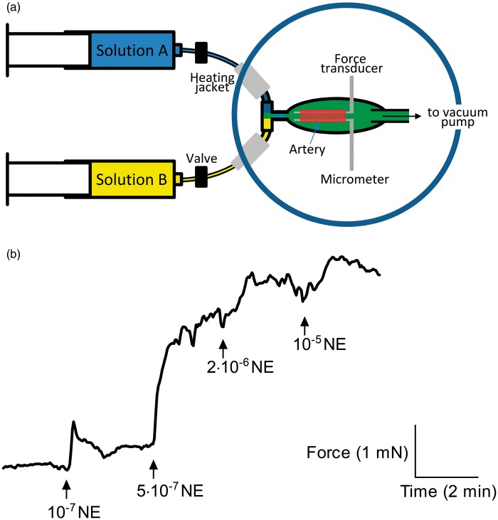
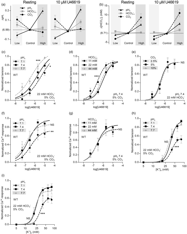
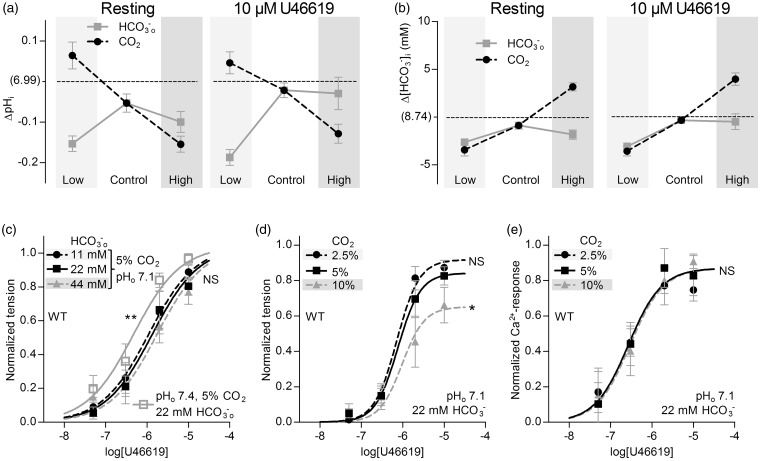
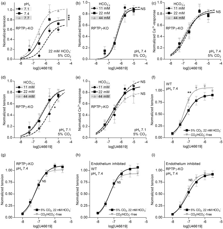
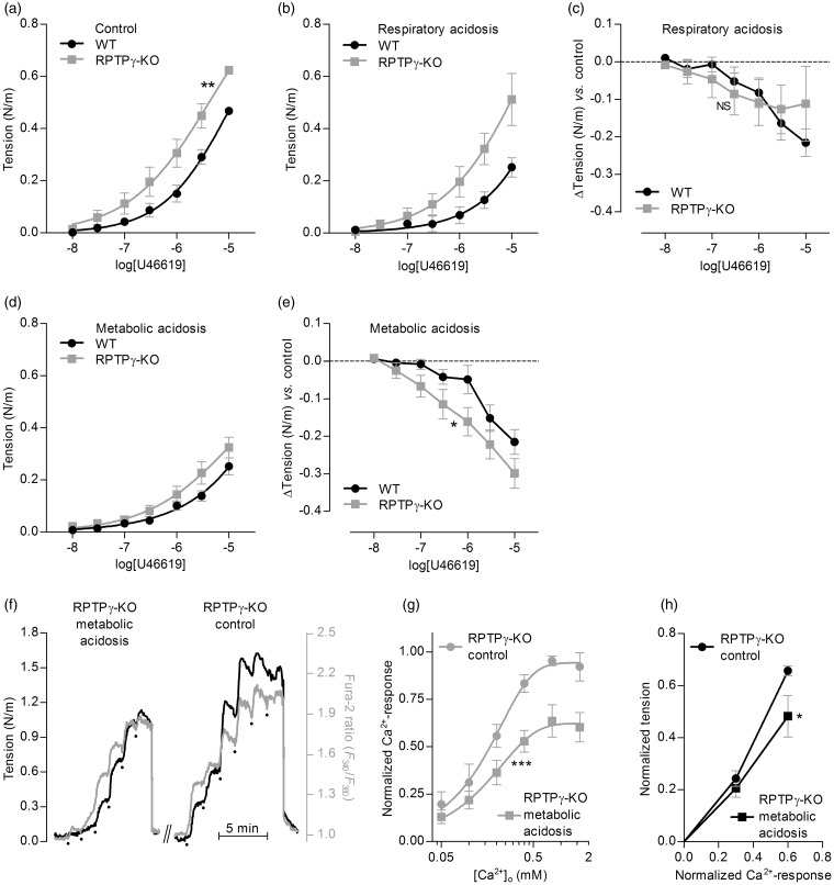
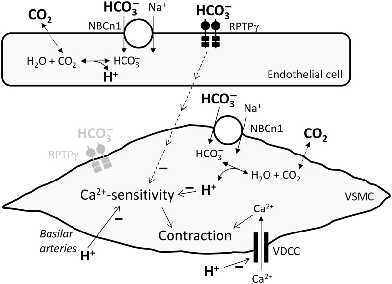
We investigate sensing and signaling mechanisms for H(+), [Formula: see text] and CO2 in basilar arteries using out-of-equilibrium solutions. Selectively varying pHo, [[Formula: see text]]o, or pCO2, we find: (a) lowering pHo attenuates vasoconstriction and vascular smooth muscle cell (VSMC) Ca(2+)-responses whereas raising pHo augments vasoconstriction independently of VSMC [Ca(2+)]i, (b) lowering [[Formula: see text]]o increases arterial agonist-sensitivity of tone development without affecting VSMC [Ca(2+)]i but c) no evidence that CO2 has direct net vasomotor effects. Receptor protein tyrosine phosphatase (RPTP)γ is transcribed in endothelial cells, and direct vasomotor effects of [Formula: see text] are absent in arteries from RPTPγ-knockout mice. At pHo 7.4, selective changes in [[Formula: see text]]o or pCO2 have little effect on pHi At pHo 7.1, decreased [[Formula: see text]]o or increased pCO2 causes intracellular acidification, which attenuates vasoconstriction. Under equilibrated conditions, anti-contractile effects of CO2/[Formula: see text] are endothelium-dependent and absent in arteries from RPTPγ-knockout mice. With CO2/[Formula: see text] present, contractile responses to agonist-stimulation are potentiated in arteries from RPTPγ-knockout compared to wild-type mice, and this difference is larger for respiratory than metabolic acidosis. In conclusion, decreased pHo and pHi inhibit vasoconstriction, whereas decreased [[Formula: see text]]o promotes vasoconstriction through RPTPγ-dependent changes in VSMC Ca(2+)-sensitivity. [Formula: see text] serves dual roles, providing substrate for pHi-regulating membrane transporters and modulating arterial responses to acid-base disturbances.

Keywords: Vascular biology; basic science; calcium imaging; confocal microscopy; electrophysiology; endothelium; experimental; pH; physiology; receptors; smooth muscle.


Figures

Figure 1.
Figure 1.
Application of OOE CO2/HCO3- solutions to small-artery myography. (a) Schematic showing generation of OOE solutions by rapid mixing and delivery of differently composed CO2/HCO3- solutions. The time delay from the point of mixing to the artery is <100 ms. See Supplemental Methods for details. (b) Force recordings from a mesenteric artery exposed to increasing concentrations of norepinephrine (NE) under OOE conditions (pHo=7.7, [HCO3-]o=22 mM, CO2=5%).
Figure 2.
Figure 2.
Selective changes in [HCO3-]o or pHo—but not pCO2—directly modulate basilar artery tone under OOE conditions. (a) Effects of selectively varying pHo, [HCO3-]o or pCO2 (maintaining other two at control levels) on VSMC pHi in resting basilar arteries (left panel) or basilar arteries contracted by 10 µM U46619 (right panel). Under “Control” conditions, CO2 is 5%, pHo 7.4, and [HCO3-]o 22 mM. Compared to “Control”, “Low” refers to selective changes in CO2 to 2.5%, [HCO3-]o to 11 mM or pHo to 7.1, and “High” refers to selective changes in CO2 to 10%, [HCO3-]o to 44 mM or pHo to 7.7. Arteries are from wild-type mice (n = 6). (b) Effects of selectively varying pHo, [HCO3-]o or pCO2 (maintaining other two at control levels) on VSMC calculated [HCO3-]i in resting (Continued) Figure 2. Continue. basilar arteries (left panel) or basilar arteries contracted by 10 µM U46619 (right panel). Arteries are from wild-type mice (n = 6). (c-e) Effects of selectively varying pHo, [HCO3-]o, or pCO2 (maintaining other two at control levels) on U46619-induced tension development in basilar arteries from wild-type mice (n = 12 or 13). (f, g) Effects of selectively varying pHo or [HCO3-]o (maintaining unvaried parameters at control levels) on U46619-induced VSMC Ca2+-responses in basilar arteries from wild-type mice (n = 6 for both). (h) Effects of selective changes in pHo ([HCO3-]o=22 mM, CO2=5%) on depolarization-induced tension development of basilar arteries from wild-type mice (n = 8). We induce depolarization by raising [K+]o. (i) Effects of selective changes in pHo ([HCO3-]o=22 mM, CO2=5%) on depolarization-induced VSMC Ca2+ responses in basilar arteries from wild-type mice (n = 7). The curves in panels (c) through (i) are the results of least-squares fits to sigmoidal functions, and we compare them using extra sum-of-squares F-tests. *P < 0.05, **P < 0.01, ***P < 0.001, NS: not significantly different vs. control conditions (pHo=7.4, [HCO3-]o=22 mM, CO2=5%).
Figure 3.
Figure 3.
Increasing pCO2 at low pHo (pH 7.1) attenuates tension production of basilar arteries under OOE conditions. (a) Effects of selectively varying [HCO3-]o or pCO2 (maintaining the other at its control level, with pHo fixed at 7.1) on VSMC pHi in resting basilar arteries (left panel) or basilar arteries contracted by 10 µM U46619 (right panel). “Low” and “High” refer to half and twice the concentration, respectively, of CO2 and HCO3- compared to “Control” levels ([HCO3-]o=22 mM, CO2=5%). The reference pHi value on the y-axis, “(6.99)”, corresponds to control levels of all three parameters (pHo=7.4, [HCO3-]o=22 mM, CO2=5%) in the same arteries from wild-type mice (n = 6). (b) Effects of selectively varying [HCO3-]o or pCO2 (maintaining the other at its control level, with pHo fixed at 7.1) on VSMC calculated [HCO3-]i in resting basilar arteries (left panel) or basilar arteries contracted by 10 µM U46619 (right panel). The reference [HCO3-]i value on the y-axis, “(8.74 mM)”, corresponds to control levels of all three parameters (pHo=7.4, [HCO3-]o=22 mM, CO2=5%) in the same arteries from wild-type mice (n = 6). (c, d) Effects of selectively varying [HCO3-]o or pCO2 (maintaining the other at its control level, with pHo fixed at 7.1) on U46619-induced tension development in basilar arteries from wild-type mice (n = 6 in panel (c), n = 11 in panel (d)). For comparison, in panel (c), we show the reference tension response (solid gray curve) in the same arteries, under conditions that correspond to control levels of all three parameters (pHo=7.4, [HCO3-]o=22 mM, CO2=5%). (e) Effects of selectively varying pCO2 (fixed pHo=7.1, [HCO3-]o=22 mM) on U46619-induced VSMC Ca2+ responses in basilar arteries from wild-type mice (n = 5). The curves in panel (c) through (e) are the results of least-squares fits to sigmoidal functions, and we compare them using extra sum-of-squares F-tests. *P < 0.05, **P < 0.01, NS: not significantly different vs. control acidic conditions (pHo=7.1, [HCO3-]o=22 mM, CO2=5%).
Figure 4.
Figure 4.
Ptprg transcriptional activity is prominent in ECs of basilar arteries. (a, b) Basilar arteries from an RPTPγ-knockout (KO, panel (a)) and a wild-type (WT, panel (b)) mouse, stained histochemically for β-galactosidase activity. Each image is representative of five experiments. (c, d) Histological sections (8 µm thick) of basilar arteries from an RPTPγ-knockout and a wild-type mouse, stained histochemically for β-galactosidase activity. Each image is representative of three experiments. The size bars in panels (a) and (b) represent 100 µm, in (c) and (d) 10 µm. Lu indicates lumen, Adv indicates adventitia and EC indicates endothelial cell.
Figure 5.
Figure 5.
Basilar arteries from RPTPγ-knockout mice are insensitive to changes in [HCO3-]o. (a, b) Effects of selectively varying pHo or [HCO3-]o (maintaining the other at its control level, with CO2 fixed at 5%) on U46619-induced tension development in basilar arteries from RPTPγ-knockout mice (n = 4 in panel (a), n = 8 in panel (b)). (c) Effects of selectively varying [HCO3-]o (maintaining pHo at 7.4, CO2 at 5%) on U46619-induced VSMC Ca2+-responses in basilar arteries from RPTPγ-knockout mice (n = 8). (d) Effects of selectively varying [HCO3-]o at low pHo (maintaining pHo at 7.1, CO2 at 5%) on U46619-induced tension development of basilar arteries from RPTPγ-knockout mice (n = 6). (e) Effects of selectively varying [HCO3-]o at low pHo (maintaining pHo at 7.1, CO2 at 5%) on U46619-induced VSMC Ca2+-responses in basilar arteries from RPTPγ-knockout mice (n = 4). (f-i). Effects of omitting CO2/HCO3- on U46619-induced tension development at constant pHo of 7.4. We perform experiments with basilar arteries from wild-type (WT, panels (f) and (h)) and RPTPγ-knockout (KO, panels (g) and (i)) mice. In panel (h) and (i), we inhibit vasorelaxant effects of the endothelium by incubation with 100 µM L-NAME, 3 µM indomethacin, 50 nM apamin and 1 µM TRAM-34. To avoid development of resting tone after inhibition of endothelium-dependent vasorelaxation, we add 600 nM SNAP to achieve an equal [NO] in the tested arteries. The curves are the results of least-squares fits to sigmoidal functions, and we compare them using extra sum-of-squares F-tests. *P < 0.05, **P < 0.01, ***P < 0.001, NS: not significantly different vs. control conditions (panels (a–c) and (f–i): pHo=7.4, [HCO3-]o=22 mM, CO2=5%) or standard acidic conditions (panels (d) and (e): pHo=7.1, [HCO3-]o=22 mM, CO2=5%).
Figure 6.
Figure 6.
Under equilibrated conditions, RPTPγ is required for HCO3o- sensing—such that basilar arteries from RPTPγ-knockout mice are hypercontractile—and vasorelaxation in response to metabolic acidosis is explained by a combination of attenuated Ca2+ uptake and reduced Ca2+-sensitivity. (a–e) Comparison of U46619-induced contractile responses of basilar arteries from RPTPγ-knockout (KO, n=8) vs. wild-type (WT, n = 10) mice. Panel (a) summarizes experiments under control conditions (pHo=7.4, [HCO3-]o=22 mM, CO2=5%); panel (b), under respiratory acidosis (pHo=7.1, [HCO3-]o=22 mM, CO2=10%); and panel (d), under metabolic acidosis (pHo=7.1, [HCO3-]o=11 mM, CO2=5%). Panels (c) and (e) summarize the average differences in U46619-induced contractile responses in basilar arteries from RPTPγ-knockout and wild-type mice during respiratory (panel c) and metabolic (panel e) acidosis compared to control conditions. (f) Representative time courses of arterial tension development and VSMC Ca2+-dependent fluorescence signal in Ca2+-depleted basilar arteries from RPTPγ-knockout mice in response to step-increases of [Ca2+]o during constant exposure to 10 µM U46619. We perform experiments under control conditions (pHo=7.4, [HCO3-]o=22 mM, CO2=5%) and during metabolic acidosis (pHo=7.1, [HCO3-]o=11 mM, CO2=5%). The dots indicate step-wise increments in [Ca2+]o from 0 to 0.05, 0.1, 0.2, 0.4, 0.8 and 1.6 mM. (g) Average Cai2+-responses from experiments (n = 5) like that in panel (f). (h) Average tension development of basilar arteries from RPTPγ-knockout mice (n = 5) as a function of the VSMC Cai2+-response under control (Continued) Figure 6. Continue. conditions (pHo=7.4, [HCO3-]o=22 mM, CO2=5%) and during metabolic acidosis (pHo=7.1, [HCO3-]o=11 mM, CO2=5%). The curves in panels (a), (b), (d) and (g) are the results of least-squares fits to sigmoidal functions; in panels (a) and (g), we compare them using extra sum-of-squares F-tests. The data points in panels (c), (e) and (h) are compared by repeated-measures two-way ANOVA; in panel (h), we report the significance level based on Bonferroni post-tests. *P < 0.05, **P < 0.01, ***P < 0.001, NS: not significantly different vs. wild-type (panels (a), (c) and (e)) or control conditions (panels (g) and (h)).
Figure 7.
Figure 7.
Schematic of proposed signaling pathways affecting cerebral vascular tone in response to acid-base disturbances. Note that in the cases of endothelium-mediated RPTPγ-dependent actions of HCO3o- and the effect of Ho+ acting on Ca2+-sensitivity (which could occur via a parallel change in pHi), we have only seen effects with reducing the concentrations. RPTPγ in the vascular smooth muscle cell is shown in gray because we have evidence for expression but no function has yet been demonstrated. VDCC, voltage-dependent Ca2+-channels; VSMC, vascular smooth muscle cell.

References

    1. Kontos HA. Regulation of the cerebral circulation. Ann Rev Physiol 1981; 43: 397–407. - PubMed
    1. Gaskell WH. On the tonicity of the heart and blood vessels. J Physiol 1880; 3: 48–92. - PMC - PubMed
    1. Zhao J, Hogan EM, Bevensee MO, et al. Out-of-equilibrium CO2/HCO3- solutions and their use in characterizing a new K/HCO3 cotransporter. Nature 1995; 374: 636–639. - PubMed
    1. Peng HL, Ivarsen A, Nilsson H, et al. On the cellular mechanism for the effect of acidosis on vascular tone. Acta Physiol Scand 1998; 164: 517–525. - PubMed
    1. Boedtkjer E, Praetorius J, Aalkjaer C. NBCn1 (slc4a7) mediates the Na+-dependent bicarbonate transport important for regulation of intracellular pH in mouse vascular smooth muscle cells. Circ Res 2006; 98: 515–523. - PubMed

Publication types

MeSH terms