. 2016 Jul;36(7):1202-11.
doi: 10.1177/0271678X15611912. Epub 2015 Oct 23.

Hemichannel-mediated release of lactate

Affiliations

Hemichannel-mediated release of lactate

Anastassios Karagiannis et al. J Cereb Blood Flow Metab. 2016 Jul.

Abstract

In the central nervous system lactate contributes to the extracellular pool of readily available energy substrates and may also function as a signaling molecule which mediates communication between glial cells and neurons. Monocarboxylate transporters are believed to provide the main pathway for lactate transport across the membranes. Here we tested the hypothesis that lactate could also be released via opening of pannexin and/or functional connexin hemichannels. In acute slices prepared from the brainstem, hippocampus, hypothalamus and cortex of adult rats, enzymatic amperometric biosensors detected significant tonic lactate release inhibited by compounds, which block pannexin/connexin hemichannels and facilitated by lowering extracellular [Ca(2+)] or increased PCO2 Enhanced lactate release triggered by hypoxia was reduced by ∼50% by either connexin or monocarboxylate transporter blockers. Stimulation of Schaffer collateral fibers triggered lactate release in CA1 area of the hippocampus, which was facilitated in conditions of low extracellular [Ca(2+)], markedly reduced by blockade of connexin hemichannels and abolished by lactate dehydrogenase inhibitor oxamate. These results indicate that lactate transport across the membranes may occur via mechanisms other than monocarboxylate transporters. In the central nervous system, hemichannels may function as a conduit of lactate release, and this mechanism is recruited during hypoxia and periods of enhanced neuronal activity.

Keywords: Astrocytes; connexin; lactate; metabolism; monocarboxylate transporters; pannexin.


Figures

Figure 1.
Figure 1.
Performance of the lactate biosensors. (a) Current (I) calibration curve and sample responses of the lactate biosensor demonstrating linearity of analyte detection in concentrations from 10 μM to 500 μM; (b) Insensitivity of the lactate biosensor detection system to pharmacological agents used in this study. Traces illustrate sample biosensor current responses to lactate (100 µM) when calibrated in control artificial cerebrospinal fluid (aCSF), and then in the presence of (i) carbenoxolone (CBX), (ii) 5-nitro-2 -(3-phenylpropylamino)-benzoic acid (NPPB, 200 µM), (iii) probenecid (Prob, 150 µM), (iv) α-cyano-4-hydroxycinnamate (4-CIN, 250 µM), (v) Ruthenium red (RuR, 20 µM), and (vi) tetrodotoxin (TTX, 1 µM) + muscimol (100 µM).
Figure 2.
Figure 2.
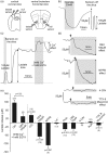
Hemichannel-mediated tonic release of lactate. (a) Schematic illustration of a dual recording configuration of lactate and null (control) biosensors placed in a direct contact with the surface of the coronal cortical slice or uncut ventral surface of the horizontal brainstem slice. Difference in current between lactate and null biosensors was used to determine the amount of lactate release. XII hypoglossal rootlets; (b) Representative example of oxamate-induced changes in lactate release detected by the biosensors placed on the surface of the brainstem slice followed by calibration with 100 µM lactate; (c) Representative example of changes in lactate biosensor current during calibration by application of 100 µM lactate, after biosensor placement in a direct contact with the surface of the brainstem slice (revealing lactate tone), and in response to lowering extracellular [Ca2+] (indicated by grey shading); (d) Representative recordings of lactate biosensor current showing changes in lactate tone in response to application of CBX (100 µM), NPPB (200 µM), 4-CIN (250 µM), or co-application of TTX (1 µM) and muscimol (100 µM). Period of drug application is indicated by grey shading; (e) Summary data illustrating peak changes in tonic lactate release in response to lowering extracellular [Ca2+] (in the absence and presence of CBX, 100 µM), increases in PCO2, or application of compounds known to inhibit functional connexin hemichannels (CBX, 100 µM; NPPB, 200 µM; La3+, 100 µM), pannexin hemichannels (CBX, 10 µM; Prob, 150 µM), monocarboxylate transporters (4-CIN, 250 µM), CALHM1 channels (RuR, 20 µM) or suppress the neuronal activity (TTX, 1 µM + muscimol, 100 µM). Mean ± SEM. p values – Wilcoxon signed-rank test. Hip: hippocampus; py: pyramidal tract.
Figure 3.
Figure 3.
Facilitated release of lactate in response to hypoxia. (a) Representative example of changes in lactate biosensor current during calibration, after biosensor placement on the surface of the brainstem slice, and in response to a hypoxic challenge (perfusion with aCSF saturated with 95% N2/5% CO2). Peak hypoxia-induced lactate release is measured upon re-oxygenation. (b) Representative examples of changes in lactate biosensor current recorded during and after hypoxia when the sensor is placed on the surface of the brain slice (left) or in the bath away from the slice (right) and on the surface of the slice during application of myxothiazol (middle). Decrease in O2 availability reduces biosensor current followed by a positive signal upon re-oxygenation (hypoxia-induced lactate release), which is only observed when the sensor is in a direct contact with the brain tissue. (c) Peak lactate efflux of a similar magnitude is triggered by hypoxia and myxothiazol.
Figure 4.
Figure 4.
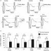
Connexin hemichannels and monocarboxylate transporters (MCTs) mediate facilitated release of lactate during hypoxia. (a) Representative recordings of changes in lactate biosensor current showing hypoxia-induced lactate release by the brainstem slices in the absence and presence of CBX (100 µM), NPPB (200 µM), 4-CIN (250 µM) or co-application of TTX (1 µM) and muscimol (100 µM); (b) Summary data illustrating peak hypoxia-induced lactate release in the absence and presence of CBX, NPPB, La3+, 4-CIN, TTX+muscimol.
Figure 5.
Figure 5.
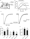
Connexin hemichannels contribute to the increases in lactate release triggered by enhanced neuronal activity. (a) Schematic drawing of the experimental design. Miniature (20 µm) carbon fiber lactate biosensors were placed in the vicinity of the patch-clamped hippocampal CA1 neurons to record lactate release in response to electrical stimulation of Schaffer collateral (SC) fibers; (b) Representative recordings of changes in biosensor current illustrating lactate release in CA1 in response to stimulation of Schaffer collaterals in control and low (0.2 mM) extracellular [Ca2+] conditions and in the presence of oxamate (20 mM). Schaffer collaterals were stimulated for 10 min; (c) Representative AMPAR EPSCs recorded in the same experiment in the absence and presence of CBX (each trace is an average of 10); (d) Summary data illustrating peak lactate release in CA1 area in response to stimulation of Schaffer collaterals in control and low extracellular [Ca2+] conditions, in the presence of oxamate or CBX; (e) Summary data illustrating amplitudes of AMPAR-mediated EPSCs normalized to the control values.

References

    1. Pellerin L, Magistretti PJ. Sweet sixteen for ANLS. J Cereb Blood Flow Metab 2012; 32: 1152–1166. - PMC - PubMed
    1. Pellerin L, Magistretti PJ. Glutamate uptake into astrocytes stimulates aerobic glycolysis: a mechanism coupling neuronal activity to glucose utilization. Proc Natl Acad Sci U S A 1994; 91: 10625–10629. - PMC - PubMed
    1. Dienel GA. Brain lactate metabolism: the discoveries and the controversies. J Cereb Blood Flow Metab 2012; 32: 1107–1138. - PMC - PubMed
    1. Ide K, Horn A, Secher N. Cerebral metabolic response to submaximal exercise. J Appl Physiol 1999; 87: 1604–1608. - PubMed
    1. Ide K, Schmalbruch IK, Quistorff B, et al. Lactate, glucose and O2 uptake in human brain during recovery from maximal exercise. J Physiol 2000; 522: 159–164. - PMC - PubMed

Publication types

LinkOut - more resources