

4D flow MRI for intracranial hemodynamics assessment in Alzheimer's disease
- PMID: 26661239
- PMCID: PMC5076787
- DOI: 10.1177/0271678X15617171
4D flow MRI for intracranial hemodynamics assessment in Alzheimer's disease
Abstract
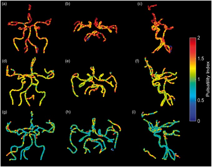
Cerebral blood flow, arterial pulsation, and vasomotion play important roles in the transport of waste metabolites out of the brain. Impaired vasomotion results in reduced driving force for the perivascular/glymphatic clearance of beta-amyloid. Noninvasive cerebrovascular characteristic features that potentially assess these transport mechanisms are mean blood flow (MBF) and pulsatility index (PI). In this study, 4D flow MRI was used to measure intra-cranial flow features, particularly MBF, PI, resistive index (RI) and cross-sectional area in patients with Alzheimer's disease (AD), mild cognitive impairment and in age matched and younger cognitively healthy controls. Three-hundred fourteen subjects participated in this study. Volumetric, time-resolved phase contrast (PC) MRI data were used to quantify hemodynamic parameters from 11 vessel segments. Anatomical variants of the Circle of Willis were also cataloged. The AD population reported a statistically significant decrease in MBF and cross-sectional area, and also an increase in PI and RI compared to age matched cognitively healthy control subjects. The 4D flow MRI technique used in this study provides quantitative measurements of intracranial vessel geometry and the velocity of flow. Cerebrovascular characteristics features of vascular health such as pulsatility index can be extracted from the 4D flow MRI data.
Keywords: 4D flow MRI; Alzheimer’s disease; circle of Willis; mean blood flow; pulsatility index.
© The Author(s) 2015.
Figures


References
-
- Jellinger KA. Alzheimer disease and cerebrovascular pathology: An update. J Neural Transm 2002; 109: 813–836. - PubMed
-
- Roher AE. Cardiovascular system participation in Alzheimer’s disease pathogenesis. J Intern Med 2015; 277: 426–428. - PubMed
-
- Carare RO, Hawkes CA, Jeffrey M, et al. Cerebral amyloid angiopathy, prion angiopathy, CADASIL and the spectrum of protein elimination failure angiopathies (PEFA) in neurodegenerative disease with a focus on therapy. Neuropathol Appl Neurobiol 2013; 39: 593–611. - PubMed
Publication types
MeSH terms
Grants and funding
LinkOut - more resources
Full Text Sources
Other Literature Sources
Medical
Research Materials
Miscellaneous
