Review
. 2016 Mar;36(3):513-38.
doi: 10.1177/0271678X15617172. Epub 2015 Nov 16.

Molecular pathophysiology of cerebral edema

Affiliations
Review

Molecular pathophysiology of cerebral edema

Jesse A Stokum et al. J Cereb Blood Flow Metab. 2016 Mar.

Abstract

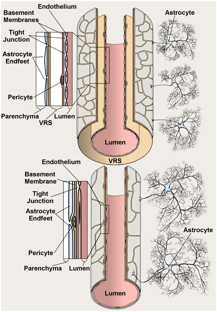
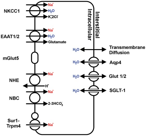
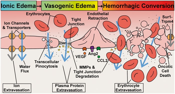
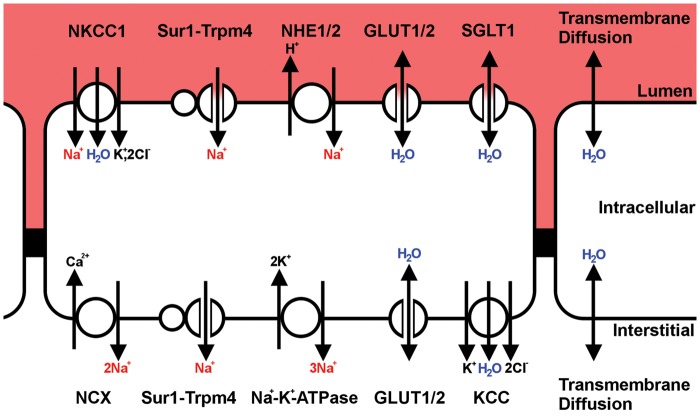
Advancements in molecular biology have led to a greater understanding of the individual proteins responsible for generating cerebral edema. In large part, the study of cerebral edema is the study of maladaptive ion transport. Following acute CNS injury, cells of the neurovascular unit, particularly brain endothelial cells and astrocytes, undergo a program of pre- and post-transcriptional changes in the activity of ion channels and transporters. These changes can result in maladaptive ion transport and the generation of abnormal osmotic forces that, ultimately, manifest as cerebral edema. This review discusses past models and current knowledge regarding the molecular and cellular pathophysiology of cerebral edema.

Keywords: Astrocytes; brain edema; capillaries; cerebrospinal fluid; endothelium.


Figures

Figure 1.
Figure 1.
Anatomy of cerebral arterioles (top) and capillary (bottom). The innermost layer of arterioles and capillaries is composed of a continuous layer of endothelial cells, linked by tight junctions, and bounded externally by a layer of basement membrane that contains pericytes; arterioles, but not capillaries, travel inside the perivascular Virchow Robin Space (VRS); the outermost layer of the blood brain barrier is composed of astrocyte endfeet, the terminal pads of large astrocyte processes.
Figure 2.
Figure 2.
Major routes for influx of ions and water in astrocytic cytotoxic edema. Schematic depiction of the major astrocytic transporters and channels that are implicated in the formation of cytotoxic edema; in regards to water transport, single-headed arrows denote water co-transport, while double-headed arrows denote passive water transport.
Figure 3.
Figure 3.
Phases and select mechanisms of endothelial dysfunction. In ionic edema, water flux (blue arrows) and ion flux (grey arrows) are mediated by plasmalemma channels and transporters; vasogenic edema, which includes extravasation of plasma proteins, but not erythrocytes, is mediated by transcellular channels, MMP degradation of tight junctions, and endothelial retraction, phenomena that are, in part, triggered by VEGF, Ang2, and CCL2 signaling; hemorrhagic transformation occurs due to structural failure of the vessel, driven by either complete degradation of tight junctions or Sur1-Trpm4-mediated oncotic cell death of endothelial cells.
Figure 4.
Figure 4.
Major routes for influx of ions and water in ionic edema. Schematic depiction of the major endothelial transporters and channels that have been implicated in the formation of ionic edema; in regards to water transport, single-headed arrows denote water co-transport, while double-headed arrows denote passive water transport.

References

    1. Kochanek KD, Xu J, Murphy SL, et al. Deaths: Final data for 2009. Natl Vital Stat Rep 2011; 60: 1–116. - PubMed
    1. Battey TW, Karki M, Singhal AB, et al. Brain edema predicts outcome after nonlacunar ischemic stroke. Stroke 2014; 45: 3643–3648. - PMC - PubMed
    1. Bernal W, Hyyrylainen A, Gera A, et al. Lessons from look-back in acute liver failure? A single centre experience of 3300 patients. J Hepatol 2013; 59: 74–80. - PubMed
    1. Donkin JJ, Vink R. Mechanisms of cerebral edema in traumatic brain injury: Therapeutic developments. Curr Opin Neurol 2010; 23: 293–299. - PubMed
    1. Abbott NJ. Comparative physiology of the blood-brain barrier. In: Bradbury MWB. (ed). Physiology and pharmacology of the blood-brain barrier, Berlin: Springer-Verlag, 1992, pp. 171–196.

Publication types

LinkOut - more resources