. 2016 Mar;245(3):414-26.
doi: 10.1002/dvdy.24376. Epub 2016 Jan 8.

Ectodermal Wnt controls nasal pit morphogenesis through modulation of the BMP/FGF/JNK signaling axis

Affiliations

Ectodermal Wnt controls nasal pit morphogenesis through modulation of the BMP/FGF/JNK signaling axis

Xiao-Jing Zhu et al. Dev Dyn. 2016 Mar.

Abstract

Background: Mutations of WNT3, WNT5A, WNT9B, and WNT11 genes are associated with orofacial birth defects, including nonsyndromic cleft lip with cleft palate in humans. However, the source of Wnt ligands and their signaling effects on the orofacial morphogenetic process remain elusive.

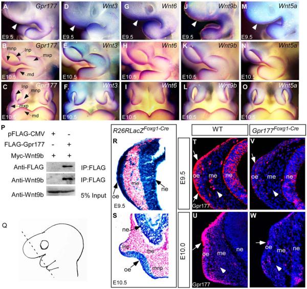
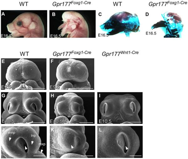
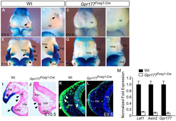
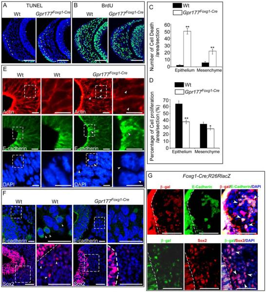
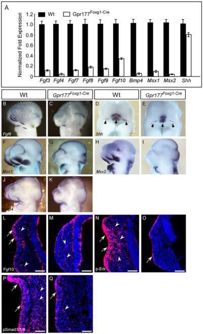
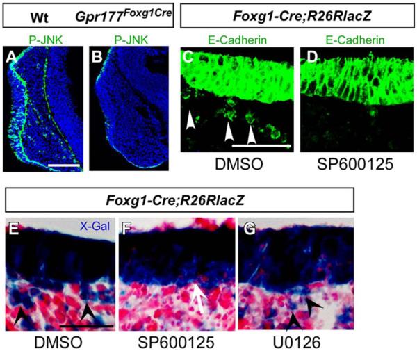
Results: Using Foxg1-Cre to impair Wnt secretion through the inactivation of Gpr177/mWls, we investigate the relevant regulation of Wnt production and signaling in nasal-facial development. Ectodermal ablation of Gpr177 leads to severe facial deformities resulting from dramatically reduced cell proliferation and increased cell death due to a combined loss of WNT, FGF and BMP signaling in the developing facial prominence. In the invaginating nasal pit, the Gpr177 disruption also causes a detrimental effect on migration of the olfactory epithelial cells into the mesenchymal region. The blockage of Wnt secretion apparently impairs the olfactory epithelial cells through modulation of JNK signaling.

Conclusions: Our study thus suggests the head ectoderm, including the facial ectoderm and the neuroectoderm, as the source of canonical as well as noncanonical Wnt ligands during early development of the nasal-facial prominence. Both β-catenin-dependent and -independent signaling pathways are required for proper development of these morphogenetic processes.

Keywords: Gpr177; JNK; Wntless; canonical Wnt; nasal pit; noncanonical Wnt.


Figures

Fig. 1
Fig. 1
Expression of Gpr177 and multiple Wnts in early facial primordial development and interaction of Gpr177. A–O: Whole-mount in situ hybridization shows that Gpr177, Wnt3, Wnt6, Wnt9b, and Wnt5a are expressed in the nasal prominences of E9.5 as lateral views (A,D,G,J,M, white arrowheads represent the nasal placode region) and E10.5 as lateral views (B,E,H,K,N) and frontal views (C,F,I,LO). P: Gpr177 coimmunoprecipitates with Wnt9b in vivo. HEK293 cells transfected with the indicated vectors were subjected to immunoprecipitation analysis with anti-FLAG antibody-coupled beads. Western blot was performed with anti-FLAG and Wnt9b antibodies. Q: A schematic showing the orientation of the tissue sections. R,S: X-gal staining shows that Foxg1-Cre activity is present in the facial epithelium and neuroepithelium at E9.5 (R) and E10.5 (S). Note the X-Gal positive stains in the mesenchymal cells underlying the thickening nasal placode at E9.5 (R). T–W: Immunofluorescence of Gpr177 expression in the facial epithelium, the neuroepithelium, and the underling mesenchyme (arrowheads) at E9.5 and E10.0. Arrows indicate positive staining of Gpr177 in the olfactory epithelium. Note that Gpr177 is almost deleted in the Gpr177Foxg1-Cre mutant at E10.0 (W). lnp: Lateral nasal process; mnp: medial nasal process; mxp: maxillary process; md: mandibular process; oe, olfactory epithelium; me, mesenchyme; ne, neuroepithelium.
Fig. 2
Fig. 2
Gpr177 is required for facial development. A,B: Foxg1-Cre mediated loss of ectodermal Gpr177 leads to the disruption of craniofacial structures as revealed by gross morphology. C,D: Alizarin Red and Alcian Blue staining of skeletons on embryos at E16.5. E–L: Scanning electron micrographs of the facial prominences at E9.5 and E10.5. Front facial views of wild-type (E,G,J) and Gpr177-deficient (F,H,K). Gpr177Foxg1-Cre embryos display dramatic defects in nasal pit invagination (arrowhead in K vs. J) and outgrowth of mnp, lnp, and mxp (K vs. J) at E10.5. In contrast, mesenchymal deletion of Gpr177 by Wnt1-Cre shows minor defects with formation of the nasal pit (arrowhead in L). lnp, lateral nasal process; mnp, medial nasal process; mx, maxillary process; md, mandibular process. Scale bars ¼ 500 mm in E–I; 200 mm in J–L.
Fig. 3
Fig. 3
Ectodermal Gpr177 loss-of-function leads to disruption of canonical Wnt signaling. A–H: Whole-mount X-gal staining shows significant reduction of TopGal activity in E9.5 and E10.5 Gpr177Foxg1-Cre embryos (p E10.5 embryos shows that TopGal signal is present in the epithelium (arrows in I). Note the loss of staining and the inhibited outgrowth of lnp, mnp, and mxp in E10.5 Gpr177Foxg1-Cre embryos (J). The olfactory epithelial invagination of mutant embryos is also significantly inhibited (arrowheads, J compared with wild-type I). K,L: Immunostaining with active β-Catenin shows that the presence of active β-catenin in facial epithelium, neuroepithelium, and facial mesenchyme (arrows and arrowhead in K) is significantly reduced upon the deletion of epithelial Gpr177 (L). The dash line indicates the boundary between olfactory epithelium and mesenchyme. M: Quantitative RT-PCR analysis of gene expression in facial prominence of E10.25 embryos. Expression of Lef1, Axin2, and Gpr177 is significantly down-regulated in Gpr177Foxg1-Cre embryos. lnp, lateral nasal process; mnp, medial nasal process; mx, maxillary process; md, mandibular process; oe, olfactory epithelium; op, olfactory primordium; me, mesenchyme; ne, neuroepithelium.
Fig. 4
Fig. 4
Deletion of ectodermal Gpr177 leads to increased cell death, reduced cell proliferation, and inhibition of nasal pit invagination. A: TUNEL staining shows that cell death is significantly increased in both the olfactory epithelium and the mesenchyme of Gpr177Foxg1-Cre embryos at E9.5. B: BrdU staining shows reduced cell proliferation in both the olfactory epithelium and the mesenchyme of E9.5 Gpr177Foxg1-Cre embryos. C,D: Comparison of percentage of cell death (C) and cell proliferation (D) in the designated area of the nasal pit in the control and mutant embryos. Standard deviation values are presented as error bars. *P < 0.05. **P < 0.01. E: Immunofluorescence analysis on the nasal pit epithelium of E10.5 embryos for Actin, E-cadherin and DAPI show defective cell shape and cell movement in the nasal pit epithelium of Gpr177Foxg1-Cre embryos. Note the irregular Actin fibers, aggregated E-cadherin, and round-shaped nuclei in the epithelium of Gpr177Foxg1-Cre mutants as indicated by arrowheads. F: We found a dramatic reduction in the number of E-cadherin positive cells in the Gpr177 mutants in the facial mesenchyme as revealed by confocal microscopy analysis at E10.5. Immunostaining of Sox2 for neural stem cells displays a migration defect of E10.5 Gpr177Foxg1-Cre olfactory epithelial cells. The magnified images of the regions within the dotted boxes in E and F are shown to the right of the original photographs. G: Immunofluorescence demonstrates colocalization of Foxg1-Cre positive epithelial cells (β-gal) with E-cadherin or Sox2 positive cells in the mesenchyme of E9.5 embryos. Cells that double-stained with ß-gal and E-cadherin or Sox2 are indicated by arrowheads. The dotted line indicates the boundary between epithelium and mesenchyme. Scale bars ¼ 100 mm in A,B; 10 mm in E; 25 mm in F upper panels,100 mm in lower panels; 50 mm in G.
Fig. 5
Fig. 5
FGF, BMP, and SHH signaling pathways are altered in the facial prominence of Gpr177Foxg1-Cre embryos. A: Quantitative RT-PCR analysis of gene expression in the facial prominences of E10.25 embryos. Expression of Fgf3, Fgf4 Fgf7, Fgf8, Fgf9, Fgf10, Bmp4, Msx1, and Msx2 is significantly down-regulated in Gpr177Foxg1-Cre embryos. By contrast, expression of Shh is mildly decreased. B–K: Whole-mount in situ analysis shows that expression of Fgf8, Shh, Msx1, Msx2, and Bmp4, in the facial prominences of E10.5 Gpr177Foxg1-Cre embryos as presented in sagittal views. Note that the expression of Fgf8, Msx1, and Msx2, is abolished completely in lnp, mnp, and mxp of Gpr177 mutant embryos. D,E: Frontal facial views of the wild-type and mutant embryos show that in Gpr177Foxg1-Cre embryos, the expression of Shh is retained in the FEZ (Arrowheads); however, its expression region in telencephalon and diencephalon becomes smaller (Arrows). Immunofluorescence analysis on sections of E10.25 facial primordia for Fgf10 (L,M), p-Erk (N,O), and pSmad1/5/8 (P,Q). Expression of Fgf10 protein is reduced in both the epithelium (arrows) and the mesenchyme (arrowheads) of embryos lacking epithelial Gpr177. Note that, in the facial primordia of wild-type embryos, distribution of Fgf10 proteins is concentrated at the sub-epithelial mesenchyme where the epithelium recesses (L). However, Fgf10 proteins are scattered in the facial mesenchyme of Gpr177Foxg1-Cre embryos (M). Phosphorylation of Erk of Gpr177Foxg1-Cre embryos is significantly reduced in the epithelium and diminished in the mesenchyme (O vs. wild-type N). Phosphorylation of Smad1/5/8 of Gpr177Foxg1-Cre embryos is reduced in the olfactory epithelium and diminished in the mesenchyme (Q vs. P). Arrows and arrowheads point out positive staining in the epithelium and the mesenchyme, respectively. Scale bars ¼ 100 mm. FEZ, frontonasal ectodermal zone.
Fig. 6
Fig. 6
A Gpr177/JNK signaling is required for nasal epithelial cell migration. A,B: Immunofluorescent staining shows that phosphorylated JNK is reduced in the epithelium and completely abolished in the mesenchyme of Gpr177Foxg1-Cre embryos (B) comparing with the wild-type (A). C–G: Immunofluorescence and X-gal staining on the sections of R26RLacZFoxg1-Cre facial explants shows that the epithelial cell migration is inhibited with treatment of SP600125 (D, F vs. C, E). Note that the migrating cells with positive staining are present in dimethyl sulfoxide group (C,E) and U0126 (G), but not in SP600125 (D,F) group. Arrowheads indicate migrating epithelial cells. Scale bars ¼ 100 mm in A,B; 50 mm in C–G.

Similar articles

Cited by

References

    1. Ahtiainen L, Lefebvre S, Lindfors PH, Renvoise E, Shirokova V, Vartiainen MK, Thesleff I, Mikkola ML. Directional cell migration, but not proliferation, drives hair placode morphogenesis. Dev Cell. 2014;28:588–602. - PubMed
    1. Al Alam D, Green M, Tabatabai Irani R, Parsa S, Danopoulos S, Sala FG, Branch J, El Agha E, Tiozzo C, Voswinckel R, Jesudason EC, Warburton D, Bellusci S. Contrasting expression of canonical Wnt signaling reporters TOPGAL, BATGAL and Axin2(LacZ) during murine lung development and repair. PLoS One. 2011;6:e23139. - PMC - PubMed
    1. Bachler M, Neubuser A. Expression of members of the Fgf family and their receptors during midfacial development. Mech Dev. 2001;100:313–316. - PubMed
    1. Banziger C, Soldini D, Schutt C, Zipperlen P, Hausmann G, Basler K. Wntless, a conserved membrane protein dedicated to the secretion of Wnt proteins from signaling cells. Cell. 2006;125:509–522. - PubMed
    1. Brugmann SA, Goodnough LH, Gregorieff A, Leucht P, ten Berge D, Fuerer C, Clevers H, Nusse R, Helms JA. Wnt signaling mediates regional specification in the vertebrate face. Development. 2007;134:3283–3295. - PubMed

Publication types

MeSH terms

LinkOut - more resources