. 2015;20(12):121312.
doi: 10.1117/1.JBO.20.12.121312.

Rapid prototyping of biomimetic vascular phantoms for hyperspectral reflectance imaging

Affiliations

Rapid prototyping of biomimetic vascular phantoms for hyperspectral reflectance imaging

Pejhman Ghassemi et al. J Biomed Opt. 2015.

Abstract

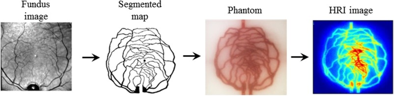
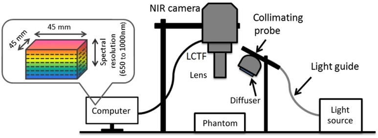
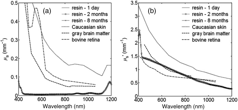
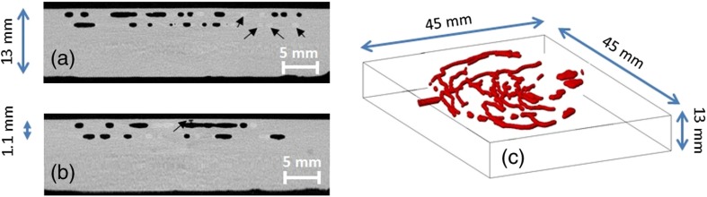
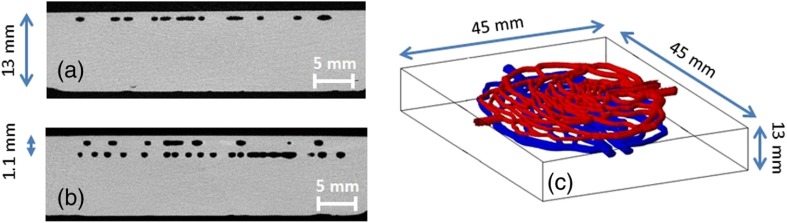
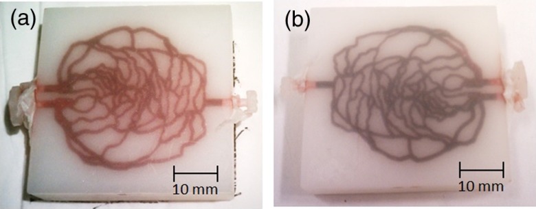
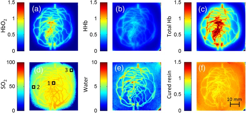
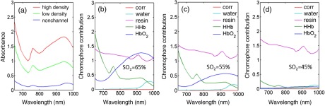
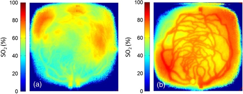
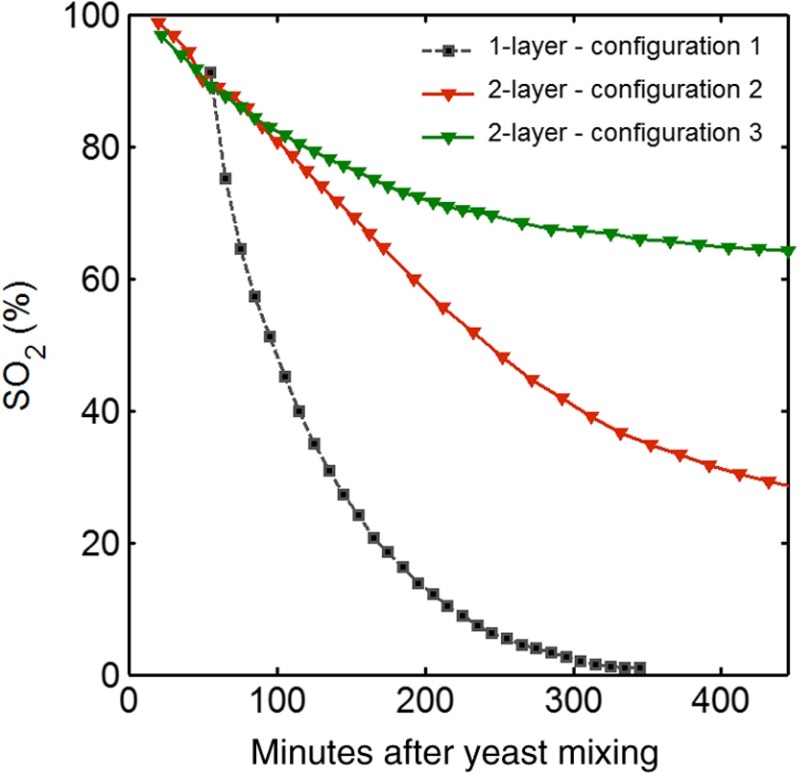
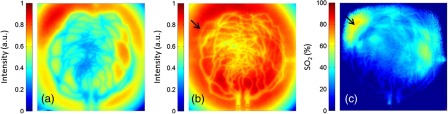
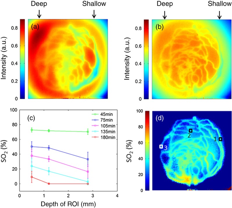
The emerging technique of rapid prototyping with three-dimensional (3-D) printers provides a simple yet revolutionary method for fabricating objects with arbitrary geometry. The use of 3-D printing for generating morphologically biomimetic tissue phantoms based on medical images represents a potentially major advance over existing phantom approaches. Toward the goal of image-defined phantoms, we converted a segmented fundus image of the human retina into a matrix format and edited it to achieve a geometry suitable for printing. Phantoms with vessel-simulating channels were then printed using a photoreactive resin providing biologically relevant turbidity, as determined by spectrophotometry. The morphology of printed vessels was validated by x-ray microcomputed tomography. Channels were filled with hemoglobin (Hb) solutions undergoing desaturation, and phantoms were imaged with a near-infrared hyperspectral reflectance imaging system. Additionally, a phantom was printed incorporating two disjoint vascular networks at different depths, each filled with Hb solutions at different saturation levels. Light propagation effects noted during these measurements—including the influence of vessel density and depth on Hb concentration and saturation estimates, and the effect of wavelength on vessel visualization depth—were evaluated. Overall, our findings indicated that 3-D-printed biomimetic phantoms hold significant potential as realistic and practical tools for elucidating light–tissue interactions and characterizing biophotonic system performance.


Figures

Fig. 1
Fig. 1
Phantom fabrication and preparation procedure, from left to right: human retinal vasculature fundus image, two-dimensional (2-D) segmented vascular map (© 2012 OSA. Reprinted with permission from the Optical Society of America), three-dimensional (3-D)-printed vascular phantom with hemoglobin (Hb)-filled channel, and hyperspectral reflectance imaging (HRI) result processed to estimate oxyhemoglobin (HbO2) concentration.
Fig. 2
Fig. 2
Diagram of the HRI system.
Fig. 3
Fig. 3
(a) Absorption coefficient and (b) reduced scattering coefficient of a white 3-D-printed slab. Results of measurements over a period of 8 months after 3-D printing are compared to biological tissues.
Fig. 4
Fig. 4
μ-Computed tomography (μ-CT) validation of a defective phantom: (a) cross-sectional image of two-layer phantom, arrows point to the unshaped channels; (b) cross-sectional image from the same phantom with a surface leakage, where the arrow points to the leaking area; and (c) 3-D view with bottom layer omitted for clarity.
Fig. 5
Fig. 5
μ-CT validation of viable phantoms: cross-sectional images of (a) one-layer and (b) two-layer vascular phantoms and (c) a 3-D view of the two-layer phantom.
Fig. 6
Fig. 6
Photographs of one-layer vascular phantom at (a) 30 min and (b) 120 min after solution injection.
Fig. 7
Fig. 7
One-layer phantom chromophore concentrations and saturation maps 75 min after adding the HbO2+yeast solution: (a) HbO2, (b) deoxyhemoglobin (HHb), (c) total Hb, (e) water, and (f) cured resin. The hemoglobin saturation levels (SO2) map (d) is marked with three regions of interest (ROIs) used to generate results in Fig. 8.
Fig. 8
Fig. 8
Sample HRI spectral data measured from the one-layer vascular phantom 75 min after solution injection, including absorbance curves for regions with (a) high- and low-channel densities and nonchannel area (the effect of phantom matrix is removed from the absorbance data for demonstration purpose) and (b–d) chromophore contributions after NNLS unmixing. Results are shown for channel regions with (b) relatively high and (c) low densities, and (d) a nonchannel area.
Fig. 9
Fig. 9
HRI-calculated SO2 data in two-layer phantoms, 4 h after yeast mixing for (a) experimental configuration 2 (Video 1, AVI, 1.2 MB) [URL: http://dx.doi.org/10.1117/1.JBO.20.12.121312.1] and (b) configuration 3 (Video 2, AVI, 1.5 MB) [URL: http://dx.doi.org/10.1117/1.JBO.20.12.121312.2].
Fig. 10
Fig. 10
Phantoms oxygen desaturation over time. In configurations 2 and 3, SO2 values are fixed at approximately 100% in the bottom and top layers, respectively.
Fig. 11
Fig. 11
Normalized reflectance images at (a) λ=650  nm and (b) λ=1000  nm (Video 3, AVI, 1.3 MB) [URL: http://dx.doi.org/10.1117/1.JBO.20.12.121312.3] and (c) SO2 map at 8 h after adding yeast to the HbO2 solution (experimental configuration 2). Arrows indicate the effect of deep channel on both reflectance image and saturation map.
Fig. 12
Fig. 12
Vascular phantom with tilted channels: normalized reflectance images at (a) 650 nm and (b) 1000 nm; (c) SO2 level versus depth for different time points after adding yeast to Hb solution; and (d) SO2 map at 105 min after adding yeast to the Hb solution [ROIs used to generate the graph (c) are marked on the map].

References

    1. Kanawade R., et al. , “In vivo monitoring of vessel density pattern in skin phantoms for the application of early sign of shock detection by using diffuse reflectance spectroscopy,” Proc. SPIE 7890, 789009 (2011).PSISDG10.1117/12.874208 - DOI
    1. Chen C., et al. , “Preparation of a skin equivalent phantom with interior micron-scale vessel structures for optical imaging experiments,” Biomed. Opt. Express 5(9), 3140–3149 (2014).BOEICL10.1364/BOE.5.003140 - DOI - PMC - PubMed
    1. Baxi J., et al. , “Retina-simulating phantom for optical coherence tomography,” J. Biomed. Opt. 19(2), 021106 (2014).JBOPFO10.1117/1.JBO.19.2.021106 - DOI - PubMed
    1. Lurie K. L., et al. , “Three-dimensional, distendable, bladder phantom for optical coherence tomography and white light cystoscopy,” J. Biomed. Opt. 19(3), 036009 (2014).JBOPFO10.1117/1.JBO.19.3.036009 - DOI - PMC - PubMed
    1. Nouvong A., et al. , “Evaluation of diabetic foot ulcer healing with hyperspectral imaging of oxyhemoglobin and deoxyhemoglobin,” Diabetes Care 32(11), 2056–2061 (2009).DICAD210.2337/dc08-2246 - DOI - PMC - PubMed

Publication types