. 2016 Jan 1;310(1):F15-26.
doi: 10.1152/ajprenal.00226.2015. Epub 2015 Oct 14.

Cell-specific regulation of L-WNK1 by dietary K

Affiliations

Cell-specific regulation of L-WNK1 by dietary K

Tennille N Webb et al. Am J Physiol Renal Physiol. .

Abstract

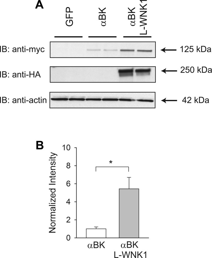
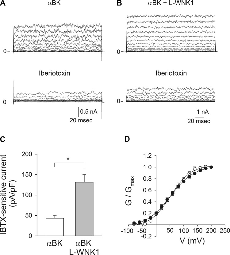
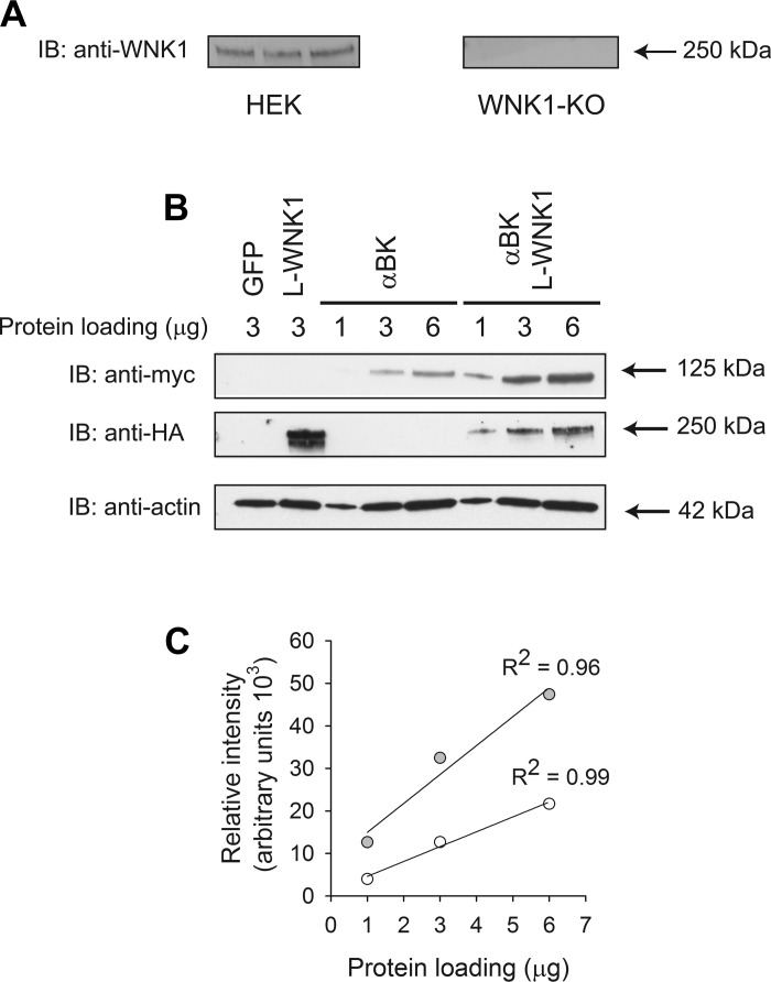
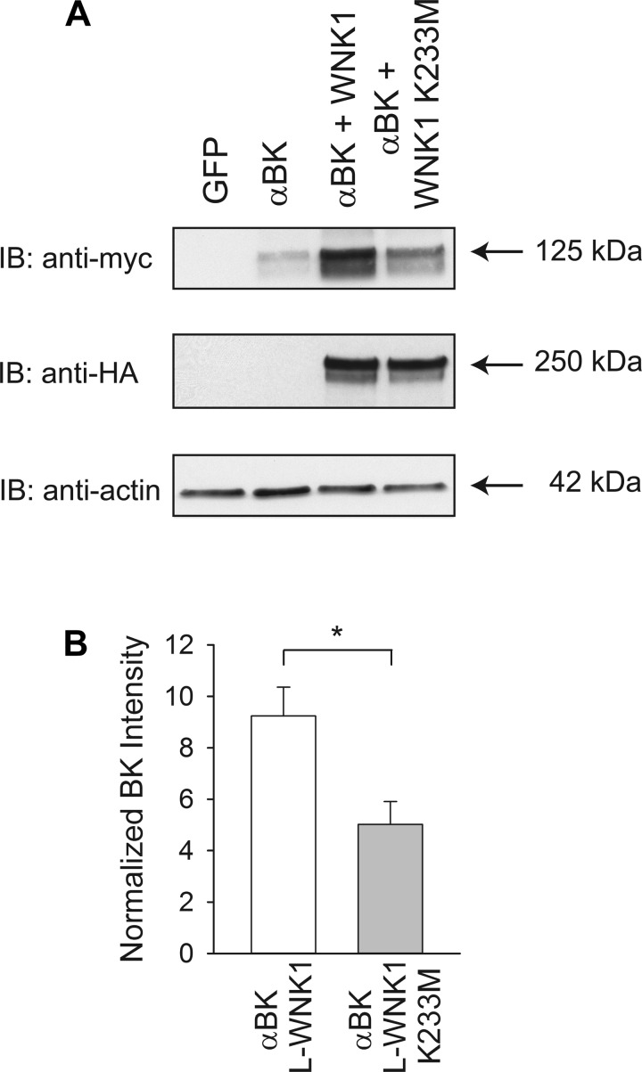
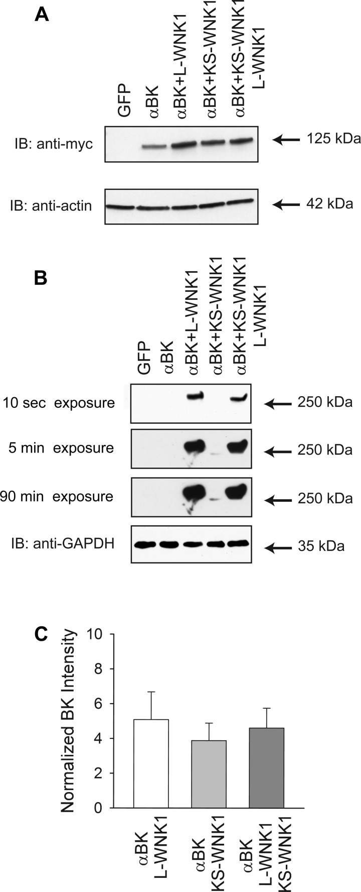
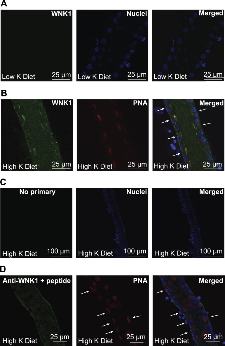
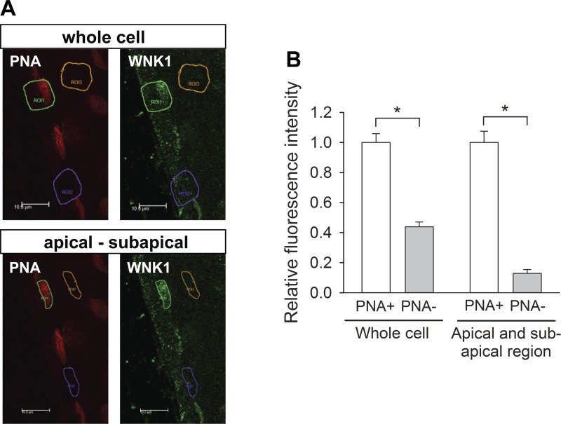
Flow-induced K(+) secretion in the aldosterone-sensitive distal nephron is mediated by high-conductance Ca(2+)-activated K(+) (BK) channels. Familial hyperkalemic hypertension (pseudohypoaldosteronism type II) is an inherited form of hypertension with decreased K(+) secretion and increased Na(+) reabsorption. This disorder is linked to mutations in genes encoding with-no-lysine kinase 1 (WNK1), WNK4, and Kelch-like 3/Cullin 3, two components of an E3 ubiquitin ligase complex that degrades WNKs. We examined whether the full-length (or "long") form of WNK1 (L-WNK1) affected the expression of BK α-subunits in HEK cells. Overexpression of L-WNK1 promoted a significant increase in BK α-subunit whole cell abundance and functional channel expression. BK α-subunit abundance also increased with coexpression of a kinase dead L-WNK1 mutant (K233M) and with kidney-specific WNK1 (KS-WNK1), suggesting that the catalytic activity of L-WNK1 was not required to increase BK expression. We examined whether dietary K(+) intake affected L-WNK1 expression in the aldosterone-sensitive distal nephron. We found a paucity of L-WNK1 labeling in cortical collecting ducts (CCDs) from rabbits on a low-K(+) diet but observed robust staining for L-WNK1 primarily in intercalated cells when rabbits were fed a high-K(+) diet. Our results and previous findings suggest that L-WNK1 exerts different effects on renal K(+) secretory channels, inhibiting renal outer medullary K(+) channels and activating BK channels. A high-K(+) diet induced an increase in L-WNK1 expression selectively in intercalated cells and may contribute to enhanced BK channel expression and K(+) secretion in CCDs.

Keywords: high-conductance calcium-activated potassium channels; kidney-specific with-no-lysine kinase 1; long with-no-lysine kinase 1; potassium adaptation; pseudohypoaldosteronism; with-no-lysine kinase 1.


Figures

Fig. 1.
Fig. 1.
Long (L) with-no-lysine kinase 1 (WNK1) expression increases high-conductance Ca2+-activated K+ (BK) α-subunit abundance. HEK293 cells were transfected with green fluorescent protein (GFP, control), BK α-subunit, or BK α-subunit and L-WNK1. A: L-WNK1 expression increases whole cell BK α-subunit abundance. Whole cell lysates were subjected to immunoblot with an anti-myc antibody to detect BK α-subunit, an anti-hemagglutinin (HA) antibody to detect L-WNK1, or an anti-actin antibody. B: summary of analyses of BK α-subunit whole cell expression. Immunoblots were quantified as described in materials and methods (n = 11, *P < 0.001) (Student's t-test). Results were normalized to BK α-subunit expression in the absence of exogenous L-WNK1.
Fig. 2.
Fig. 2.
L-WNK1 expression increases whole cell BK channel activity. HEK293 cells were transfected with BK α-subunit alone or with L-WNK1. Patch-clamp studies were performed with the amphotericin B perforated whole cell technique (A–C) or with conventional whole cell patch-clamp (D), as described in materials and methods. BK currents were evoked by voltage steps from −80 to 100 mV in 10-mV increments from a holding potential of −60 mV (A–C) or by voltage steps from −80 to 200 mV in 20-mV increments from a holding potential of −80 mV (D). A and B: representative tracings obtained in the absence or presence of 100 nM iberiotoxin (IBTX) from cells transfected with myc-tagged BK α-subunit alone (A) or with L-WNK1 (B). C: summary of the effect of L-WNK1 on IBTX-sensitive whole cell currents measured at +80 mV (n = 6–7, *P < 0.01) (Student's t-test). D: normalized steady-state conductance-voltage (G–V) relationships for cells transfected with BK α-subunit alone (open circles) or with L-WNK1 (closed circles) (n = 9–11). Data were fitted to a Boltzmann function as described in materials and methods.
Fig. 3.
Fig. 3.
L-WNK1 increases whole cell BK expression in HEK293 L-WNK1 knockout (KO) cells. A: lack of endogenous L-WNK1 expression in HEK293 L-WNK1 KO cells. Lysates obtained from HEK293 and HEK293 L-WNK1 KO cells were subjected to immunoblot with an anti-L-WNK1 antibody as described in materials and methods. Each lane was loaded with 12 μg of protein. B: L-WNK1 increases whole cell BK expression in HEK293 L-WNK1 KO cells. Whole cell lysates from HEK293 L-WNK1 KO cells transfected with GFP (control), L-WNK1, BK α-subunit, or BK α-subunit with L-WNK1 were subjected to immunoblot with an anti-HA antibody to detect L-WNK1, an anti-myc antibody to detect BK α-subunit, or an anti-actin antibody. Lanes were loaded with 1–6 μg of protein. Representative of 4 independent experiments. C: linearity of the Western blot detection system for BK α-subunit abundance. The relative intensity of the bands in the immunoblot shown in B was calculated with ImageJ. Data from cells transfected with BK α-subunit alone are shown as open symbols while those from cells transfected with the BK α-subunit and L-WNK1 are shown as gray symbols. Data were analyzed by linear regression.
Fig. 4.
Fig. 4.
L-WNK1 increases whole cell BK expression in the absence of kinase activity. A: kinase dead L-WNK1 (K233M) increases whole cell BK α-subunit abundance. Whole cell lysates from HEK293 L-WNK1 KO cells transfected with GFP (control), BK α-subunit, or BK α-subunit with either wild-type L-WNK1 or L-WNK1 K233M were subjected to immunoblot with an anti-HA antibody to detect L-WNK1, an anti-myc antibody to detect BK α-subunit, or an anti-actin antibody. B: summary of analyses of BK α-subunit whole cell expression. Immunoblots were quantified as described in materials and methods (n = 8, *P < 0.01) (Student's t-test). Results were normalized to BK α-subunit expression in the absence of exogenous L-WNK1 or L-WNK1 K233M. Levels of expression of BK α-subunit were significantly increased by coexpression of L-WNK1 (P < 0.001) or by coexpression of the K233M mutant (P < 0.05) (Kruskal-Wallis test followed by Dunn's multiple-comparisons test).
Fig. 5.
Fig. 5.
kidney-specific (KS) WNK1 increases whole cell BK expression. A: both L-WNK1 and KS-WNK1 increase whole cell BK α-subunit expression. Lysates from HEK293 L-WNK1 KO cells transfected with GFP (control), BK α-subunit, or BK α-subunit with either wild-type L-WNK1, KS-WNK1, or both L-WNK1 and KS-WNK1 were subjected to immunoblot with an anti-myc antibody to detect BK α-subunit, an anti-HA antibody to detect L-WNK1 or KS-WNK1, or an anti-actin antibody. B: reduced levels of KS-WNK1 compared with levels of L-WNK1 in HEK293 L-WNK1 knockout cells. Whole cell lysates from cells transfected as indicated above were subjected to immunoblot with an anti-HA antibody to detect L-WNK1 or KS-WNK1 or an anti-GAPDH antibody. Note that the detection of KS-WNK1 required significantly longer exposure times than were required to detect L-WNK1. For this experiment, cells were harvested 24 h posttransfection. C: summary of analyses of BK α-subunit whole cell expression. Immunoblots were quantified as described in materials and methods (n = 9). Results were normalized to BK α-subunit expression in the absence of exogenous L-WNK1 or KS-WNK1. Levels of expression of BK α-subunit were significantly increased by coexpression of L-WNK1 (P < 0.001), KS-WNK1 (P < 0.01), or both L-WNK1 and KS-WNK1 (P < 0.001) (Kruskal-Wallis test followed by Dunn's multiple-comparisons test).
Fig. 6.
Fig. 6.
High-K+ (HK) diet increases L-WNK1 expression in rabbit cortical collecting ducts (CCDs). Single CCDs isolated from New Zealand White (NZW) rabbits fed a HK or a low-K+ (LK) diet for 10 days were microperfused and fixed for immunofluorescence microscopy. Immunolabeling was performed as indicated in materials and methods. A: L-WNK1 expression in CCDs from LK-fed animals. Immunostaining for L-WNK1 was difficult to visualize in CCDs from rabbits fed a LK diet. B: expression of L-WNK1 in rabbit CCD intercalated cells after 10 days on a HK diet. Intercalated cells (arrows) were identified by their apical labeling with rhodamine-conjugated peanut agglutinin (PNA). C: negative control incubated with Alexa488-conjugated goat anti-rabbit IgG antibody but not with primary antibody. D: antigen competition. Only modest basal membrane labeling was seen when the anti-L-WNK1 antibody was preincubated with a WNK1 peptide fragment (arrows indicate PNA+ cells).
Fig. 7.
Fig. 7.
Quantitation of L-WNK1 expression in the apical and subapical regions of PNA-positive (PNA+) and PNA-negative (PNA) cells. A: representative confocal photomicrographs of L-WNK1 immunolabeling (green) in immunoperfused CCD isolated from a HK-fed rabbit. Type B intercalated cells are labeled with apical PNA (red). Rectangles identify PNA-positive β-intercalated cells (green), PNA-negative cells (purple), and lumen (background, yellow). B: relative fluorescence intensity of PNA+ and PNA cells. The L-WNK1 immunofluorescence intensity of PNA+ cells (1.00 ± 0.06) was 2.3-fold greater than that of PNA cells (0.44 ± 0.03). The relative fluorescence intensity of the apical and subapical region of PNA+ cells (1.00 ± 0.07) was 7.8-fold greater than that of PNA cells (0.13 ± 0.02). Fluorescence intensity was normalized to the average signal in PNA+ cells. Median comparisons between cell types made using a Mann-Whitney test. *P ≤ 0.001; n = 20 (whole cell) and n = 29 (apical-subapical region) (PNA+ and PNA, each) in 4 tubules.

References

    1. Alessi DR, Zhang J, Khanna A, Hochdorfer T, Shang Y, Kahle KT. The WNK-SPAK/OSR1 pathway: master regulator of cation-chloride cotransporters. Sci Signal 7: 3, 2014. - PubMed
    1. Atkinson NS, Robertson GA, Ganetzky B. A component of calcium-activated potassium channels encoded by the Drosophila slo locus. Science 253: 551–555, 1991. - PubMed
    1. Beck FX, Dorge A, Blumner E, Giebisch G, Thurau K. Cell rubidium uptake: a method for studying functional heterogeneity in the nephron. Kidney Int 33: 642–651, 1988. - PubMed
    1. Boim MA, Ho K, Shuck ME, Bienkowski MJ, Block JH, Slightom JL, Yang Y, Brenner BM, Hebert SC. ROMK inwardly rectifying ATP-sensitive K+ channel. II. Cloning and distribution of alternative forms. Am J Physiol Renal Fluid Electrolyte Physiol 268: F1132–F1140, 1995. - PubMed
    1. Boyden LM, Choi M, Choate KA, Nelson-Williams CJ, Farhi A, Toka HR, Tikhonova IR, Bjornson R, Mane SM, Colussi G, Lebel M, Gordon RD, Semmekrot BA, Poujol A, Valimaki MJ, De Ferrari ME, Sanjad SA, Gutkin M, Karet FE, Tucci JR, Stockigt JR, Keppler-Noreuil KM, Porter CC, Anand SK, Whiteford ML, Davis ID, Dewar SB, Bettinelli A, Fadrowski JJ, Belsha CW, Hunley TE, Nelson RD, Trachtman H, Cole TR, Pinsk M, Bockenhauer D, Shenoy M, Vaidyanathan P, Foreman JW, Rasoulpour M, Thameem F, Al-Shahrouri HZ, Radhakrishnan J, Gharavi AG, Goilav B, Lifton RP. Mutations in kelch-like 3 and cullin 3 cause hypertension and electrolyte abnormalities. Nature 482: 98–102, 2012. - PMC - PubMed

Publication types

MeSH terms

Substances

LinkOut - more resources