Review
. 2016 Jul;283(14):2676-89.
doi: 10.1111/febs.13624. Epub 2016 Jan 6.

The deadly landscape of pro-apoptotic BCL-2 proteins in the outer mitochondrial membrane

Affiliations
Review

The deadly landscape of pro-apoptotic BCL-2 proteins in the outer mitochondrial membrane

Mark P A Luna-Vargas et al. FEBS J. 2016 Jul.

Abstract

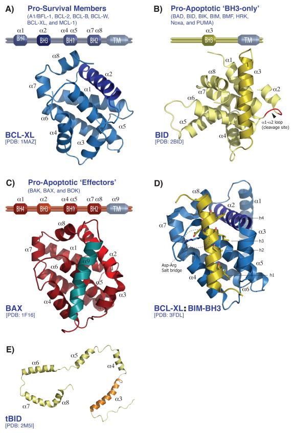
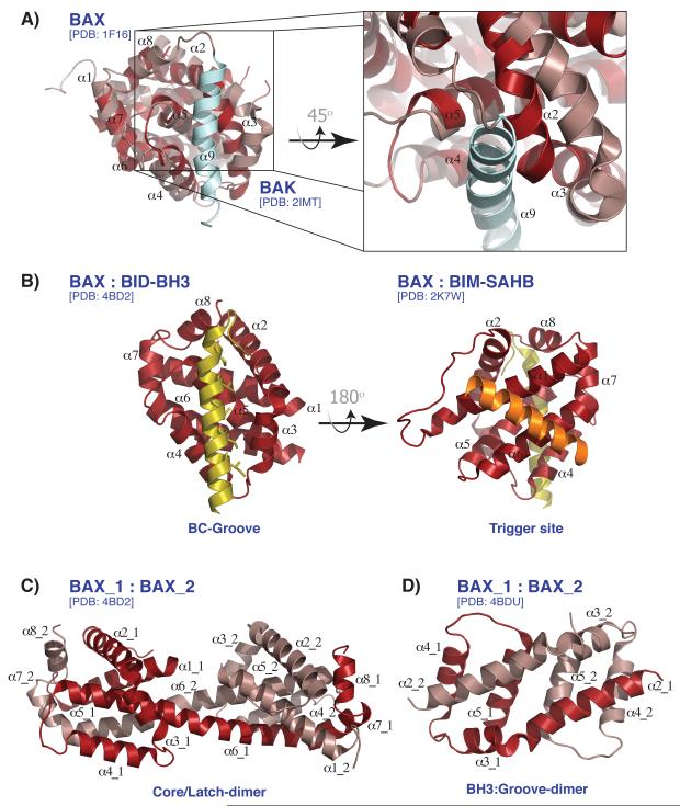
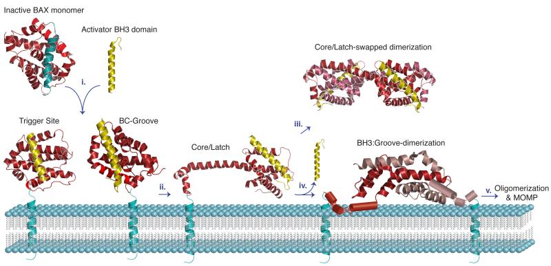
Apoptosis is a biological process that removes damaged, excess or infected cells through a genetically controlled mechanism. This process plays a crucial role in organismal development, immunity and tissue homeostasis, and alterations in apoptosis contribute to human diseases including cancer and auto-immunity. In the past two decades, significant efforts have focused on understanding the function of the BCL-2 proteins, a complex family of pro-survival and pro-apoptotic α-helical proteins that directly control the mitochondrial pathway of apoptosis. Diverse structural investigations of the BCL-2 family members have broadened our mechanistic understanding of their individual functions. However, an often over-looked aspect of the mitochondrial pathway of apoptosis is how the BCL-2 family specifically interacts with and targets the outer mitochondrial membrane to initiate apoptosis. Structural information on the relationship between the BCL-2 family and the outer mitochondrial membrane is missing; likewise, knowledge of the biophysical mechanisms by which the outer mitochondrial membrane affects and effects apoptosis is lacking. In this mini-review, we provide a current overview of the BCL-2 family members and discuss the latest structural insights into BAK/BAX activation and oligomerization in the context of the outer mitochondrial membrane and mitochondrial biology.

Keywords: BAK; BAX; BCL-2 family; MOMP; Structure; apoptosis; lipids; membrane; mitochondria; mitochondrial landscape.


Figures

Figure 1
Figure 1. Structural overview of BCL-2 family
(A-C) Domain architecture of the pro-survival members (A), the pro-apoptotic ‘BH3-only’ (B), and the pro-apoptotic ‘Effectors’ (C) in blue, yellow and red, respectively. The corresponding three-dimensional structures are depicted (BCL-XL, BID, and BAX). (D) Cartoon representation of BCL-XL bound to BIM-BH3 peptide showing the four hydrophobic residues (h1-h4) of the BIM-BH3 domain and the Asp-Arg salt bridge involved in the interaction with the BC-groove of BCL-XL. (E) The C-shape conformation of the NMR structure of tBID (pale yellow) presented with the BH3 domain (α3) colored orange. The tBID structure is determined in the presence of micelles and presumably represents an activated conformation of tBID, in which each α helix interacts with the micelles but no longer with each other, as shown in (B).
Figure 2
Figure 2. Activation, rearrangement, and dimerization of BAX
(A) BAK and BAX structures superimposed shown in dark red and salmon, respectively. The transmembrane of BAX (α9, colored in light blue) sits in the BC-groove. Zoomed in, the superposition shows a narrow BAK BC-groove restricting the docking of the α9 helix. (B) BAX activation sites are shown. BAX bound to BID-BH3 peptide (yellow) in the BC-groove (left) and bound to BIM-SAHB peptide (orange) at the ‘trigger site’ of BAX on the opposite side of the BC-groove (right). (C) The ‘Core/Latch-dimer’ of BAX is shown with α6-α8 being interchanged between two BAX monomers. (D) The ‘BH3:Groove-dimer’ of BAX is shown with α2 of each monomer engaged with the BC-groove of the other.
Figure 3
Figure 3. Model for the structural transitions of BAX activation, rearrangement, and dimerization leading to oligomerization and MOMP
The BAX structures are shown in cartoon representation and color coded as in Figure 2. In step i. the inactivated BAX monomer interacts with the BH3 domain of an activator BH3-only protein via the BC-groove or the α1-α6 the trigger site. ii. BAX activation leads to major structural rearrangement in which the latch domain (α6-α8) detaches from the core. iii. In solution, two BAX monomers can engage in the core/latch swapped dimerization. iv. At the OMM, BAX engages in BH3:groove dimerization, after disassociation of the activator BH3 domain. The structure of the symmetrical α2-α5 core dimer of BAX revealed an extensive hydrophobic surface defined by residues from the α4 and α5 helices that stabilize the hydrophobic core of the inactivated BAX monomer. v. The formation of BAX oligomers remains unclear, although an interface involving helix α6 has been proposed. One of the emerging models suggests that the α2-α5 core dimers perturb the OMM through their hydrophobic α4-α5 surface, thereby inducing MOMP.

References

    1. Kerr JF, Wyllie AH, Currie AR. Apoptosis: a basic biological phenomenon with wide-ranging implications in tissue kinetics. Br J Cancer. 1972;26:239–57. - PMC - PubMed
    1. Hotchkiss RS, Strasser A, McDunn JE, Swanson PE. Cell death in disease: mechanisms and emerging therapeutic concepts. N Engl J Med. 2009;361:1670–83. - PMC - PubMed
    1. Riedl SJ, Galvesen GS. The apoptosome: signaling platform of cell death. Nat Rev Mol Cell Biol. 2007;8:405–13. - PubMed
    1. Tait SW, Green DR. Mitochondria and cell death: outer membrane permeabilization and beyond. Nat Rev Mol Cell Biol. 2010;11:621–32. - PubMed
    1. Yuan S, Akey CW. Apoptosome structure, assembly, and procaspase activation. Structure. 2013;21:501–15. - PMC - PubMed

Publication types

MeSH terms

Substances