

Longitudinal assessment of white matter abnormalities following sports-related concussion
- PMID: 26663463
- PMCID: PMC6867335
- DOI: 10.1002/hbm.23072
Longitudinal assessment of white matter abnormalities following sports-related concussion
Abstract
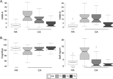
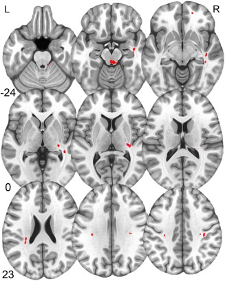
There is great interest in developing physiological-based biomarkers such as diffusion tensor imaging to aid in the management of concussion, which is currently entirely dependent on clinical judgment. However, the time course for recovery of white matter abnormalities following sports-related concussion (SRC) is unknown. We collected diffusion tensor imaging and behavioral data in forty concussed collegiate athletes on average 1.64 days (T1; n = 33), 8.33 days (T2; n = 30), and 32.15 days post-concussion (T3; n = 26), with healthy collegiate contact-sport athletes (HA) serving as controls (n = 46). We hypothesized that fractional anisotropy (FA) would be increased acutely and partially recovered by one month post-concussion. Mood symptoms were assessed using structured interviews. FA differences were assessed using both traditional and subject-specific analyses. An exploratory analysis of tau plasma levels was conducted in a subset of participants. Results indicated that mood symptoms improved over time post-concussion, but remained elevated at T3 relative to HA. Across both group and subject-specific analyses, concussed athletes exhibited increased FA in several white matter tracts at each visit post-concussion with no longitudinal evidence of recovery. Increased FA at T1 and T3 was significantly associated with an independent, real-world outcome measure for return-to-play. Finally, we observed a nonsignificant trend for reduced tau in plasma of concussed athletes at T1 relative to HA, with tau significantly increasing by T2. These results suggest white matter abnormalities following SRC may persist beyond one month and have potential as an objective biomarker for concussion outcome. Hum Brain Mapp 37:833-845, 2016. © 2015 Wiley Periodicals, Inc.
Keywords: diffusion tensor imaging; fractional anisotropy; mTBI; tau.
© 2015 Wiley Periodicals, Inc.
Figures


Similar articles
-
A longitudinal diffusion tensor imaging study assessing white matter fiber tracts after sports-related concussion.J Neurotrauma. 2014 Nov 15;31(22):1860-71. doi: 10.1089/neu.2014.3368. Epub 2014 Sep 23. J Neurotrauma. 2014. PMID: 24786666 Free PMC article.
-
Acute white matter changes following sport-related concussion: A serial diffusion tensor and diffusion kurtosis tensor imaging study.Hum Brain Mapp. 2016 Nov;37(11):3821-3834. doi: 10.1002/hbm.23278. Hum Brain Mapp. 2016. PMID: 27237455 Free PMC article.
-
Serial Assessment of Gray Matter Abnormalities after Sport-Related Concussion.J Neurotrauma. 2017 Nov 15;34(22):3143-3152. doi: 10.1089/neu.2017.5002. Epub 2017 Aug 17. J Neurotrauma. 2017. PMID: 28665173
-
Diffusion tensor imaging (DTI) findings in adult civilian, military, and sport-related mild traumatic brain injury (mTBI): a systematic critical review.Brain Imaging Behav. 2018 Apr;12(2):585-612. doi: 10.1007/s11682-017-9708-9. Brain Imaging Behav. 2018. PMID: 28337734
-
Diffusion Tensor Imaging in Sport-Related Concussion: A Systematic Review Using an a priori Quality Rating System.J Neurotrauma. 2021 Nov 15;38(22):3032-3046. doi: 10.1089/neu.2021.0154. Epub 2021 Aug 26. J Neurotrauma. 2021. PMID: 34309410
Cited by
-
Concussions in young adult athletes: No effect on cerebral white matter.Front Hum Neurosci. 2023 Mar 1;17:1113971. doi: 10.3389/fnhum.2023.1113971. eCollection 2023. Front Hum Neurosci. 2023. PMID: 36936617 Free PMC article.
-
Concussion Biomarkers Assessed in Collegiate Student-Athletes (BASICS) I: Normative study.Neurology. 2018 Dec 4;91(23):e2109-e2122. doi: 10.1212/WNL.0000000000006613. Epub 2018 Nov 7. Neurology. 2018. PMID: 30404785 Free PMC article.
-
Increased brain age and relationships with blood-based biomarkers following concussion in younger populations.J Neurol. 2023 Dec;270(12):5835-5848. doi: 10.1007/s00415-023-11931-8. Epub 2023 Aug 18. J Neurol. 2023. PMID: 37594499 Free PMC article.
-
Neurophysiological Biomarkers of Persistent Post-concussive Symptoms: A Scoping Review.Front Neurol. 2021 Sep 9;12:687197. doi: 10.3389/fneur.2021.687197. eCollection 2021. Front Neurol. 2021. PMID: 34566837 Free PMC article.
-
Chronic differences in white matter integrity following sport-related concussion as measured by diffusion MRI: 6-Month follow-up.Hum Brain Mapp. 2018 Nov;39(11):4276-4289. doi: 10.1002/hbm.24245. Epub 2018 Jul 2. Hum Brain Mapp. 2018. PMID: 29964356 Free PMC article.
References
-
- Arun P, Abu‐Taleb R, Oguntayo S, Tanaka M, Wang Y, Valiyaveettil M, Long JB, Zhang Y, Nambiar MP (2013): Distinct patterns of expression of traumatic brain injury biomarkers after blast exposure: Role of compromised cell membrane integrity. Neurosci Lett 552:87–91. - PubMed
-
- Bazarian JJ, Zhong J, Blyth B, Zhu T, Kavcic V, Peterson D (2007): Diffusion tensor imaging detects clinically important axonal damage after mild traumatic brain injury: A pilot study. J Neurotrauma 24:1447–1459. - PubMed
-
- Beaulieu C (2002): The basis of anisotropic water diffusion in the nervous system—A technical review. NMR Biomed 15:435–455. - PubMed
-
- Borich M, Makan N, Boyd L, Virji‐Babul N (2013): Combining whole‐brain voxel‐wise analysis with in vivo tractography of diffusion behavior after sports‐related concussion in adolescents: A preliminary report. J Neurotrauma 30:1243–1249. - PubMed
Publication types
MeSH terms
LinkOut - more resources
Full Text Sources
Other Literature Sources
Medical
Miscellaneous
