. 2016 Jan;24(1):172-83.
doi: 10.1002/oby.21354. Epub 2015 Dec 10.

Macrophage and adipocyte IGF1 maintain adipose tissue homeostasis during metabolic stresses

Affiliations

Macrophage and adipocyte IGF1 maintain adipose tissue homeostasis during metabolic stresses

Hye Rim Chang et al. Obesity (Silver Spring). 2016 Jan.

Abstract

Objective: Insulin-like growth factor-1 (IGF1) regulates differentiation and growth of tissues and reduces stress and injury. IGF1 also in a tissue-specific manner modulates the differentiation and lipid storage capacity of adipocytes in vitro, but its roles in adipose tissue development and response to stress are not known.

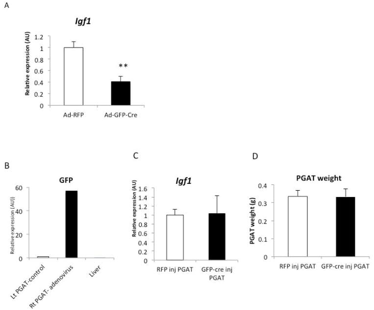
Methods: To study IGF1 in vivo, the cellular sources of adipose tissue Igf1 expression were identified and mice were generated with targeted deletion in adipocytes and macrophages. The effects of adipocyte and macrophage deficiency of IGF1 on adipose tissue development and the response to chronic (high-fat feeding) and acute (cold challenge) stress were studied.

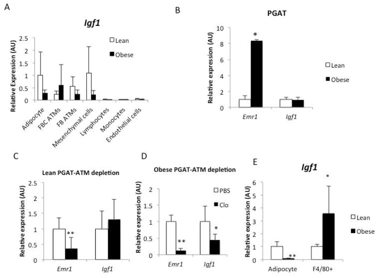
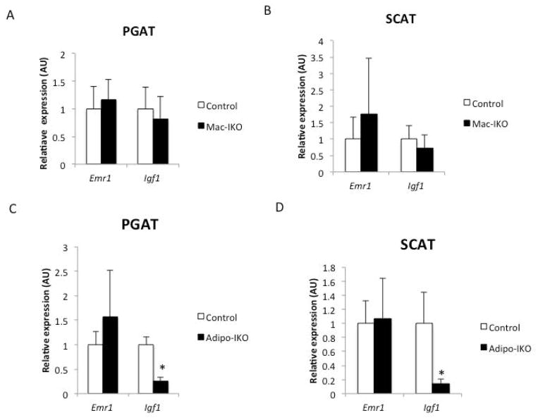
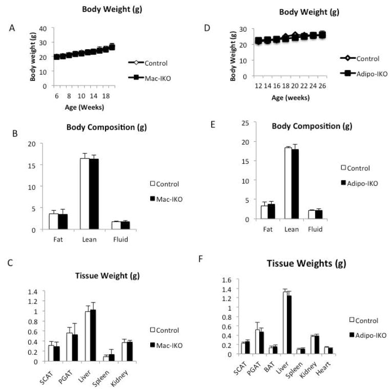
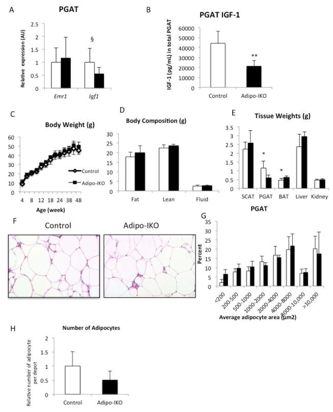
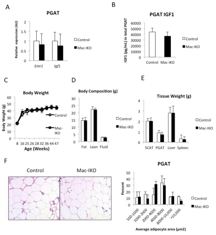
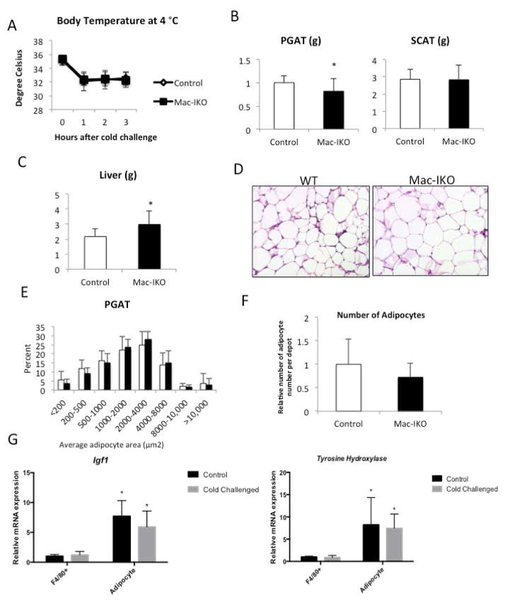
Results: The expression of Igf1 by adipose tissue was derived from multiple cell types including adipocytes and macrophages. In lean animals, adipocytes were the primary source of IGF1, but in obesity expression by adipocytes was reduced and by macrophages increased, so as to maintain overall adipose tissue Igf1 expression. Genetic deletion studies revealed that adipocyte-derived IGF1 regulated perigonadal but not subcutaneous adipose tissue mass during high-fat feeding and the development of obesity. Conversely, macrophage-derived IGF1 acutely modulated perigonadal adipose tissue mass during thermogenic challenges.

Conclusions: Local IGF1 is not required in lean adipose tissue development but is required to maintain homeostasis during both chronic and acute metabolic stresses.


Conflict of interest statement

Conflict of Interest: None of the authors have any conflicts of interest.

Figures

Figure 1
Figure 1. Regulation of local Igf1 expression is dynamically maintained during the development of obesity
(A) Expression of Igf1 in purified cell populations from PGAT of lean (Lep +/+) and obese (Lep ob/ob) mice (n=5). (B) Gene expression of F4/80 (Emr1) and Igf1 in both lean and obese PGAT (n=3). Expression of F4/80 (Emr1) and Igf1 in PGAT explants from (C) lean mice (n=5) and (D) high fat fed obese animals (n=3). (E) Expression of Igf1 in purified adipocytes and purified adipose tissue macrophages (ATMs) population from lean and obese PGAT. Fluorescence activated cell sorting was used to purify cells from adipose tissue (n=5). All data are presented as mean ± SD *p-value <0.05 **p-value <0.01.
Figure 2
Figure 2. Compensation in vivo but not ex vivo of adipose tissue IGF1 deletion
(A) Igf1 expression in Ad-RFP injected and Ad-GFP-Cre injected PGAT explants from 18-week-old obese Igf1 fl/fl mice. (n=4) (B) GFP expression in Ad-RFP treated (left side) and Ad-GFP-Cre treated (right side) and liver of 14-week-old lean Igf1fl/fl mice (n=3). (C) Igf1 gene expression in Ad-RFP treated (left side) and Ad-GFP-Cre treated (right side) PGAT of 14-week-old lean Igf1fl/fl mice (n=3) (D) Ad-RFP treated (left side) and Ad-GFP-Cre treated (right side) weight of 14-week-old lean Igf1fl/fl mice (n=3). All data are presented as mean + SD **p-value <0.01.
Figure 3
Figure 3. In lean animals adipose tissue IGF1 is primarily adipocyte-derived
F4/80 (Emr1) and Igf1 expression in (A) perigonadal and (B) subcutaneous adipose tissue of 24-week-old lean mice and those in which IGF1 had been genetically deleted from myeloid cells (Mac-IKO) (n=9–16). F4/80 (Emr1) and Igf1 expression in (C) perigonadal or (C) subcutaneous adipose tissue of 26-week-old lean control mice and those in which IGF1 had been genetically deleted from adipocytes (Adipo-IKO) (n=5). All data are presented as mean + SD *p-value <0.05.
Figure 4
Figure 4. Neither myeloid nor adipocyte IGF1 is required for normal development in lean animals
(A) Body weights of 6- to 24-week-old lean control and Mac-IKO male mice (n=9–16). (B) Body compositions (fat, lean, and fluid mass) of 24-week-old lean control and Mac-IKO male mice as measured by MRI (n=9–16). (C) Organ weights of 24-week-old lean control and Mac-IKO male mice (n=9–16). All data are presented as mean + SD. (D) Body weights of 12- to 26-week old lean control and Adipo-IKO male mice. n=5/genotype. (E) Body compositions of 26-week-old lean control and Adipo-IKO male mice. n=5/genotype. (F) Organ weights of 26-week-old lean control and Adipo-IKO mice. n=5/genotype. All data are presented as mean + SD.
Figure 5
Figure 5. Adipose tissue develops normally in the absence of adipocyte or myeloid IGF1 in lean animals
(A) PGAT histology (hemotoxylin and eosin stained) and (B) adipocyte size (area um2) distribution of 24-week-old lean control and Mac-IKO male mice (n=9–16). (C) PGAT histology (hemotoxylin and eosin stained) and (D) adipocyte size (area um2) distribution of 26-week-old lean control and Adipo-IKO male mice. All data are presented as mean + SD.
Figure 6
Figure 6. Adipocyte-derived IGF-1 is required to maintain perigonadal adipose tissue mass during chronic high fat feeding
(A) F4/80 (Emr1) and Igf1 expression in PGAT of 48-week-old control (white bars) and Adipo-IKO (black bars) obese male mice (n=5–7). (B) IGF1 protein concentration in PGAT of 48-week-old control and Adipo-IKO obese male mice. (n=5–7) (C) Body weights of 4- to 48-week-old control (empty circles) and Adipo-IKO (filled circles) high fat fed male mice. (D)Body compositions of 48-week-old male mice (n=5–7). (E) SCAT, PGAT, BAT, liver, and kidney weights of 45-week-old control (white bars) and Adipo-IKO (black bars) high fat fed mice (n=5–7). (F)PGAT histology (H&E stained) of 48-week-old male obese mice. (G) Quantification of perigonadal adipocyte size from figure 8F. (H) Relative adipocyte number in PGAT of 45-week-old control (white bars) and Adipo-IKO (black bars) high fat fed mice (n=5–7) p-value § =0.08, * <0.05 **<0.01. All data are presented as mean + SD.
Figure 7
Figure 7. ATM-derived IGF-1 is not required for adipose tissue development during the onset of obesity
(A) F4/80(Emr1) and Igf1 mRNA gene expression of PGAT in 45-week-old control and Mac-IKO obese male mice (n=5–6/genotype) (B) Total IGF1 protein level (as measured by ELISA) of PGAT in 45-week-old control and Mac-IKO obese male mice (n=5–6). (C) Body weights of 4- to 45-week-old control and Mac-IKO obese male mice. Mice were fed high-fat diet from 5-week of age. (n=8–15) (D) Body compositions of 45-week-old control and Mac-IKO obese male mice. n=5–6 per genotype. (E) Tissue weights of 45-week-old control and Mac-IKO obese male mice (n=5–6) (F) PGAT histology (H&E stained) of 45-week-old control and Mac-IKO obese male mice (n=5–6). All data are presented as mean ± SD.
Figure 8
Figure 8. ATM-derived IGF1 is required to maintain perigonadal adipose tissue during a cold challenge in obese animals
(A) Body temperature of 46-week-old control (white diamonds) and Mac-IKO mice (filled circles) during a cold challenge at 4 °C (n= 8–15). (B) PGAT, and SCAT weights of cold challenged 46-week-old male obese mice. (C) Liver weight of cold challenged 46-week-old obese mice (D) PGAT histology, (E) adipocyte size distribution and (F) adipocyte number of PGAT from 46-week-old male obese mice following a cold challenged for 3 hours at 4 ° C. (G) 9-week old lean male mice were placed at either 25°C or 4°C for 72 hours and F4/80+ macrophage and adipocytes were purified. Expression of Igf1 and Tyrosine Hydroxylase (Th) were measured qPCR (n=10 per genotype), *p-value<0.05 relative to F4/80+ cells. All data are presented as means + SD. *p-value <0.05.

Comment in

  • IGF1 and adipose tissue homeostasis.
    Stephens JM. Stephens JM. Obesity (Silver Spring). 2016 Jan;24(1):10. doi: 10.1002/oby.21372. Epub 2015 Dec 5. Obesity (Silver Spring). 2016. PMID: 26638118 No abstract available.

References

    1. Le Roith D, Bondy C, Yakar S, Liu JL, Butler A. The somatomedin hypothesis: 2001. Endocrine reviews. 2001;22(1):53–74. doi: 10.1210/edrv.22.1.0419. - DOI - PubMed
    1. Baker J, Liu JP, Robertson EJ, Efstratiadis A. Role of insulin-like growth factors in embryonic and postnatal growth. Cell. 1993;75(1):73–82. - PubMed
    1. Liu JP, Baker J, Perkins AS, Robertson EJ, Efstratiadis A. Mice carrying null mutations of the genes encoding insulin-like growth factor I (Igf-1) and type 1 IGF receptor (Igf1r) Cell. 1993;75(1):59–72. - PubMed
    1. Powell-Braxton L, Hollingshead P, Warburton C, Dowd M, Pitts-Meek S, Dalton D, et al. IGF-I is required for normal embryonic growth in mice. Genes & development. 1993;7(12B):2609–17. - PubMed
    1. Sjogren K, Liu JL, Blad K, Skrtic S, Vidal O, Wallenius V, et al. Liver-derived insulin-like growth factor I (IGF-I) is the principal source of IGF-I in blood but is not required for postnatal body growth in mice. Proceedings of the National Academy of Sciences of the United States of America. 1999;96(12):7088–92. - PMC - PubMed

Publication types

Substances