

Enzymatic vitreolysis with recombinant tissue plasminogen activator for vitreomacular traction
- PMID: 26664047
- PMCID: PMC4669920
- DOI: 10.2147/DDDT.S88361
Enzymatic vitreolysis with recombinant tissue plasminogen activator for vitreomacular traction
Abstract
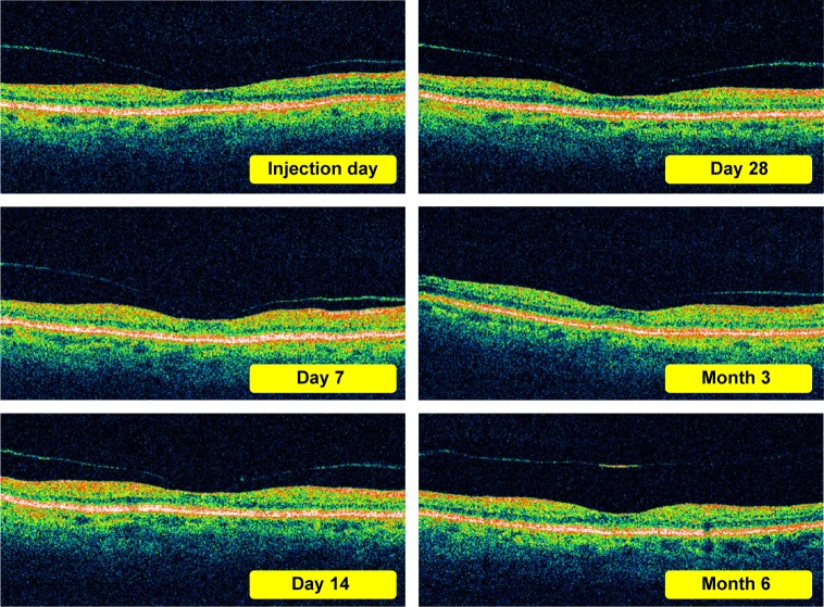
Aims: The aim of our research was to gain data about the efficacy of intravitreal injections of a recombinant tissue plasminogen activator (rTPA) in dissolving vitreoretinal tractions (VRTs).
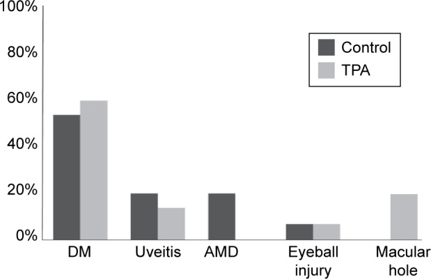
Materials and methods: The study group consisted of patients of our Ophthalmology Clinic who had received an injection of rTPA (TPA Group) for an existent vitreomacular traction confirmed by optical coherence tomography and stereoscopic examinations. The control group consisted of patients who had declined treatment despite the existence of a vitreomacular traction confirmed by the same diagnostic methods. Each group consisted of 30 people (30 eyes). The observation period was 6 months.
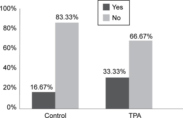
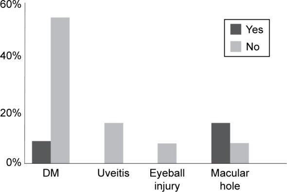
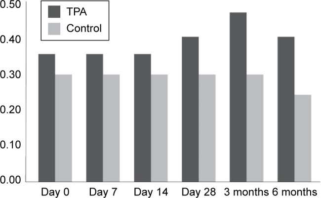
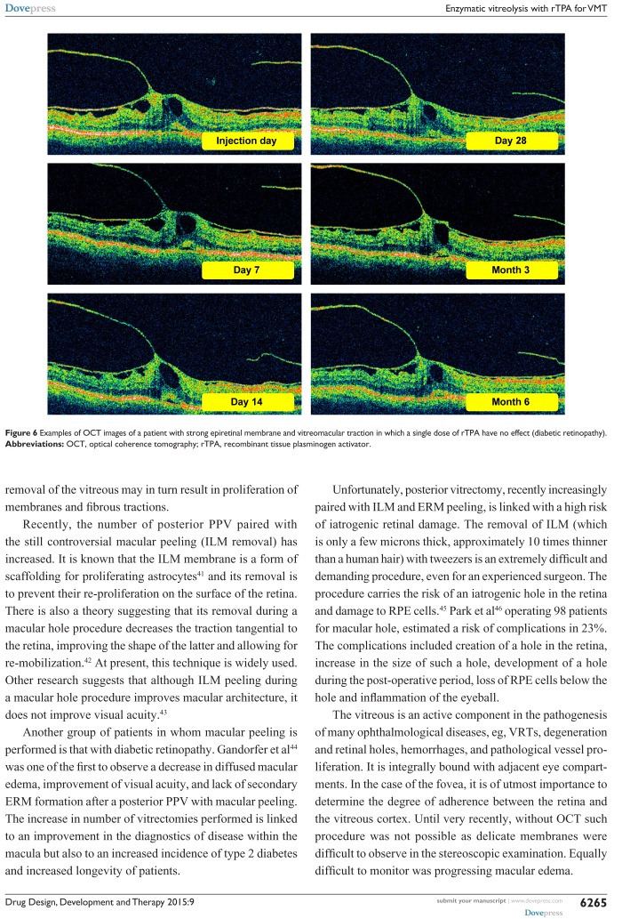
Conclusion: In both groups some of the VRTs had dissolved. In the TPA group the traction dissolved in 10 patients (33.33%) and in the control group only in 5 (16.67%). It is also important to point out that the mean baseline membrane thickness was higher in the TPA group than in the control group. Observing patients in both groups we noticed that the dissolution of vitreoretinal membrane occurred most frequently in those cases where the membrane was thin. In the TPA group, the mean membrane thickness after 6 months decreased considerably. At the same time, no significant change in the membrane thickness could be observed in the control group. Observation of the retinal thickness allows us to draw the following conclusion: in the TPA group, the retinal thickness in the macular area (edema) had decreased over the study period, whereas in the control group it had increased. In those cases where the traction had dissolved, the edema of the retina decreased by the end of the 6-month period in both groups. In the TPA group, the dissolution of the membrane occurred most often within 3 months from the primary injection. Based on statistics, we can confirm that in the control group there was a decrease in visual acuity during the 6 months of the study period. At the same time, visual acuity in the TPA group underwent a small improvement. A 6-month observation had shown that in patients with strong VRTs, and in particular with VRTs accompanied by epiretinal membranes, a single intraocular injection is not enough to achieve posterior vitreous detachment. We have also shown that rTPA is a safe drug, with no adverse effects observed during the study period.
Keywords: diabetic retinopathy; epiretinal membrane; macular hole; tissue plasminogen activator (rTPA); vitrectomy; vitreous body.
Figures
Similar articles
-
Intravitreal tissue plasminogen activator to treat refractory diabetic macular edema by induction of posterior vitreous detachment.Retina. 2011 Nov;31(10):2065-70. doi: 10.1097/IAE.0b013e31820f49ff. Retina. 2011. PMID: 21983248 Clinical Trial.
-
Enzymatic Vitreolysis for Vitreomacular Traction in Diabetic Retinopathy.Dev Ophthalmol. 2017;60:160-164. doi: 10.1159/000460275. Epub 2017 Apr 20. Dev Ophthalmol. 2017. PMID: 28427074 Review.
-
Induction of Posterior Vitreous Detachment in Pediatric Vitrectomy by Preoperative Intravitreal Injection of Tissue Plasminogen Activator.J Pediatr Ophthalmol Strabismus. 2016 Mar-Apr;53(2):113-8. doi: 10.3928/01913913-20160209-01. J Pediatr Ophthalmol Strabismus. 2016. PMID: 27018884
-
[Ocriplasmin as a treatment option for symptomatic vitreomacular traction with and without macular hole. First clinical experiences].Ophthalmologe. 2015 Dec;112(12):990-4. doi: 10.1007/s00347-015-0073-z. Ophthalmologe. 2015. PMID: 26062717 Clinical Trial. German.
-
[Vitreomacular traction: diagnostics, natural course, treatment decision and guideline recommendations].Ophthalmologie. 2024 Jun;121(6):470-475. doi: 10.1007/s00347-024-02042-4. Epub 2024 May 29. Ophthalmologie. 2024. PMID: 38809382 Review. German.
Cited by
-
Towards smart self-clearing glaucoma drainage device.Microsyst Nanoeng. 2018 Nov 5;4:35. doi: 10.1038/s41378-018-0032-3. eCollection 2018. Microsyst Nanoeng. 2018. PMID: 31057923 Free PMC article.
-
Enzymatic vitreolysis using reengineered Vibrio mimicus-derived collagenase.Mol Vis. 2021 Apr 1;27:125-141. eCollection 2021. Mol Vis. 2021. PMID: 33907368 Free PMC article.
References
-
- Zdenek G, Kański J. Retinal Detachment: A Colour Manual of Diagnosis and Treatment. 1995
-
- Jaffe NS. Vitreous traction at the posterior pole of the fundus due to alterations in the vitreous posterior. Trans Am Acad Ophthalmol Otolaryngol. 1967;71:642–652. - PubMed
-
- Kański J. Clinical Ophthalmology. Polish edition 2009. p. 705.
-
- Brown DJ, Bishop P, Hamdi H, Kenney MC. Cleavage of structural components of mammalian vitreous by endogenous matrix metalloproteinase-2. Curr Eye Res. 1996;15:439–445. - PubMed
-
- William E, Smiddy MD, Ronald G, et al. Morphology, pathology and surgery of idiopathic vitreoretinal macular disorders. A review. Retina. 1990;10(4):288–296. - PubMed
MeSH terms
Substances
LinkOut - more resources
Full Text Sources
Medical
