Review
. 2015 Oct-Dec;10(4):231-42.
doi: 10.4103/1817-1737.160365.

Pictorial essay of radiological features of benign intrathoracic masses

Affiliations
Review

Pictorial essay of radiological features of benign intrathoracic masses

Syahminan Suut et al. Ann Thorac Med. 2015 Oct-Dec.

Abstract

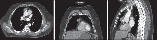
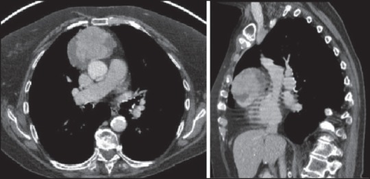
With increased exposure of patients to routine imaging, incidental benign intrathoracic masses are frequently recognized. Most have classical imaging features, which are pathognomonic for their benignity. The aim of this pictorial review is to educate the reader of radiological features of several types of intrathoracic masses. The masses are categorized based on their location/origin and are grouped into parenchymal, pleural, mediastinal, or bronchial. Thoracic wall masses that invade the thorax such as neurofibromas and lipomas are included as they may mimic intrathoracic masses. All examples are illustrated and include pulmonary hamartoma, pleural fibroma, sarcoidosis, bronchial carcinoid, and bronchoceles together with a variety of mediastinal cysts on plain radiographs, computed tomography (CT) and magnetic resonance imaging (MRI). Sometimes a multimodality approach would be needed to confirm the diagnosis in atypical cases. The study would include the incorporation of radionuclide studies and relevant discussion in a multidisciplinary setting.

Keywords: Intrathoracic masses; pulmonary mass; radiology.


Conflict of interest statement

Conflicts of interest: None declared.

Figures

Figure 1
Figure 1
CXR of a 27-year-old female (nonsmoker) showing a solitary well-defined nodule in the left upper zone (arrow). CT further characterizes the opacity by demonstrating fat content within the mass highly suggested of a hamartoma. No biopsy was taken. The nodule remains stable 3 years later. CXR = Chest X-ray, CT = computed tomography
Figure 2
Figure 2
CXRs from two different patients showing the characteristics popcorn calcification of a hamartoma
Figure 3
Figure 3
A 35-year-old with known sarcoidosis. Her chest radiograph demonstrates the “Garland sign”—Bilateral hilar and right paratracheal lymphadenopathy. Coronal CT confirms the findings. No lung parenchymal changes are seem
Figure 4
Figure 4
Sarcoidosis in a 56-year-old female patient demonstrating classical eggshell hilar (1) calcification. Note the reticulonodular parenchymal changes of perilymphatic distribution in the middle and upper lobes (2)
Figure 5
Figure 5
Silicosis massive pulmonary fibrosis (MPF) symmetrical bilateral opacities of more than 1 cm, formed by nodule coalescence forming mass that may even cavitate (*)
Figure 6
Figure 6
Intrapulmonary lymph node: Incidental finding of a small, oval-shaped parenchymal opacity in the right lower lobe. The appearances are classically those of intrapulmonary lymph node and no further actions are required
Figure 7
Figure 7
Rheumatoid nodules: Long standing solitary pulmonary nodule in the periphery of the left lower lobe of a 60-year-old male patient with known rheumatoid arthritis. This is likely a rheumatoid nodule
Figure 8
Figure 8
Rheumatoid nodules: PA CXR of a 53-year-old man with seropositive rheumatoid arthritis being treated with rituximab showing a nodule in the right upper lobe (red arrow) and a cavitating nodule (yellow arrow) in the left upper lobe on a background of multinodularity suggestive of Kaplan's syndrome. PA = Posteroanterior
Figure 9
Figure 9
Note the solid pleural-based nodule, associated with a small pleural effusion (pulmonary infarct). The patient had a travel-related deep venous thrombosis that presented with pleuritic, left-sided chest pain
Figure 10
Figure 10
Septic emboli. Gas gangrene of the foot in a 49-year-old woman with type II diabetes mellitus, showing gas within the soft tissues of the foot (1), also elegantly shown by gas within the soft tissues of the foot on ultrasound examination. There are multiple pleural-based nodules on the CXR suggestive of septic emboli (3)
Figure 11
Figure 11
Axial CT images from the same patient as in Figure 10 with a gas gangrene of the foot showing multiple, pleural-based nodules due to septic emboli with evidence of early cavitation in one of the nodules (arrow)
Figure 12
Figure 12
CXR showing a thick-walled cavity with an air-fluid level due to a cavitating pulmonary infarct. The cavity is indistinguishable from other cavities such as cavitating neoplasia. To achieve a diagnosis, the history and the radiological evolution is important. This patient was a known pulmonary infarct, which subsequently cavitated
Figure 13
Figure 13
Wegener's granulomatosis: CXR in a patient with Wegener's granulomatosis showing a thick-walled cavity in the left upper lobe and patchy bilateral basal shadowing. CT confirmed multiple ground glass changes in keeping with pulmonary hemorrhage secondary to the disease
Figure 14
Figure 14
Pleural fibroma: Incidental pleural-based mass in the right costophrenic angle in a 65-year-old male presenting with shortness of breath. CT confirms the presence of a well-defined homogenous right lower parietal pleural mass measuring 6 cm × 3 cm with focal calcification
Figure 15
Figure 15
A pleural fibroma should not be confused with a mesothelioma, note the calcified pleural plaque, a pleural-based mass invading the intercostal space (arrow) in this patient with previous asbestos exposure
Figure 16
Figure 16
Splenosis: Incidental finding on a chest radiograph of a left-sided, pleural-based mass. CT confirms this and also other pleural nodularities, with the absence of a spleen. Further history was obtained and this patient was noted to have been involved in a blast injury. Splenic injuries were therefore suspected
Figure 17
Figure 17
Splenic scintigraphy SPECT/CT study scans confirm intense uptake in the left hemithorax pleural nodularities. The appearances are those of post-traumatic abdominal and intrathoracic splenosis. SPECT = Single-photon emission CT
Figure 18
Figure 18
Soft and calcified pleural plaques, due to asbestos exposures. Soft plaques cannot be reliably differentiated from a mesothelioma without a biopsy
Figure 19
Figure 19
Loculated interlobular pleural effusion or pulmonary pseudotumor; also called vanishing tumor
Figure 20
Figure 20
Loculated pleural effusion: A smoothly-defined, right, pleural-based mass is demonstrated on the chest radiograph. On CT, this “mass” was noted to be of fluid density. Appearances are in keeping with a loculated pleural effusion
Figure 21
Figure 21
Round atelectasis occurs when there is unfolding of redundant pleura. This may give a false, mass-like appearance. It is most commonly associated with asbestos-related disease, but it may arise from a variety of chronic pleural conditions such as infection, uraemia, therapeutic pneumothorax in the treatment of tuberculosis, pulmonary infarction, or heart failure and is usually asymptomatic. The images show a rounded atelectasis in association with pleural thickening secondary to asbestoses exposure
Figure 22
Figure 22
Chest wall lipoma: Examples of chest wall lipomas on differing imaging modalities. The chest radiograph (1) demonstrates a well-defined, smooth, right lateral chest wall mass. Ultasound scan (2) demonstrates a homogeneous mildly echogenic ovoid structure and T1-weighted MR scan (3) demonstrated a homogeneous. MR = Magnetic resonance
Figure 23
Figure 23
Carcinoid: CT in a breathless 70-year=old gentleman demonstrates the presence of an endobronchial lesion in the right bronchus intermedius. An endbronchial ultrasound (EBUS) was performed and samples obtained. This was later shown to be a bronchial carcinoid
Figure 24
Figure 24
On a CXR bronchogenic cysts usually appear as soft-tissue density rounded structures. As the cysts may contain calcium oxalate, dependent layering of calcific density material may occasionally be seen as seen on the CXR. CT is better able to detect calcium oxalate (milk of calcium) layering dependently as shown
Figure 25
Figure 25
Bronchogenic cyst: Well-defined, left hilar shadows seen on the chest radiograph. CT shows well-defined 3.5 × 24 cm uniformly low attenuation (HU 34) lesion arising from the posterior mediastinum in keeping with a bronchogenic cyst
Figure 26
Figure 26
Bronchocele: Chest radiograph performed in asthmatic 50-year-old male demonstrated branching tubular structures in the left lower zone. An incidental left upper zone nodule has remained unchanged for many years
Figure 27
Figure 27
Bronchocele: CT confirms the presence of dilated and ectatic left lower lobe bronchioles with evidence of mucus impaction. Appearances are suggestive of bronchocele secondary to asthma and increased mucus production
Figure 28
Figure 28
This patient presented with severe right shoulder pain. The CXR and T1- and T2-weighted axial images show features of a hydatid cyst in the right superior sulcus (Images courtesy: Shyam Sunder)
Figure 29
Figure 29
Retrosternal goiter: Axial and sagittal CT images show heterogeneous, well-defined, 6.5 × 24.0 cm, right retrosternal goiter with speckles of calcification
Figure 30
Figure 30
Retrosternal thyroid: Chest radiograph demonstrates a soft tissue mass in the superior right mediastinum causing deviation of the trachea to the contralateral left. CT confirms a heterogeneous 5 cm × 4 cm right retrosternal thyroid gland
Figure 31
Figure 31
Retrosternal thyroid: CXR demonstrates a large soft tissue mass in the superior right mediastinum causing deviation and compression of the trachea and displacement of the mediastinum to the contralateral left. CT confirms a heterogeneous right retrosternal thyroid gland. The patient presented with stridor at surgery; a partial thyroidectomy confirmed benign retrosternal goiter
Figure 32
Figure 32
Pericardial cyst: Axial and sagittal views of an arterial contrast-enhanced scan through the thorax of another patient demonstrated a 3.5 cm × 2.0 cm left pericardial cyst in the region of the lingual
Figure 33
Figure 33
Pericardial cyst: Axial T1- and T2-weighted MRI showing typical fluid signal in the pericardial cyst with occasional high signal on T1-weighted images (not shown). MRI = MR imaging
Figure 34
Figure 34
A 45-year-old man recently diagnosed with myasthenia gravis undergoes CT thorax. This demosntrated a 2.5 cm × 3.5 cm, well-defined homogenous anterior mediastinal mass in keeping with a thymoma
Figure 35
Figure 35
Thymoma: Axial CT shows a well-defined homogenous anterior mediastinal mass in a patient that presented with myasthenia gravis. FDG-PET showing uptake within the tumor. A thymoma was confirmed at surgery. FDG-PET = Fluorodeoxygenase positron emission tomography
Figure 36
Figure 36
This male patient in the mid-30s presented with clinical features of myasthenia gravis. The axial and sagittal CT showed a mass in the anterior mediastinum, with slight surface tumor irregularity and areas of necrosis. At surgery a malignant thymoma was confirmed
Figure 37
Figure 37
STIR sequence and T2-weighted axial and coronal images through the mid mediastinum showing diffuse symmetric and smooth enlargement of the thymus gland due to thymic hyperplasia. STIR = Short TI inversion recovery
Figure 38
Figure 38
Axial CT through the anterior mediastinum shows a lobulated thymic mass with low attenuation areas in keeping with thymic cysts. FDG-PET shows no uptake within the cysts
Figure 39
Figure 39
Cystic thymoma: Axial T1, T2, and post-gadolinium MRI showing characteristic features of a cystic thymoma without intrinsic contrast enhancement
Figure 40
Figure 40
CXR shows a well-defined, smooth, lower right paravertebral mass with close association with spinal canal (axial CT) in keeping with a benign neurogenic tumor
Figure 41
Figure 41
Extramedullary hematopoiesis usually occurs in association with hematological disorders and normally involves the reticuloendothelial system, including the liver, spleen, and lymph nodes. This patient had a beta thalassemia. Note the paravertebral location of the extramedullary hematopoiesis
Figure 42
Figure 42
Noncontrast axial CT, T2-weighted MR, and post-contrast CT chest shows features of a chronic calcific hematoma

Similar articles

Cited by

References

    1. Leader JK, Warfel TE, Fuhrman CR, Golla SK, Weissfeld JL, Avila RS, et al. Pulmonary nodule detection with low-dose CT of the lung: Agreement among radiologists. AJR Am J Roentgenol. 2005;185:973–8. - PubMed
    1. Salahudeen HM, Hoey ET, Robertson RJ, Darby MJ. CT appearances of pleural tumours. Clin Radiol. 2009;64:918–30. - PubMed
    1. Helm EJ, Matin TN, Gleeson FV. Imaging of the pleura. J Magn Reson Imaging. 2010;32:1275–86. - PubMed
    1. Gill RS, Perry T, Abele JT, Bedard EL, Schiller D. The clinical significance of incidental intra-abdominal findings on positron emission tomography performed to investigate pulmonary nodules. World J Surg Oncol. 2012;10:25. - PMC - PubMed
    1. Maffione AM, Grassetto G, Rampin L, Chondrogiannis S, Marzola MC, Ambrosini V, et al. Molecular imaging of pulmonary nodules. AJR Am J Roentgenol. 2014;202:W217–23. - PubMed