. 2015 Oct 9;5(11):e00401.
doi: 10.1002/brb3.401. eCollection 2015 Nov.

Molecular neuropathology of the synapse in sheep with CLN5 Batten disease

Affiliations

Molecular neuropathology of the synapse in sheep with CLN5 Batten disease

Inês S Amorim et al. Brain Behav. .

Abstract

Aims: Synapses represent a major pathological target across a broad range of neurodegenerative conditions. Recent studies addressing molecular mechanisms regulating synaptic vulnerability and degeneration have relied heavily on invertebrate and mouse models. Whether similar molecular neuropathological changes underpin synaptic breakdown in large animal models and in human patients with neurodegenerative disease remains unclear. We therefore investigated whether molecular regulators of synaptic pathophysiology, previously identified in Drosophila and mouse models, are similarly present and modified in the brain of sheep with CLN5 Batten disease.

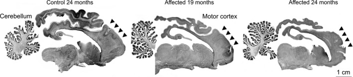
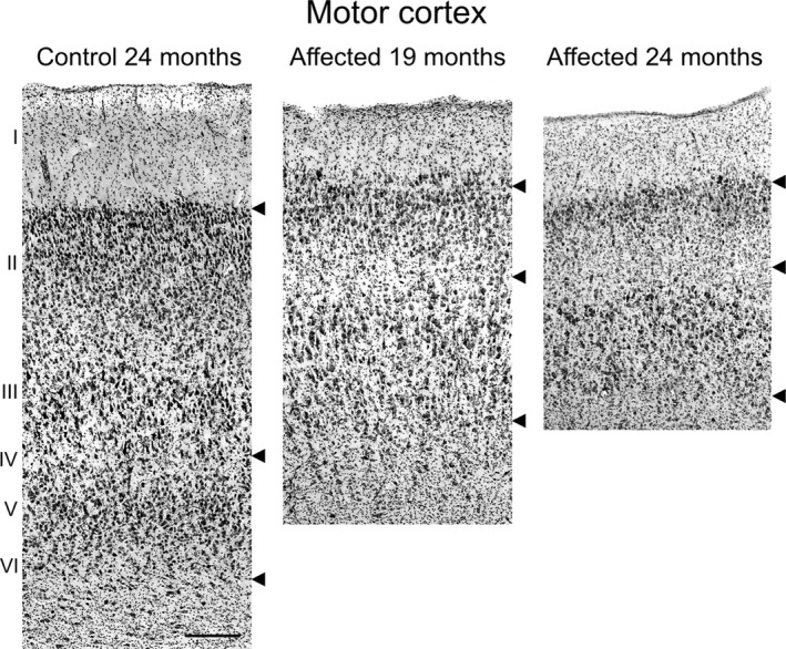
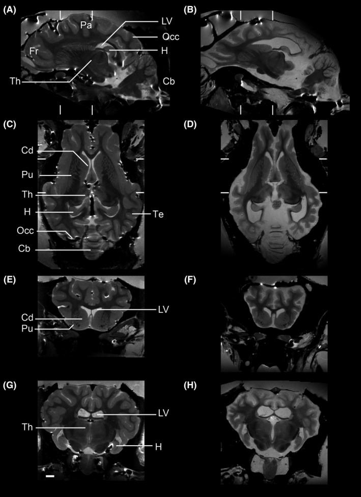
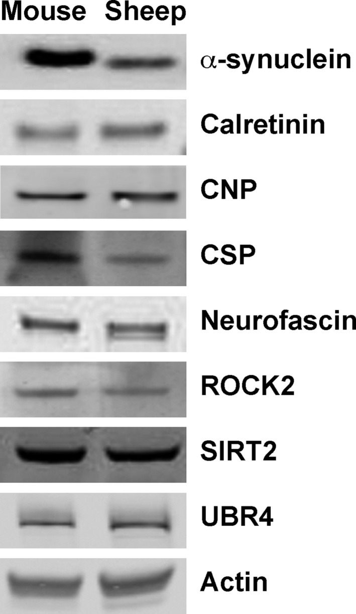
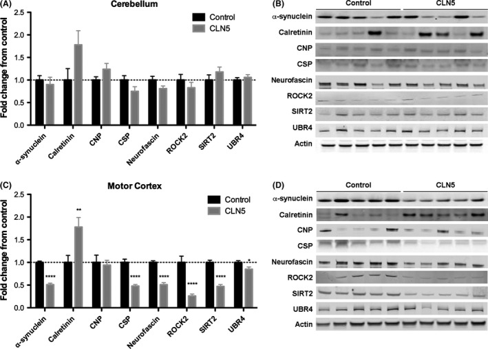
Methods: Gross neuropathological analysis of CLN5 Batten disease sheep and controls was used alongside postmortem MRI imaging to identify affected brain regions. Synaptosome preparations were then generated and quantitative fluorescent Western blotting used to determine and compare levels of synaptic proteins.

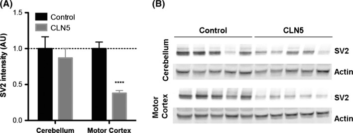
Results: The cortex was particularly affected by regional neurodegeneration and synaptic loss in CLN5 sheep, whilst the cerebellum was relatively spared. Quantitative assessment of the protein content of synaptosome preparations revealed significant changes in levels of seven out of eight synaptic neurodegeneration proteins investigated in the motor cortex, but not cerebellum, of CLN5 sheep (α-synuclein, CSP-α, neurofascin, ROCK2, calretinin, SIRT2, and UBR4).

Conclusions: Synaptic pathology is a robust correlate of region-specific neurodegeneration in the brain of CLN5 sheep, driven by molecular pathways similar to those reported in Drosophila and rodent models. Thus, large animal models, such as sheep, represent ideal translational systems to develop and test therapeutics aimed at delaying or halting synaptic pathology for a range of human neurodegenerative conditions.

Keywords: Animal model; lysosomal storage disorders; neurodegeneration; neuronal ceroid lipofuscinoses; neuronal ceroid lipofuscinosis; sheep; synapse; synaptic vulnerability.


Figures

Figure 1
Figure 1
Gross cortical atrophy in CLN5 sheep brains. Nissl‐stained sections showed marked atrophy of the cortical mantle in affected sheep at 19 months of age, which was more pronounced by 24 months of age. In contrast, the cerebellum and subcortical structures were relatively spared.
Figure 2
Figure 2
Atrophy of motor cortex in CLN5 sheep brains. Microscopic comparison of the motor cortex of Nissl‐stained sections (shown in Fig. 1). Upper arrows mark the layer I/II boundary, the middle arrows indicate layer IV, and the lower arrow marks the layer VI/white matter boundary. Scale bar 200 μm.
Figure 3
Figure 3
MRI analyses of CLN5 Batten sheep brain confirms targeting of the cortex and sparing of the cerebellum. Representative MR images of control (A, C, E, G) and CLN5 sheep (B, D, F, H). Sections are shown in sagittal (A–B), horizontal (C–D) and two coronal cuts (E–F and G–H at the levels marked in A–D). The scale bar is 2 cm. Label abbreviations: Fr frontal lobe; Pa parietal lobe; Occ occipital lobe; Te temporal lobe; LV lateral ventricle, Th thalamus, H hippocampus; Cb cerebellum; Cd caudate; Pu putamen.
Figure 4
Figure 4
Loss of synapses in affected brain regions from CLN5 Batten disease sheep. A. Quantitative fluorescent Western blotting in preparations of whole tissue revealed relatively stable levels of SV2, a core synaptic protein, in the cerebellum of control and CLN5 sheep. In contrast, in the motor cortex of CLN5 Batten disease sheep, the levels of SV2 are reduced by 60%, indicating there is severe synapse loss restricted to this brain area of the Batten sheep. (N = 5 animals, n = 10 distinct tissue samples from control sheep; N = 8, n = 16 from CLN5 sheep; ****P < 0.0001, unpaired two‐tailed t‐test). B. Representative fluorescent Western blots (N = 5 animals per genotype) showing a consistent decrease in SV2 levels in the motor cortex of CLN5 Batten disease sheep when compared to controls. Note the relatively stable levels of SV2 in the cerebellum of CLN5 sheep. Actin was used as loading control.
Figure 5
Figure 5
Characterization of synaptosome preparations from control and CLN5 Batten disease sheep brains. Representative fluorescent Western blots showing levels of a core synaptic protein (SV2), a core nuclear protein (HDAC2) and a loading control (actin) in synaptic (S) and nonsynaptic (NS) fractions generated from the cerebellum and motor cortex of control and CLN5 Batten disease sheep brains. Note the enrichment of synaptic protein and exclusion of nuclear protein in the synaptic fractions.
Figure 6
Figure 6
Conserved expression and Western blot detection of synaptic proteins in synaptosome preparations from mice and sheep. Representative fluorescent Western blots showing levels of eight synaptic proteins implicated in the regulation of synaptic stability and degeneration, as well as a loading control (actin), in synaptosome preparations from mouse and sheep brain. Note how all synaptic proteins previously identified in mouse synapses were similarly present, and detectable using the same primary antibodies, in sheep synapses.
Figure 7
Figure 7
Modified expression levels of synaptic proteins in motor cortex from CLN5 Batten disease sheep. A–B. Levels of the eight synaptic “degeneration” proteins (actin was used as a loading control) in synaptosome preparations generated from the unaffected cerebellum of control and CLN5 Batten disease sheep are shown. Data in panel A (N = 5, n = 10 control; N = 8, n = 16 CLN5) are shown as mean ± SEM. Statistical analyses revealed no significant differences (P > 0.05 in t‐test) in the level of any protein between control and CLN5 samples. Representative Western blots from a single experimental run (N = 5 animals per genotype) are shown in panel B. C–D. Levels of the eight synaptic “degeneration” proteins (actin was used as a loading control) in synaptosome preparations generated from the affected motor cortex of control and CLN5 Batten sheep are shown. Data in panel C (N = 5, n = 10 control; N = 8, n = 16 CLN5) are shown as mean ± SEM. Statistical analyses revealed significant differences in the levels of seven proteins between control and CLN5 samples (*P < 0.05; **P < 0.01; ****P < 0.0001; in t‐test). Representative Western blots from one experimental run (N = 5 animals per genotype) are shown in panel D.

References

    1. Aigner, B. , Renner S., Kessler B., Klymiuk N., Kurome M., Wünsch A., et al. 2010. Transgenic pigs as models for translational biomedical research. J. Mol. Med. 88:653–664. - PubMed
    1. Benitez, B. A. , Alvarado D., Cai Y., Mayo K., Chakraverty S., Norton J., et al. 2011. Exome‐sequencing confirms DNAJC5 mutations as cause of adult neuronal ceroid‐lipofuscinosis. PLoS One 6:e26741. - PMC - PubMed
    1. Blom, T. , Schmiedt M. L., Wong A. M., Kyttälä A., Soronen J., Jauhiainen M., et al. 2013. Exacerbated neuronal ceroid lipofuscinosis phenotype in Cln1/5 double‐knockout mice. Dis. Mod. Mech. 6:342–357. - PMC - PubMed
    1. Bond, M. , Holthaus S. M., Tammen I., Tear G., and Russell C.. 2013. Use of model organisms for the study of neuronal ceroid lipofuscinosis. Biochim. Biophys. Acta 1832:1842–1865. - PubMed
    1. Chandra, S. , Gallardo G., Fernández‐Chacón R., Schlüter O. M., and Südhof T. C.. 2005. Alpha‐synuclein cooperates with CSPalpha in preventing neurodegeneration. Cell 123:383–396. - PubMed

LinkOut - more resources