. 2015 Oct 1:11:21-30.
doi: 10.1016/j.cois.2015.08.001.

Nutrition-dependent control of insect development by insulin-like peptides

Affiliations

Nutrition-dependent control of insect development by insulin-like peptides

Naoki Okamoto et al. Curr Opin Insect Sci. .

Abstract

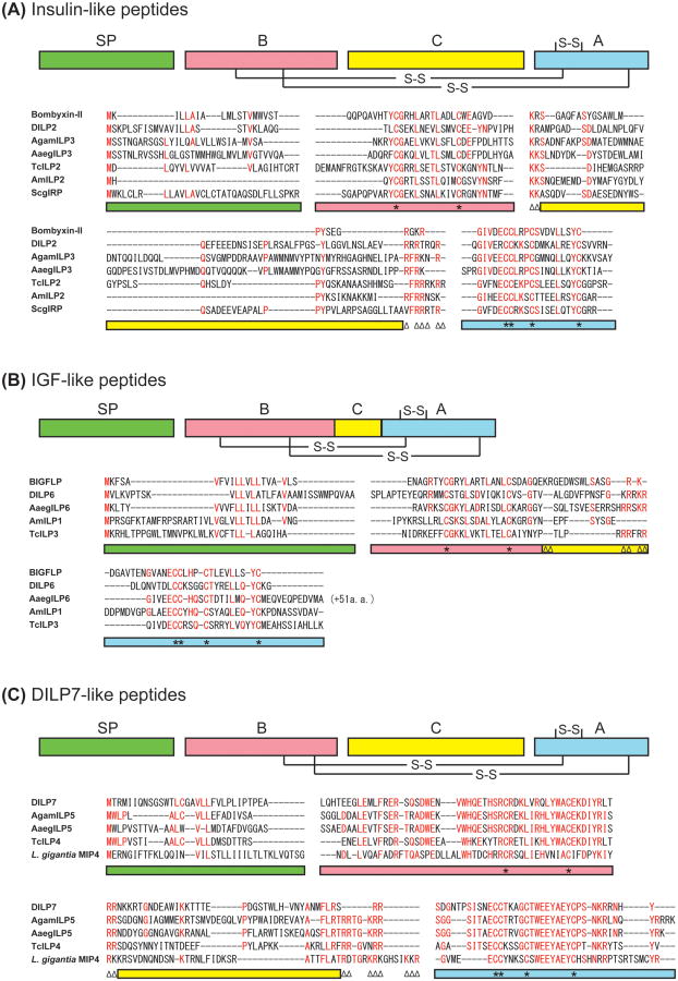
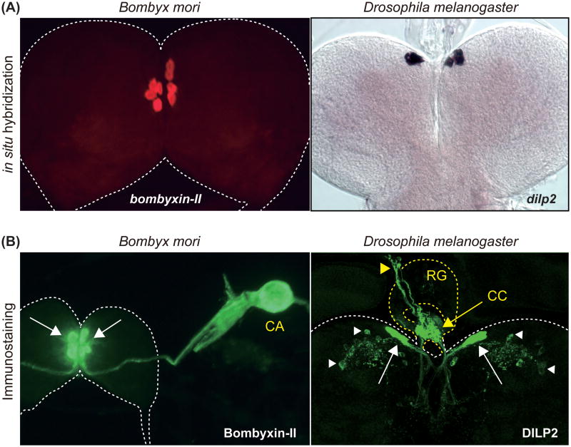
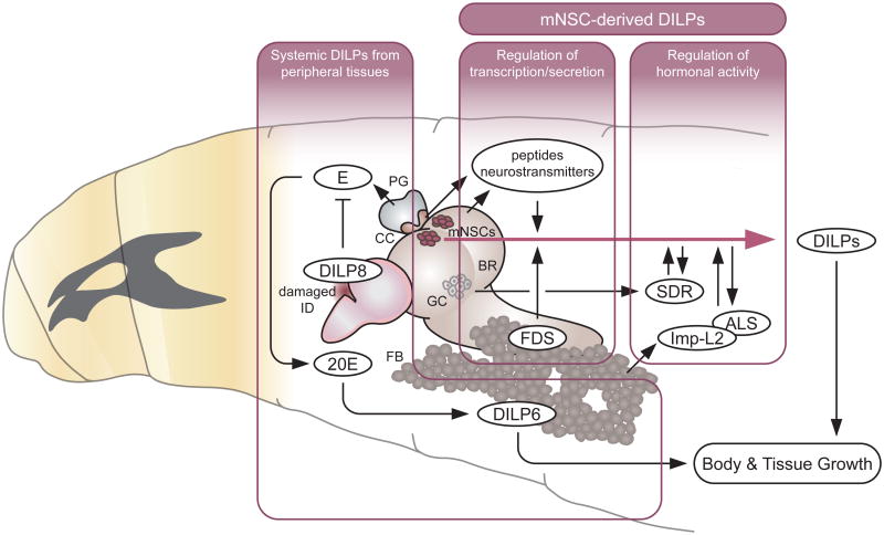
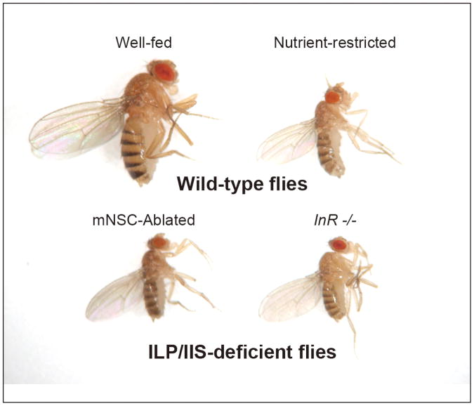
In metazoans, members of the insulin-like peptide (ILP) family play a role in multiple physiological functions in response to the nutritional status. ILPs have been identified and characterized in a wide variety of insect species. Insect ILPs that are mainly produced by several pairs of medial neurosecretory cells in the brain circulate in the hemolymph and act systemically on target tissues. Physiological and biochemical studies in Lepidoptera and genetic studies in the fruit fly have greatly expanded our knowledge of the physiological functions of ILPs. Here, we outline the recent progress of the structural classification of insect ILPs and overview recent studies that have elucidated the physiological functions of insect ILPs involved in nutrient-dependent growth during development.


Figures

Figure 1
Figure 1
Predicted insulin-like, IGF-like and DILP7-like peptides in insects. (A) Amino acid sequences of the representatives of predicted insulin-like peptides from Bombyx (bombyxin-II), Drosophila (DILP2), Anopheles (AgamILP3), Aedes (AaegILP3), Apis (AmILP2), Tribolium (TcILP2), and Schistocerca (ScgIRP) are aligned. (B) Amino acid sequences of the representatives of predicted IGF-like peptides from B. mori (BIGFLP), Drosophila (DILP6), Aedes (AaegILP6), Apis (AmILP1), and Tribolium (TcILP3) are aligned. (C) Amino acid sequences of the representatives of predicted highly conserved ILP group (DILP7-like peptides) from Drosophila (DILP7), Anopheles (AgamILP5), Aedes (AaegILP5), Tribolium (TcILP4), and Lottia (molluscan insulin-related peptide 4, MIP4) are aligned. Highly conserved amino acid residues are shown in red. Color bars indicate the predicted domains in the precursor peptides: green, signal peptide; red, B-chain; yellow, C-peptide; blue, A-chain. Asterisks on the color bars below the alignment denote Cys residues, and paired triangles denote potential cleavage sites (dibasic amino acids).
Figure 2
Figure 2
Insulin-like peptides are mainly produced by brain mNSCs in insects. (A) Detection of bombyxin-II and dilp2 mRNA in the larval brain by in situ hybridization. bombyxin-II and dilp2 expression is observed in four and seven pairs of mNSCs in Bombyx and Drosophila, respectively. (B) Detection of Bombyxin-II and DILP2 localization in the larval brain by immunostaining. Bombyxin-II produced by mNSCs (white arrows) are axonally transported to the CA. DILP2 produced by mNSCs (white arrows) are axonally transported to the CC (yellow arrow) on the ring gland, and further transported to the dorsal vessel (yellow arrowhead). DILP2 signal can also be detected in specific sets of neurons within the brain (white arrowhead). CA, corpora allata; CC, corpora cardiaca; RG, ring gland.
Figure 3
Figure 3
Systemic function of DILPs during larval development and its regulation by multiple factors (see text for details). DILP, Drosophila insulin-like peptide; E, ecdysone; 20E, 20-hydroxyecdysone (active form of ecdysone); FDS, fat body-derived signal; SDR, secreted decoy of insulin receptor; Imp-L2, ecdysone-inducible gene L2; ALS, acid-labile subunit; mNSCs, median neurosecretory cells; BR, brain; GC, glial cells; PG, prothoracic gland; CC, corpora cardiaca; ID, imaginal discs; FB, fat body.
Figure 4
Figure 4
Nutrient-restricted or ILP/IIS-deficient flies show severe growth defect. Wild-type flies, wild-type adult female flies raised either on a nutrient-rich diet (Well-fed) or low-protein diet (Nutrient-restricted). ILP/IIS-deficient flies, a brain mNSC-ablated female fly (DILP-producing mNSCs in the brain were genetically ablated using a dilp2 promoter to express the pro-apoptotic gene, reaper) and an InR hypomorphic mutant female fly.

References

    1. Nakae J, Kido Y, Accili D. Distinct and overlapping functions of insulin and IGF-I receptors. Endocr Rev. 2001;22:818–835. - PubMed
    1. Saltiel AR, Kahn CR. Insulin signaling and the regulation of glucose and lipid metabolism. Nature. 2001;414:799–806. - PubMed
    1. Stewart CE, Rotwein P. Growth, differentiation, and survival: multiple physiological functions for insulin-like growth factors. Physiol Rev. 1996;76:1005–26. - PubMed
    1. Taguchi A, White MF. Insulin-like signaling, nutrient homeostasis, and life-span. Annu Rev Physiol. 2008;70:191–212. - PubMed
    1. Prentki M, Matschinsky FM, Madiraju SR. Metabolic signaling in fuel-induced insulin secretion. Cell Metab. 2013;18:162–185. - PubMed