Review
. 2014 Dec 29:1:15.
doi: 10.3389/fcvm.2014.00015. eCollection 2014.

Balloon Angioplasty - The Legacy of Andreas Grüntzig, M.D. (1939-1985)

Affiliations
Review

Balloon Angioplasty - The Legacy of Andreas Grüntzig, M.D. (1939-1985)

Matthias Barton et al. Front Cardiovasc Med. .

Abstract

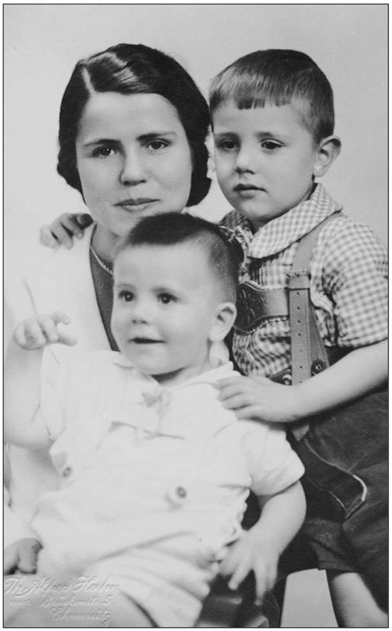
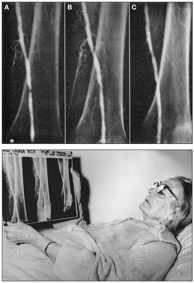
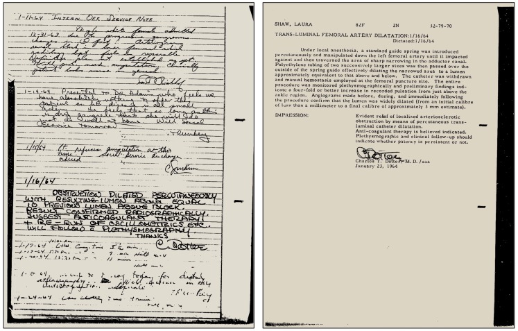
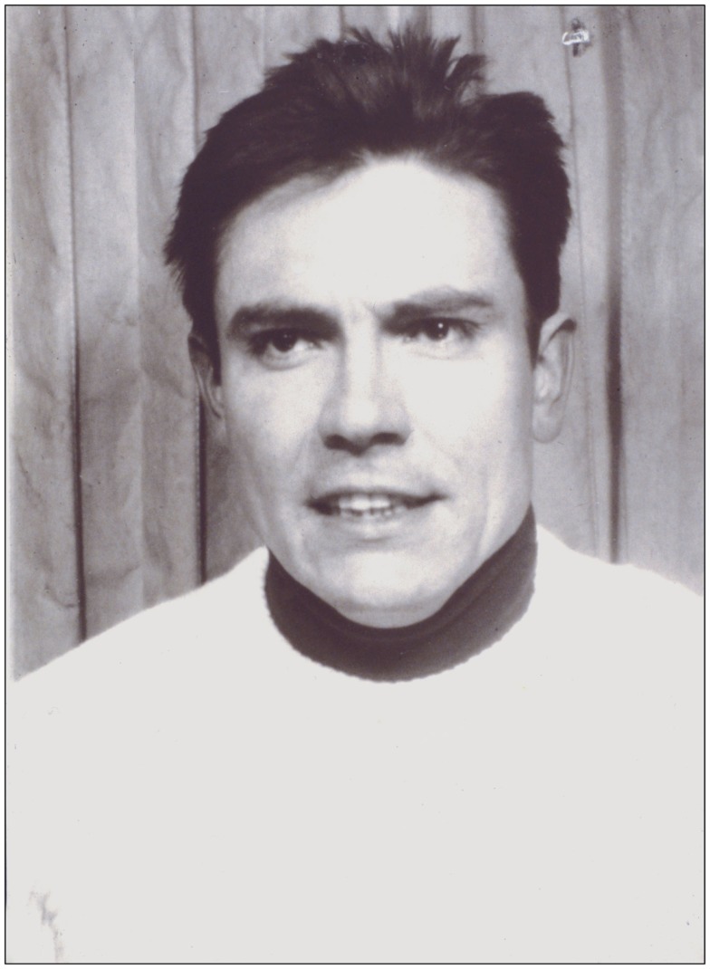
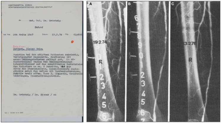
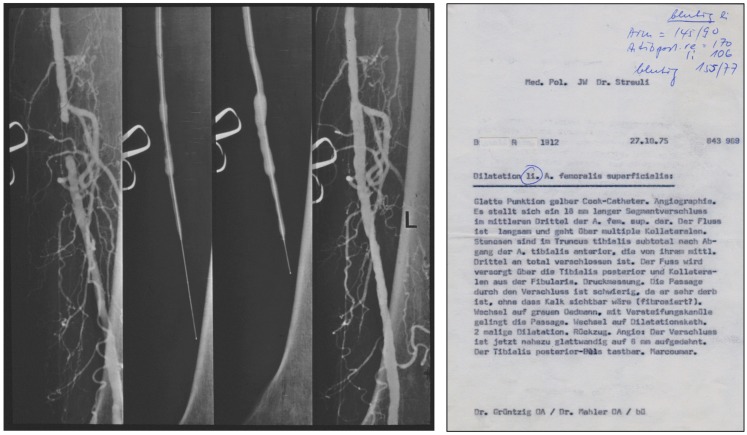
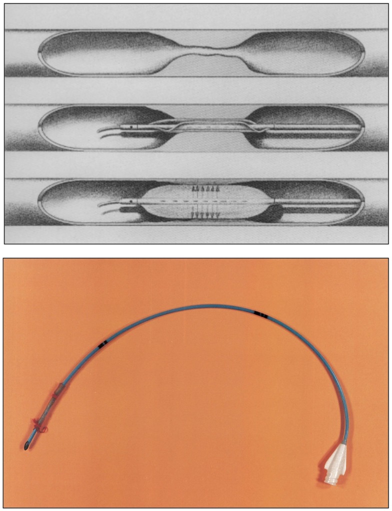
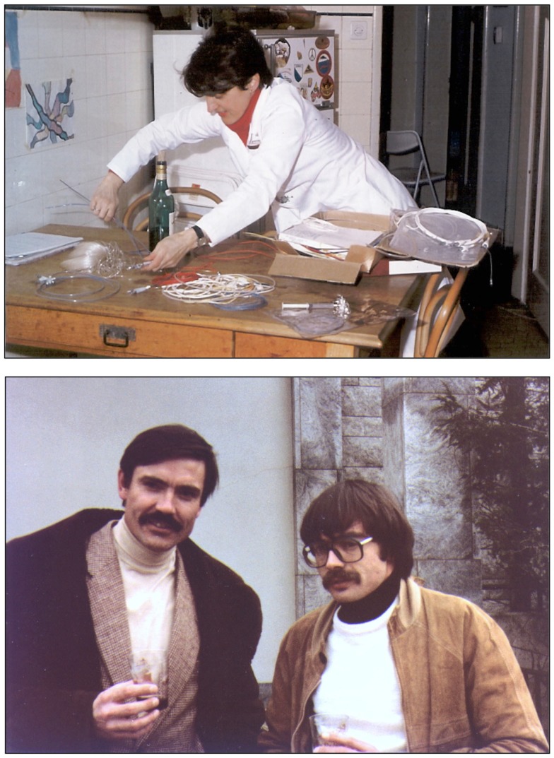
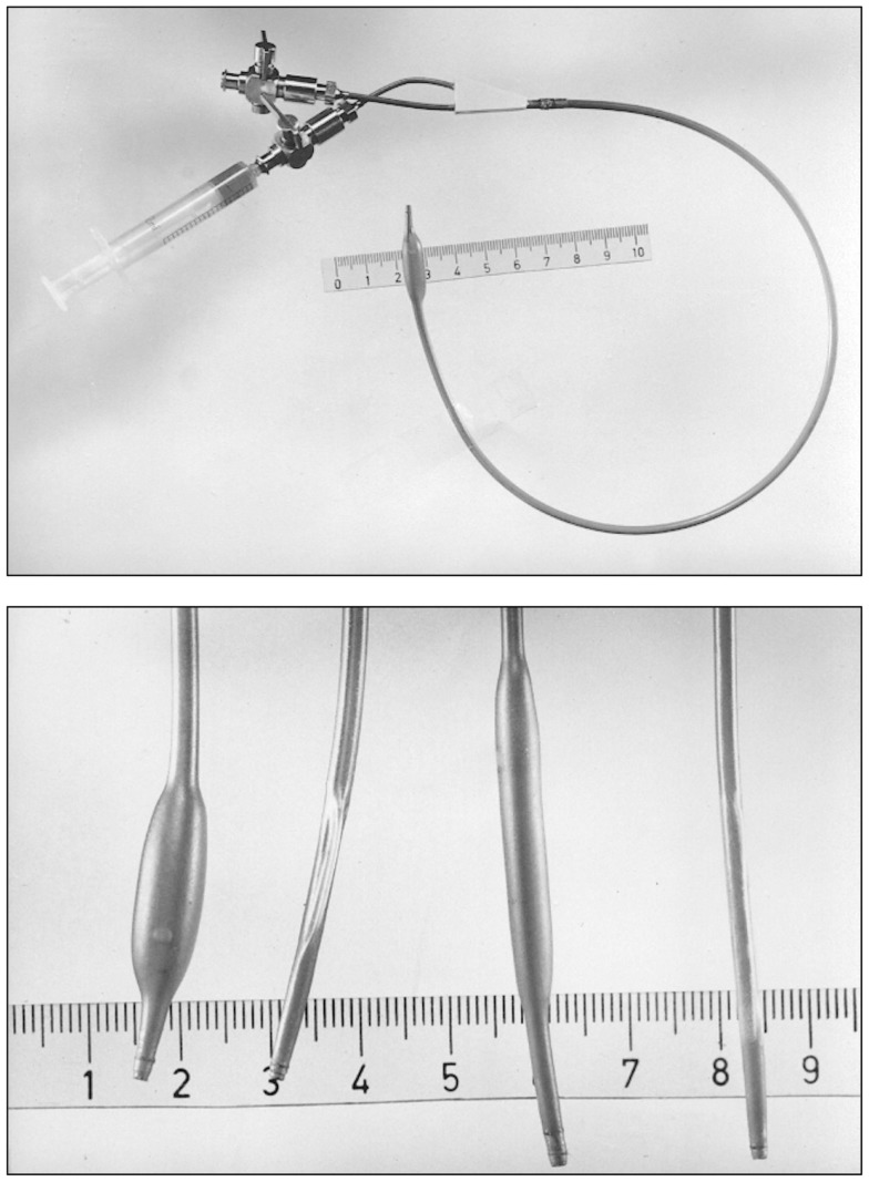
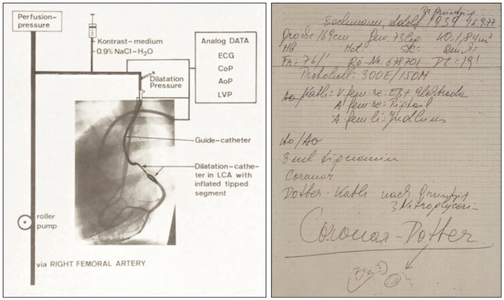
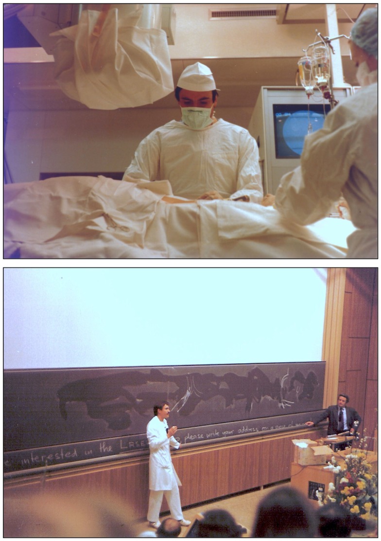
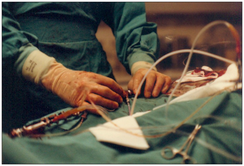
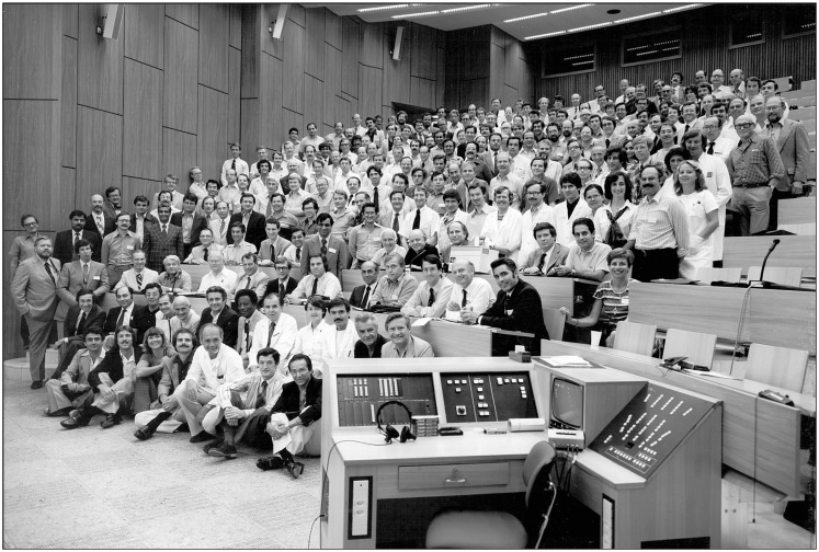
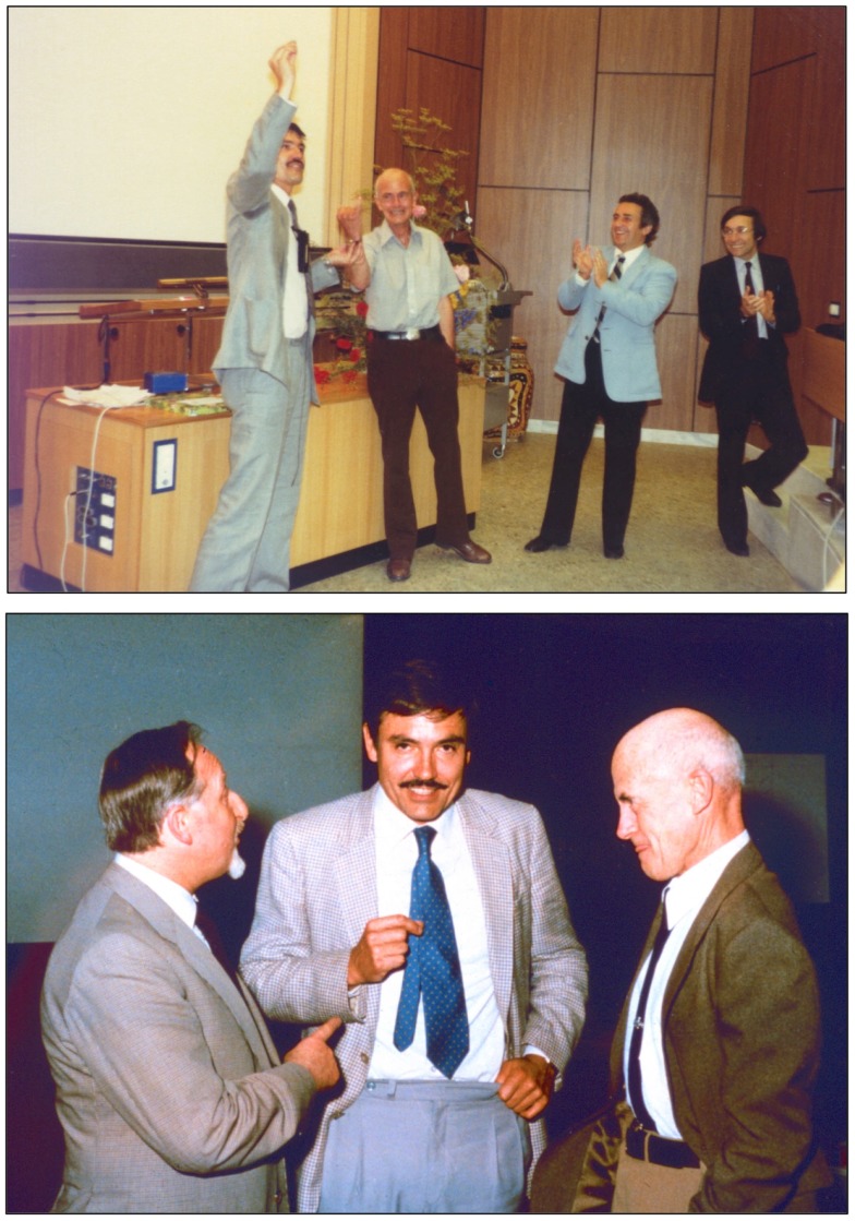
In 1974, at the Medical Policlinic of the University of Zürich, German-born physician-scientist Andreas Grüntzig (1939-1985) for the first time applied a balloon-tipped catheter to re-open a severely stenosed femoral artery, a procedure, which he initially called "percutaneous transluminal dilatation". Balloon angioplasty as a therapy of atherosclerotic vascular disease, for which Grüntzig and Charles T. Dotter (1920-1985) received a nomination for the Nobel Prize in Physiology or Medicine in 1978, became one of the most successful examples of translational medicine in the twentieth century. Known today as percutaneous transluminal angioplasty (PTA) in peripheral arteries or percutaneous transluminal coronary angioplasty (PTCA) or percutaneous coronary intervention (PCI) in coronary arteries, balloon angioplasty has become the method of choice to treat patients with acute myocardial infarction or occluded leg arteries. On the occasion of the 40(th) anniversary of balloon angioplasty, we summarize Grüntzig's life and career in Germany, Switzerland, and the United States and also review the developments in vascular medicine from the 1890s to the 1980s, including Dotter's first accidental angioplasty in 1963. The work of pioneers of catheterization, including Pedro L. Fariñas in Cuba, André F. Cournand in France, Werner Forssmann, Werner Porstmann and Eberhard Zeitler in Germany, António Egas Moniz and Reynaldo dos Santos in Portugal, Sven-Ivar Seldinger in Sweden, and Barney Brooks, Thomas J. Fogarty, Melvin P. Judkins, Richard K. Myler, Dickinson W. Richards, and F. Mason Sones in the United States, is discussed. We also present quotes by Grüntzig and excerpts from his unfinished autobiography, statements of Grüntzig's former colleagues and contemporary witnesses, and have included hitherto unpublished historic photographs and links to archive recordings and historic materials. This year, on June 25, 2014, Andreas Grüntzig would have celebrated his 75(th) birthday. This article is dedicated to his memory.

Keywords: Alexis Carrel; Andreas Gruentzig; Nobel Prize; Robert Hegglin; atherosclerosis; autobiography; biography; coronary artery disease; peripheral vascular disease.


Figures

Figure 1
Figure 1
Portrait of Charlotte Grüntzig with Andreas (sitting) and Johannes Grüntzig (right). The photograph is embossed by a mark of Photostudio Th. Alfred Hahn in Chemnitz, Germany, where the photograph was taken in 1942. Photograph reproduced with permission of Johannes Grüntzig, M.D., Düsseldorf.
Figure 2
Figure 2
Top: Angiograms of the first intentional percutaneous transluminal angioplasty (PTA) performed on January 16, 1964 by Charles T. Dotter, M.D. and Melvin P. Judkins, M.D., in patient Laura Shaw. (A) The initial control angiogram reveals a tight, thread-like stenosis of the left distal superficial femoral artery. (B) Angiogram of the same artery immediately after the angioplasty procedure, which was performed using a 3.2 mm co-axial catheter (C) Follow-up angiogram of the same artery 3 weeks after the PTA procedure demonstrates luminal patency, indicating successful angioplasty. Bottom: Charles Dotter’s first angioplasty patient, 83-year-old Laura Shaw in February 1964 three weeks after the first PTA. The patient holds the radiographic films with the same three angiograms shown in the top panel of this figure. Top figure reproduced from Dotter and Judkins (60), with permission of the American Heart Association. Bottom figure reproduced with permission of the Dotter Interventional Institute, Oregon Health and Science University, Portland, OR, USA.
Figure 3
Figure 3
Left: Case notes by Charles T. Dotter, M.D., hand-written on January 16, 1964, after the first successful superficial femoral artery PTA was performed in patient Laura Shaw. Right: The typed PTA intervention report of January 21, 1964, signed by Dotter. Note: already in 1964 Dotter was using the metric system to indicate lesion size, although in the article reported the same year he was still using inches. Figure reproduced with permission of the Dotter Interventional Institute, Oregon Health and Science University, Portland, OR, USA.
Figure 4
Figure 4
Photograph of Andreas Grüntzig taken in 1969 at the time when he was recruited by Robert Hegglin, M.D., to join the Medical Policlinic at the Kantonspital of the University of Zürich. Photograph reproduced with permission of Ernst Schneider, M.D., Zürich.
Figure 5
Figure 5
Left: A copy of Grüntzig’s report written in German of the first balloon angioplasty performed on February 12, 1974, utilizing his hand-made, single-lumen balloon catheter. In this report, Grüntzig describes “Dottern, linkes Bein” [Dottering, left leg], and summarizes the procedure. Grüntzig refers to a “neuer Dehnungskatether” [new dilatation catheter], which is the only hint that a balloon angioplasty was performed. To specifically indicate this, Maria Schlumpf added “Dilation” in red ink above the description of the procedure right after the typed report had become available (39). Right: Femoral artery dilatation by Grüntzig performed on February 19, 1974 using a single-lumen balloon catheter. (A) Severe atherosclerotic disease resulting in several stenoses of the right superficial femoral artery, note severe wall irregularities. (B) The same artery after balloon dilation: Grüntzig described the PTA procedure as follows: “We dilated the entire artery down to the knee. The step-by-step dilatation of the superficial femoral artery resulted in a hemodynamically sufficient lumen, but severe wall irregularities are visible after dilation. After 2 years the vessel lumen is completely clear. Since the intervention, the patient has been symptom free.” (81) (C) In the dilated artery, patency was confirmed two years later. Photographs reproduced with permission of Maria Schlumpf, Zürich.
Figure 6
Figure 6
Top: Photograph of the inflated, original balloon-dilating catheter hand-made by Andreas Grüntzig, which he used for the first iliac artery balloon angioplasty on March 6, 1974. For size comparison, a centimeter tape measure is shown. Angiograms obtained during this PTA procedure are available online in another article of the Journal (26). Bottom: Part of the first page of the U.S. patent application by Andreas Grüntzig and Hans Gleichner, also showing a cross-section drawing of the balloon catheter, which resembles the original device shown in the upper panel of this figure. Top photograph reproduced with permission of Ernst Schneider, M.D., Zürich.
Figure 7
Figure 7
Left: Pre-, inter-, and post-interventional angiograms of balloon PTA of an occluded left superficial femoral artery in a 63-year-old male patient, performed by Grüntzig and Mahler on October 27, 1975. Right: A copy of the report summarizing the PTA procedure of October 27, 1975. Femoral artery angiography demonstrates an occluded segment measuring 18 mm in length. Passage of the occlusion is difficult, but possible after changing to a special catheter (gray Ödman catheter). The operator changes to the balloon catheter [“Wechsel auf Dilatationskatheter”]; subsequently, the occluded segment is dilated twice and blood flow successfully restored. Photographs reproduced with permission of Maria Schlumpf, Zürich.
Figure 8
Figure 8
Top: the Grüntzig balloon catheter principle. The double-lumen dilatation catheter (120) now contains a main lumen and an additional lumen. The main lumen allows insertion of the guide wire, pressure measurements, and injection of contrast dye. The balloon segment at the catheter tip can be positioned in the stenosed or occluded vascular segment and is filled with liquid via the additional lumen. By applying an equally distributed and constant pressure between 4 and 6 atmospheres, the atherosclerotic plaque occluding the artery is pressed against the vessel wall for 10–30 s. The maximum diameter of the inflated balloon is 4 mm for femoral arteries and 8 mm for iliac arteries. Once the balloon is deflated, the newly formed vascular lumen opens, and blood flow is restored. In its deflated form, the balloon will adhere to the catheter like an umbrella and can be pulled back, and the procedure is completed. Figure legend and drawing according to a sketch by Andreas Grüntzig (36). Bottom: the very first balloon catheter constructed by Grüntzig in 1971, long before balloon catheters were fully functional for the use in patients. The tip of this single-lumen catheter was sealed to allow inflation and extension of the balloon, which was glued and fixed on both sides with a thread onto the catheter tube. Balloons were fixed with surgical sutures to all hand-made catheters until mid-1975 when Cook and later the Schneider company began manufacturing Grüntzig balloon catheters. Photographs reproduced with permission of Maria Schlumpf, Zürich.
Figure 9
Figure 9
Top: Maria Schlumpf at the Grüntzig kitchen table sorting out materials used for building the hand-made balloon catheters. Catheters were built by both of them with the help of their spouses Michaela Grüntzig and Walter Schlumpf. Photograph ca. 1975. Bottom: Andreas Grüntzig and Pierre Levis in Atlanta, GA, USA, during the 1981 angioplasty course held at Emory University. Photographs reproduced with permission of Maria Schlumpf, Zürich.
Figure 10
Figure 10
Top: A double-lumen catheter for iliac artery balloon angioplasty, connected to a three port stop-cock, with one port connected to a syringe containing liquid to inflate the distensible balloon portion of the catheter. Tape measure indicates centimeters. Bottom: Hand-made double-lumen balloon catheters assembled by Grüntzig and his associates on his kitchen table shown in Figure 9. Shown on the left is an inflated iliac artery balloon catheter (with a similar, deflated catheter to its right) and an inflated femoral artery catheter (with a similar, deflated catheter to its right). Note that the balloons are tied to the catheters with fine surgical threads and that catheter tips have been conically tapered, which was done by hand using sandpaper. Inflated balloon diameter of femoral catheters is 4 and 8 mm in iliac artery catheters. Tape measure indicates centimeters. Photographs reproduced with permission of Maria Schlumpf, Zürich.
Figure 11
Figure 11
Left: Schematic representation of the coronary double-lumen balloon dilatation catheter used for PTCA procedures of stenosed canine coronary arteries, which Grüntzig published in 1976 (74, 121, 124). The preformed guiding catheter is sealed by an adaptor against the dilatation catheter localized inside. The main channel of the double-lumen catheter is connected to a three port stop-cock, which can be used to either measure coronary pressure, to inject contrast dye, or to perfuse the coronary artery with arterialized blood. Connected to the neighboring channel of the dilatation catheter is a syringe, which is used to inflate the distensible segment of the dilatation catheter. Pressure is applied by filling the distensible segment, which exhibits only minimal compliance. To demonstrate perfusion with arterialized blood, contrast dye was injected via the main channel, resulting in visible filling of the circumflex branch of the left coronary artery. Additional catheters positioned in the left ventricle and the aorta are also connected to the central recording unit. AoP, aortic pressure; ECG, electrocardiogram; CoP, distal coronary artery pressure; LCA, left coronary artery; LVP, left ventricular pressure. Right: Grüntzig’s notes taken by the nurse assisting him with the first coronary balloon angioplasty performed a conscious, awake patient on September 16, 1977, in Zürich, Switzerland. Four months earlier, Grüntzig had already performed several coronary balloon angioplasties after coronary arteriotomy during open heart surgery together with Richard K. Myler, M.D. and Elias Hanna, M.D. at St. Mary’s Medical Center in San Francisco. At the bottom of the note, there is a small drawing of the aortic root with the coronary arteries, indicating with an arrow the portion of the proximal LAD where Grüntzig performed what he refers to as the first “Coronar-Dotter,” paying tribute to the inventor of endovascular therapy. On this note, the catheter used for the procedure is referred to as “Coronar-Dotter-Kath. nach Grüntzig” [“Coronar-Dotter catheter according to Grüntzig”]. Left photograph reproduced with permission of Maria Schlumpf, Zürich, right photograph reproduced with permission of Ernst Schneider, M.D., Zürich.
Figure 12
Figure 12
Left: Andreas Grüntzig on his Italian Piaggio Vespa scooter in Zürich. Sitting in the front is Richard K. Myler, M.D. with whom Andreas Grüntzig performed the first coronary angioplasties intra-operatively during coronary bypass surgery in May of 1977 in San Francisco. Grüntzig kept this photograph framed on his office desk at Emory University until his death. Shown standing in the back is Simon H. Stertzer, M.D. Right: Andreas Grüntzig during discussion with a course attendee of one of the angioplasty demonstration courses held at Emory University, Atlanta, GA, USA. Photographs reproduced with permission of Ernst Schneider, M.D., Zürich.
Figure 13
Figure 13
Photographs of Andreas Grüntzig in the late 1970 during the balloon angioplasty courses at the University of Zürich. Top: Grüntzig in the catheterization laboratory of the Medical Policlinic during a PTCA procedure at the Kantonspital, which was transmitted live into the lecture hall. Bottom: Grüntzig discussing with course attendees in the lecture hall (Grosser Hörsaal Nord, Figure 15) during the 1980 balloon angioplasty course. Shown standing next to blackboard is Richard K. Myler, M.D. Photographs reproduced with permission of Maria Schlumpf, Zürich.
Figure 14
Figure 14
Photograph of the hands of Andreas Grüntzig during a PTCA procedure in the catheterization laboratory during the 1979 angioplasty course held in Zürich. The procedure was transmitted live and shown on screens located in the lecture hall shown in Figure 15. Photograph reproduced with permission of Maria Schlumpf, Zürich.
Figure 15
Figure 15
Photograph of attendees of the last angioplasty demonstration course held at the Grosser Hörsaal Nord at the Kantonspital of the University of Zürich in August 1980. Most of the pioneers of transluminal and coronary angiography and angioplasty were present, including Charles Dotter, M.D. (second row, 8th from right), Andreas Grüntzig, M.D. (second row, 3rd from right), Melvin P. Judkins, M.D. (third row, 9th from right), and F. Mason Sones, M.D. (fourth row, 4th from right). Also shown are Maria Schlumpf (second row, 4th from right), Richard K. Myler, M.D. (second row, 7th from right), Simon H. Sterzer, M.D. (second row, 12th from right), Grüntzig’s mother, Charlotte (fifth row, 3rd from right), as well as Wilhelm Rutishauser, M.D. (third row, 8th from right) and Hans-Peter Krayenbühl, M.D. (second row, 5th from right), who directed the cardiology division at the Medical Policlinic between 1969 and 1980. Photograph reproduced with permission of Maria Schlumpf, Zürich.
Figure 16
Figure 16
Top: Andreas Grüntzig, M.D. presenting Charles T. Dotter, M.D. with a ring as a token of appreciation for his work in the field of interventional vascular medicine. Shown applauding are Richard K. Myler, M.D. and Simon H. Sterzer, M.D. (right). Bottom: Three vascular medicine pioneers and founders of endovascular therapy (from left to right): Eberhard Zeitler, M.D. (1930–2011), Andreas Grüntzig, M.D. (1939–1985), and Charles Dotter, M.D. (1920–1985), discussing at a symposium held in 1982 in Nürnberg, Germany. Top photograph reproduced with permission of Maria Schlumpf, Zürich, bottom photograph reproduced with permission of Ernst Schneider, M.D., Zürich.
Figure 17
Figure 17
(A) Examples of coil-spring, endarterial tube graft, and its percutaneous placement as introduced by Charles T. Dotter, M.D., in 1969. (B) Balloon-expandable stents developed by Julio Palmaz, M.D. (A) reproduced from Ref. (178) and (B) reproduced from Ref. (183), with permission of the publishers.

References

    1. Ross R. Atherosclerosis – an inflammatory disease. N Engl J Med (1999) 340(2):115–26. 10.1056/NEJM199901143400207 - DOI - PubMed
    1. Bornfeldt KE. 2013 Russell Ross memorial lecture in vascular biology: cellular and molecular mechanisms of diabetes mellitus-accelerated atherosclerosis. Arterioscler Thromb Vasc Biol (2014) 34(4):705–14. 10.1161/ATVBAHA.113.301928 - DOI - PMC - PubMed
    1. Grüntzig A. Ein Fragebogentest zur Diagnose der koronaren Herzkrankheit. Schweiz Rundsch Med Prax (1972) 61(34):1068–70. - PubMed
    1. Grüntzig A, Schönbeck M, Winzeler A, Lichtlen P, Rutishauser W. Die Treffsicherheit einer Fragebogendiagnose bei coronarer Herzkrankheit mit der selektiven Coronarangiographie als Referenztest. Verh Dtsch Ges Inn Med (1972) 78:1017–20. - PubMed
    1. Barton M. Mechanisms and therapy of atherosclerosis and its clinical complications. Curr Opin Pharmacol (2013) 13(2):149–53. 10.1016/j.coph.2013.05.001 - DOI - PubMed

LinkOut - more resources