. 2016 Feb;75(2):134-45.
doi: 10.1111/aji.12458. Epub 2015 Dec 15.

Loss of Intra-Epithelial Endocervical Gamma Delta (GD) 1 T Cells in HIV-Infected Women

Affiliations

Loss of Intra-Epithelial Endocervical Gamma Delta (GD) 1 T Cells in HIV-Infected Women

Natasa Strbo et al. Am J Reprod Immunol. 2016 Feb.

Abstract

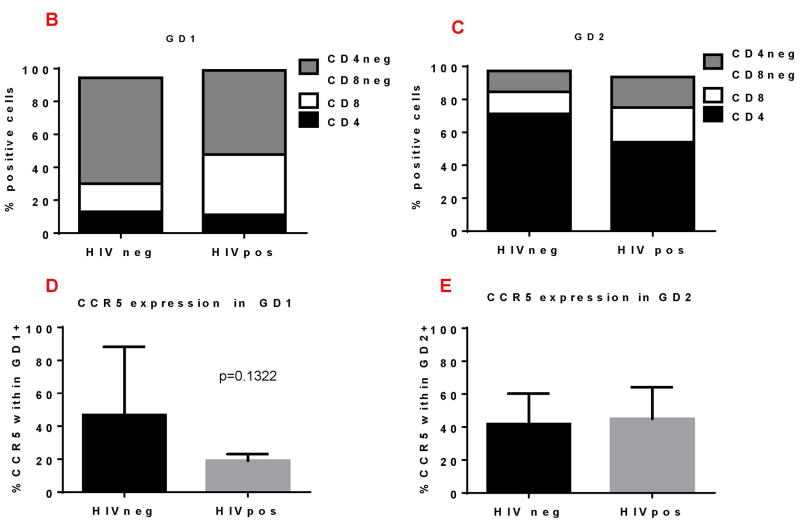
Problem: Human gamma delta (GD) T cells play a well-documented role in epithelial barrier surveillance and protection. Two subsets of GD T cells, defined by the use of either the Vdelta2 (GD2) or Vdelta1 (GD1) TCR, predominate. We hypothesized that endocervical GD T cells play important role in lower genital tract anti-HIV immune responses.

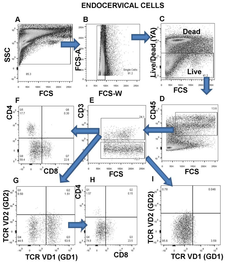
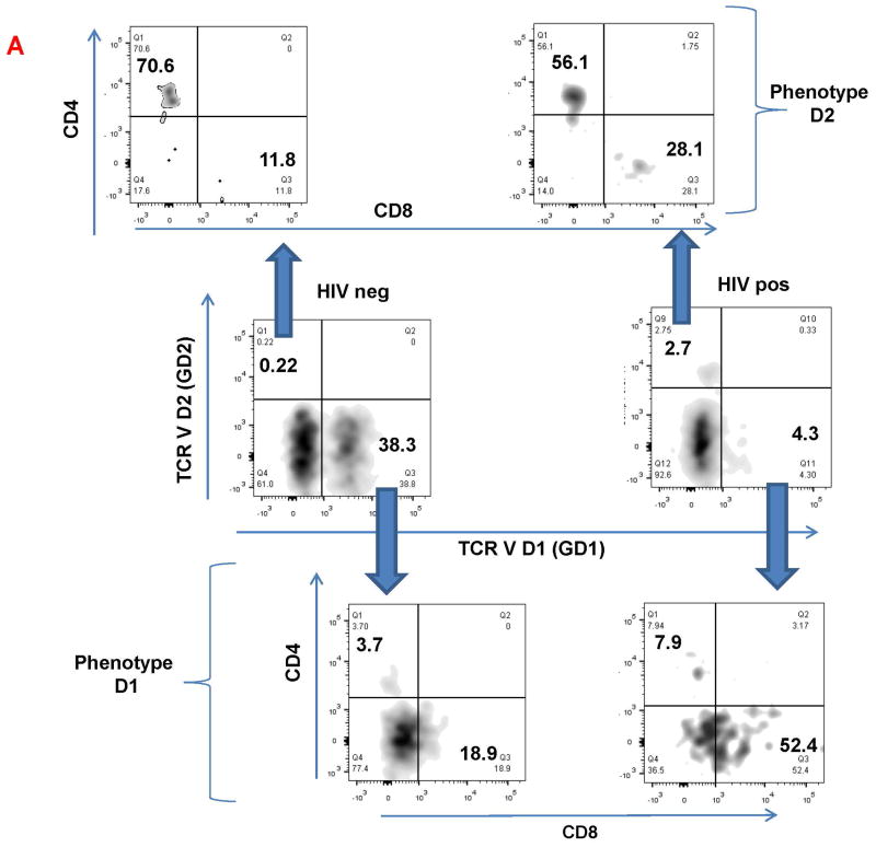
Method of study: HIV-infected (n = 18) and HIV-uninfected (n = 19) pre-menopausal women participating in the WIHS cohort were recruited. Frequency and phenotype of GD T cells were determined in endocervical cytobrush samples and peripheral blood by multicolor flow cytometry.

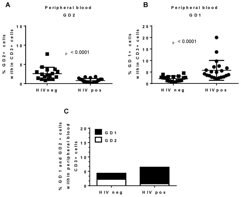
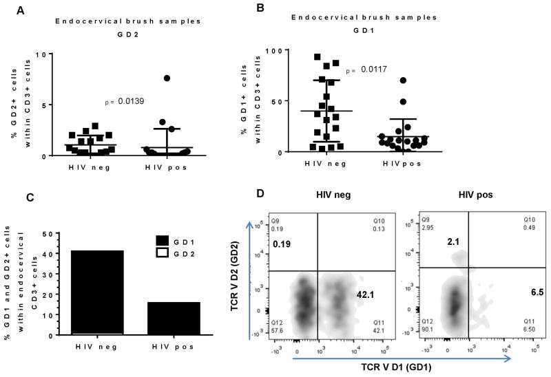
Results: We found depletion of GD2 cells in the blood of HIV-infected women as well as significant decrease in the frequency of endocervical GD1 cells compared to uninfected women.

Conclusion: We report for the first time, the GD1 cells are a predominant endocervical T-cell subset that is significantly decreased in HIV-infected women.

Keywords: Biomarker; HIV; female reproductive tract; gamma delta T cells.


Conflict of interest statement

Disclosure: Authors do not have commercial or other association that might pose a conflict of interest (e.g., pharmaceutical stock ownership, consultancy, advisory board membership, relevant patents, or research funding).

Figures

Figure 1
Figure 1. Representative flow cytometry gating strategy for enumeration of gamma delta (GD) T cell subpopulation in endocervical mucosa
Identification of endocervical T cell populations by following gating strategy: A) Large gate is set on endocervical cytobrush cells B) Exclusion of cell doublets C) Live cells are viability dye-negative population D) CD45 expression on live cells E) CD3 expression on CD45+ cells F) CD4 and CD8 expression on CD3+ cells G) TCRVD1 (GD1) and TCRVD2 (GD2) expression on CD3+ cells H) CD4 and CD8 expression on GD1+ cells I) TCRVD1 expression on gated CD3 negative cells.
Figure 2
Figure 2. Increased GD1 / GD2 T cells ratio in the blood of HIV infected women
PBMC samples from HIV infected and uninfected women were analyzed by flow cytometry A) frequency of GD2+ cells and GD1+ (B) within endocervical CD3+ cells C) combined data from A and B
Figure 3
Figure 3. Endocervical GD1 are predominant GD T cell subset in HIV infected and uninfected women
Endocervical brush samples form HIV infected and uninfected women were analyzed by flow cytometry A) frequency of GD2+ cells and GD1+ (B) within endocervical CD3+ cells C) combined data from A and B D) representative flow cytometry data for HIV negative and HIV positive endocervical CD3+ cells showing expression of TCR VD2 (GD2) and TCR VD1 (GD1).
Figure 4
Figure 4. Phenotypic characteristics of endocervical GD1+ and GD2+ T cells in HIV positive (+) and negative (-) women
Endocervical GD1+ and GD2+ cells were analyzed for expression of surface molecules: CD4, CD8 and CCR5. A) Representative flow cytometry data of gated endocervical CD3+ cells expressing TCR VD2 (GD2+) and TCR VD1 (GD1+) (middle panels). Expression of CD4 and CD8 on GD2+cells (upper panels) and GD1+cells (lower panels). Numbers in the quadrants are showing percent of positive cells. B) Percent of CD4, CD8, double negative (CD4-CD8-) and CCR5+ cells within the GD1 (B and D) and GD2 cells (C and E) in HIV+ and HIV- women.

References

    1. Wira CR, Fahey JV. A new strategy to understand how HIV infects women: identification of a window of vulnerability during the menstrual cycle. Aids. 2008;22:1909–1917. - PMC - PubMed
    1. Wira CR, Fahey JV, Rodriguez-Garcia M, Shen Z, Patel MV. Regulation of mucosal immunity in the female reproductive tract: the role of sex hormones in immune protection against sexually transmitted pathogens. American journal of reproductive immunology. 2014;72:236–258. - PMC - PubMed
    1. Rodriguez-Garcia M, Patel MV, Wira CR. Innate and adaptive anti-HIV immune responses in the female reproductive tract. Journal of reproductive immunology. 2013;97:74–84. - PMC - PubMed
    1. Hayday A, Gibbons D. Brokering the peace: the origin of intestinal T cells. Mucosal immunology. 2008;1:172–174. - PMC - PubMed
    1. Hayday A, Tigelaar R. Immunoregulation in the tissues by gammadelta T cells. Nature reviews Immunology. 2003;3:233–242. - PubMed

Publication types