. 2015;91(10):560-76.
doi: 10.2183/pjab.91.560.

Structure-function correlations of rat trigeminal primary neurons: Emphasis on club-like endings, a vibrissal mechanoreceptor

Affiliations

Structure-function correlations of rat trigeminal primary neurons: Emphasis on club-like endings, a vibrissal mechanoreceptor

Sotatsu Tonomura et al. Proc Jpn Acad Ser B Phys Biol Sci. 2015.

Erratum in

Abstract

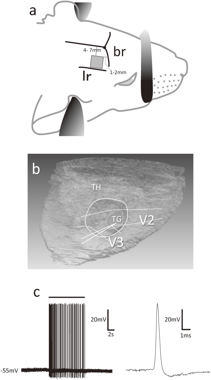
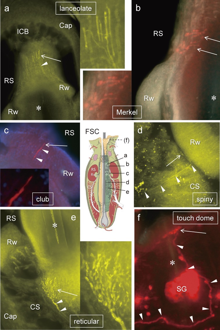
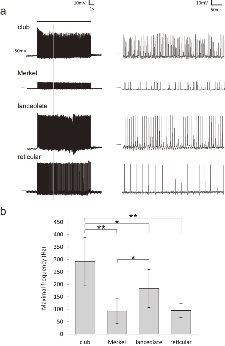
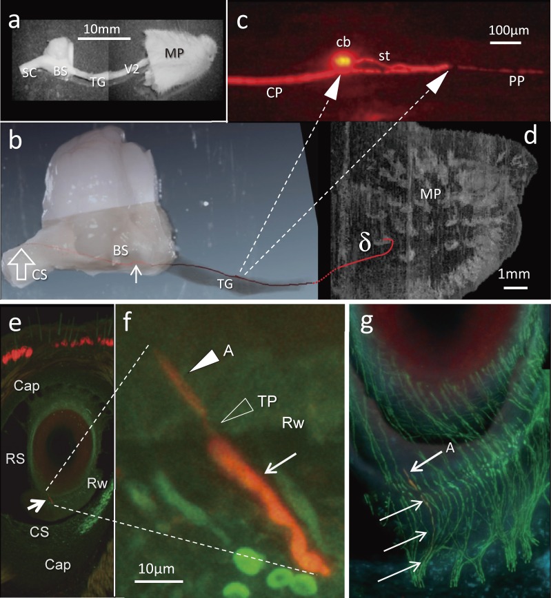
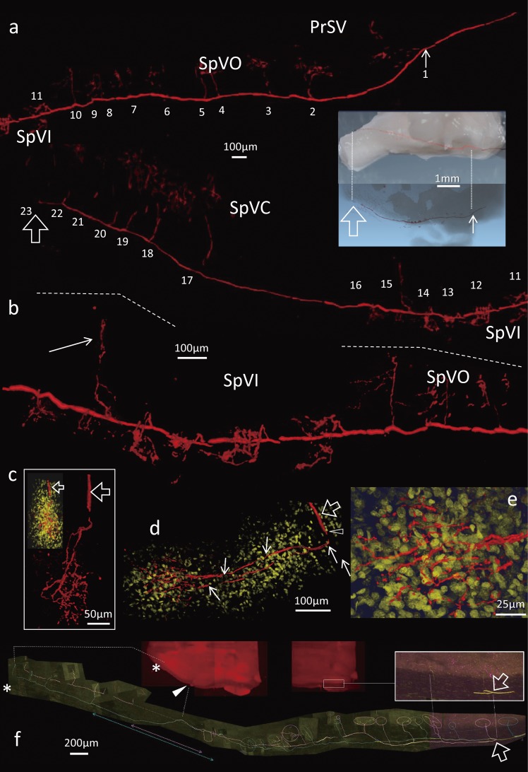
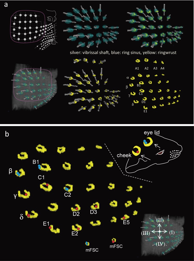
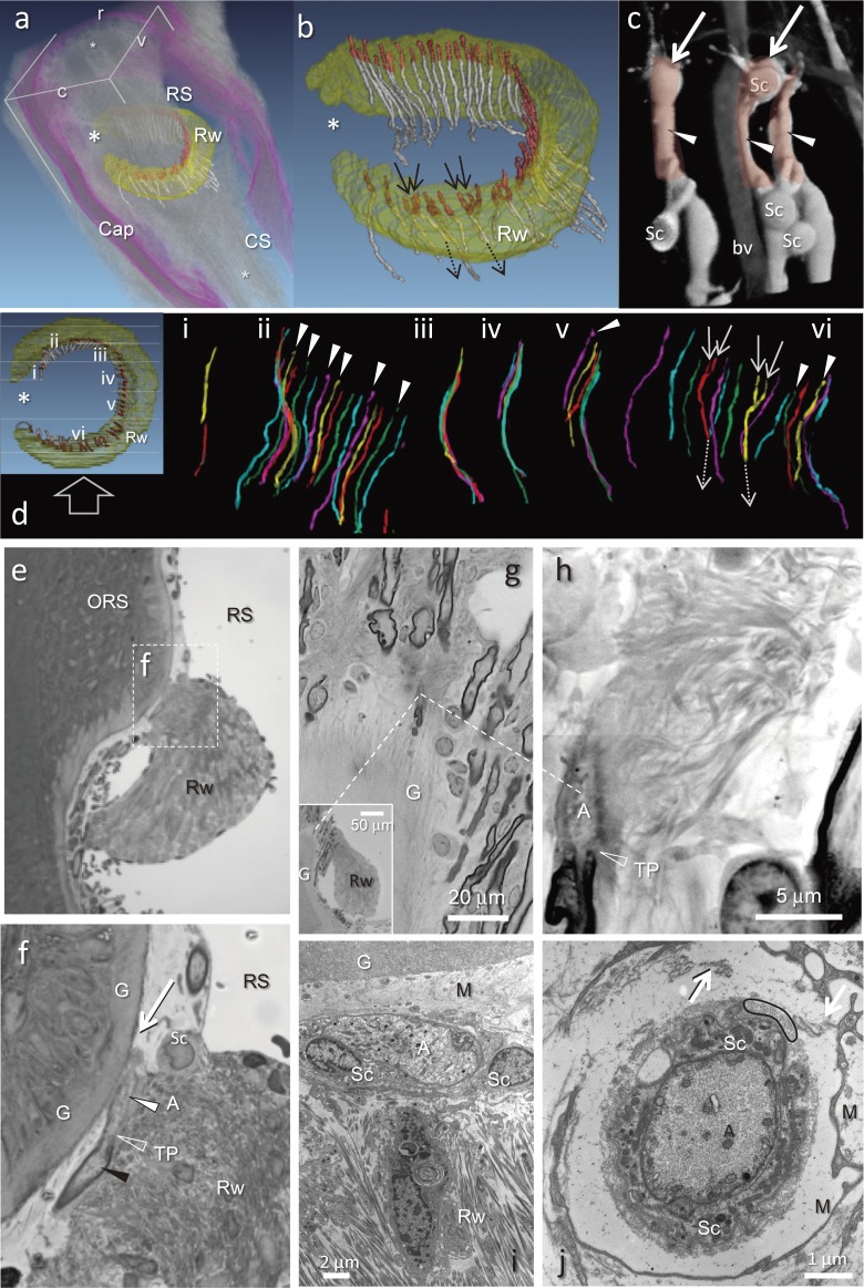
This study focuses on the structure and function of the primary sensory neurons that innervate vibrissal follicles in the rat. Both the peripheral and central terminations, as well as their firing properties were identified using intracellular labelling and recording in trigeminal ganglia in vivo. Fifty-one labelled neurons terminating peripherally, as club-like, Merkel, lanceolate, reticular or spiny endings were identified by their morphology. All neurons responded robustly to air puff stimulation applied to the vibrissal skin. Neurons with club-like endings responded with the highest firing rates; their peripheral processes rarely branched between the cell body and their terminal tips. The central branches of these neurons displayed abundant collaterals terminating within all trigeminal nuclei. Analyses of three-dimensional reconstructions reveal a palisade arrangement of club-like endings bound to the ringwulst by collagen fibers. Our morphological findings suggest that neurons with club-like endings sense mechanical aspects related to the movement of the ringwulst and convey this information to all trigeminal nuclei in the brainstem.


Figures

Figure 1.
Figure 1.
a) Shaded square area of illustration of the temporal bone (5 mm2) excised during our approach to the trigeminal ganglion. br; Bregma, lr; Lateral ridge of parietal bone. b) Three-dimensional reconstruction of the brain illustrating the location of the trigeminal ganglion (TG) with respect to the thalamus (TH), maxillary (V2) and mandibular (V3) nerves. Area circumscribed in white indicates the region of grey matter removed prior to recording from TG neurons. c) Representative trace of a TG neuron. A bunch of action potentials induced by stimulation (bar) of whiskers during recording from a TG neuron. Spontaneous firing was not observed. An enlargement of one action potential is shown at the right.
Figure 2.
Figure 2.
All labelled neurons were recognized and differentiated according to their morphologies. a) Longitudinal lanceolate endings distributed at the level of the ring sinus (RS), b) Merkel endings at the level of the RS, c) club-like ending at the ringwulst (Rw), d) spiny endings at the level of the cavernous sinus (CS), e) reticular endings at the revel of the CS, f) Merkel endings at a touch dome on the eyelid. arrowheads; labelled axon, arrow; mechanoreceptor, asterisk; hair shaft, Cap; capsule of FSC, ICB; inner conical body, SG; sebaceous gland. A schematic drawing of vibrissal follicle-sinus complex (FSC), a modified image from our previous study (Ebara et al., JCN, 2002),6) indicates each position of those mechanoreceptors. Only f is out of FSC. Fig. a, d and e are pseudo-color images taken by a monochrome camera. b, c and f are real color images. Only c showed nuclei stained (purple) with DAPI.
Figure 3.
Figure 3.
a) Response potentials of labelled four neurons evoked by 10-second air puff stimulation (0.1–0.03 MPa). At the right, about 400 ms of the 3rd second (square) were enlarged for each tracing. b) The maximal frequency (mean ± SD) during every 1 second of the air puff stimulation in labelled neurons with club-like endings compared to other labelled mechanoreceptor neurons. *; P < 0.05, **; P < 0.01 (Student’s t-test).
Figure 4.
Figure 4.
a) Dissected tissue including Mystacial Pad (MP). BS; brainstem, SC; spinal cord, TG; trigeminal ganglion, V2; maxillary nerve. b) A visualized and reconstructed trigeminal ganglion (TG) neuron (red) combined with a higher magnification image of a. The first collateral emitted at a distance of approximately 5 mm from the ganglion cell body (small arrow). Open arrow; the end of the central branch of the neuron. c) A higher magnification of the TG neuron in b. Two arrows in dashed line; a round cell body (cb) and the branching point of the stalk (st) into the peripheral (PP) and central (CP) branches are indicated in relation with b. d) A reconstructed three-dimensional image of a mystacial pad evenly enlarged to b. Lower magnification photos of serial 100 µm-thick sections were used. Red line; the peripheral branch labelled neuron in b terminated the delta FSC. e) A microphotograph of a section of the delta FSC including the club-like ending (arrow) of the labelled neuron indicated in b. Cap; capsule, RS; ringsinus, Rw; ringwulst, CS; cavernous sinus. f) A higher magnification image of the club-like ending in e with guidance by two dashed lines. Arrow; a labelled axon, TP; terminal point (open arrowhead), a filled white arrowhead; axon terminal. g) A photomontage of confocal images of 4 serial sections indicating the labelled axon (arrows) and the club-like ending (A) in f (red). Non labelled axons were visualized in green by immunohistochemistry focused on PGP9.5, a pan axonal marker.
Figure 5.
Figure 5.
A collateral extended toward medial part of the nucleus (arrow). c) Montage of three-dimensional reconstructions produced from higher resolution confocal images of one of collaterals of labelled central branch (open arrow). In the upper left insertion, non-specific nuclei of cell bodies were visualized by DAPI (yellow) at the same time. d) A dorsal-ventral view of the c. The central branch (open arrow) produced one collateral (open arrowhead) prior to totally 4 divisions into two respectively. e) A higher magnification of the glomerular-like terminal arborization in d. f) An instance of a bifurcated central parent afferent in a labelled neuron with club-like ending. The bifurcated labelled afferents in addition to their collaterals were traced (in pink and yellow respectively) to the end of the central branches of the neuron (asterisk) through the level of obex (arrowhead on a horizontal section of the brainstem). Collaterals terminated respectively in the trigeminal nuclei (open circles). Labelled both parent afferents emitted no collaterals at a level across the obex (double-headed arrows).
Figure 6.
Figure 6.
a) At upper left, schematic drawings showing the arrangements of mystacial vibrissal follicles on the right snout. Next are 5 sets of reconstructed images showing the distributions of vibrissal follicles with sausage-like ringwulsts (yellow) surrounding each vibrissal shaft (silver) at the bottom level of the ring sinus (blue). These images were provided by tracing each structure in serial 1 µm thick plastic sections and all were reconstructed in three dimensions. Only 4 vibrissae of row A were presented in a different angle of view at the top of the figure. All ringwulsts display an open area dorso-caudally. b) Locations of each 16 club-like endings of labelled 16 TG neurons (β, γ, δ (2), B1, C1, C2, D2, D3, E1, E2, E5 and labial small FSCs (micro FSC (mFSC); 2), the rostral FSC on the eye lid, and the FSC on the cheek skin). Each dot indicates the position on the Rw of the ending. Five neurons (β, B1, C2 and caudal labial mFSC, and eye lid) showed bifurcated endings (blue dots). The other neurons terminated in only one club-like ending (red dot). Arrows on the lower right insertion indicate rostral (I), dorsal (II), caudal (III) and ventral (IV) directions respectively on the C2 whisker.
Figure 7.
Figure 7.
a) A reconstruction of serial 1 µm plastic sections showing the position of the ringwulst rendered in yellow (Rw) within a follicle sinus complex (FSC) displayed in an oblique view from caudal (c) to rostral (r); ventral plane (v). Club-like endings shown in red, small asterisks indicate the vibrissal shaft. RS; ring sinus, Cap; capsule, CS; cavernous sinus. b) Higher magnification of the ringwulst (Rw) reconstructed from individual club-like endings (red) with their preterminal axons (white). All but 4 endings (arrows) were attached to a single axon. The exceptions converged as pairs of endings (solid arrows) into a single axon (dotted arrows). No club-like endings were observed at the open portion of the Rw (large asterisk. Also in a and d). c) Reconstruction of confocal images illustrating club-like endings; arrows point to spherical enlargements at their tips (see d, f). Schwann cells (Sc) envelope club-like endings (arrowheads). bv; blood vessel. d) Reconstruction of all 52 club-like endings in the ringwulst (Rw). Club-like endings were divided into 6 groups (i–vi) and colored as an aid to counting them. Typically, each club-like ending is associated with a single peripheral afferent, but in two instances, afferents bifurcated (solid white arrow; branch, dashed white arrow; parent fiber). Open arrow indicates view direction on the Rw for the colorful nerves. Nine of the 52 endings terminated in a ball-like appendage (arrowhead). e, f) A photomicrograph depicting within the ring sinus (RS), the ringwulst (Rw) attached to the outer root sheath (ORS). f) A High magnification of a square area in e. Black arrowhead; myelinated portion of a peripheral process, a filled white arrowhead; axon terminal of a peripheral process, TP; terminal point of myelination, G; glassy membrane, Sc; Schwann cell, RS; ring sinus, Rw; ringwulst, White arrow; a spherical enlargement at the tip of the club-like ending (see c). g, h) Observations of one of serial 1 µm-thick sections by a scanning electron microscope. Axon terminal (A) of club-like ending carried dense collagen fibrils connecting with the ringwulst (Rw) at a part of the opposite side to the glassy membrane (G). Dashed lines indicate the locations of these photographs at different magnifications. TP; terminal point of myelination. i) Electron micrograph depicting an axon terminal (A) adjacent to two Schwann cells (Sc), together these comprise a club-like ending. The Schwann cells are separated from the glassy membrane (G) by a mesenchymal sheath (M). j) Electron micrograph of a cross-sectioned axon terminal (A) enveloped by a Schwann cell (Sc) sheath, a layer of cross-sectioned collagen fibrils (e.g., within black oval), and finally, a layer of mesenchyme (M), arrows; fine collagen bundles connected in places between inner capsular collagen fibrils and the thin cell layer.

Similar articles

Cited by

References

    1. Retzius, G. (1880) Untersuchungen uber die- Norvensellen der cerebrospinalen. Ganglien und der ubrigen periferschen. Kopfganglien mit besonderer. Berucksichtigung auf die zellenaus laufer. In Arch. Anat. Physiol., Antomiche Abtheilung (eds. His, W., Braune, W. and Bois-Reymond, E.D.). Verlag von Veit & Comp., Leipzig, pp. 369–402.
    1. Rice F.L., Munger B.L. (1986) A comparative light microscopic analysis of the sensory innervation of the mystacial pad. I. Innervation of vibrissal follicle-sinus complexes. J. Comp. Neurol. 252, 186–205. - PubMed
    1. Rice F.L., Kinnman E., Aldskogius H., Johansson O., Arvidsson J. (1993) The innervation of the mystacial pad of the rat as revealed by PGP 9.5 immunofluorescence. J. Comp. Neurol. 337, 366–385. - PubMed
    1. Fundin B.T., Arvidsson J., Rice F.L. (1995) Innervation of nonmystacial vibrissae in the adult rat. J. Comp. Neurol. 357, 501–512. - PubMed
    1. Rice F.L., Fundin B.T., Arvidsson J., Aldskogius H., Johansson O. (1997) Comprehensive immunofluorescence and lectin binding analysis of vibrissal follicle sinus complex innervation in the mystacial pad of the rat. J. Comp. Neurol. 385, 149–184. - PubMed