. 2016 Feb;54(2):91-8.
doi: 10.1002/dvg.22913. Epub 2016 Jan 14.

Fgf3-Fgf4-cis: A new mouse line for studying Fgf functions during mouse development

Affiliations

Fgf3-Fgf4-cis: A new mouse line for studying Fgf functions during mouse development

Matthew J Anderson et al. Genesis. 2016 Feb.

Abstract

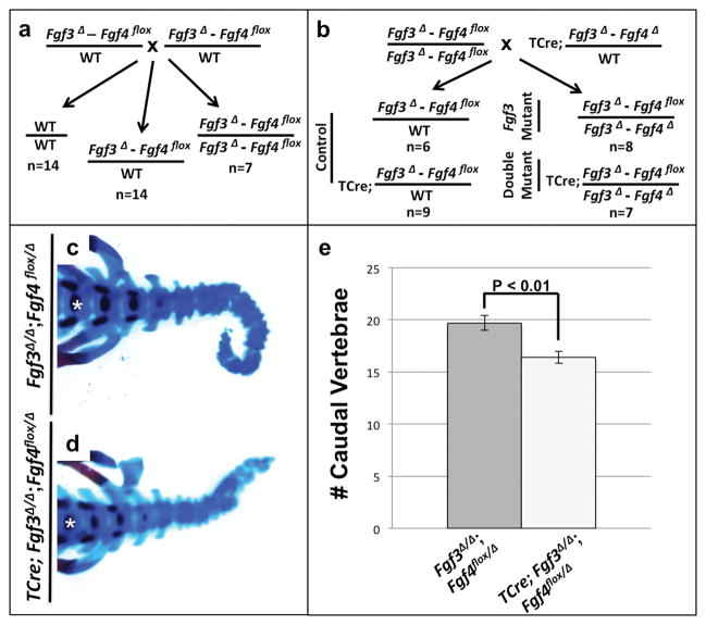
The fibroblast growth factor (FGF) family consists of 22 ligands in mice and humans. FGF signaling is vital for embryogenesis and, when dysregulated, can cause disease. Loss-of-function genetic analysis in the mouse has been crucial for understanding FGF function. Such analysis has revealed that multiple Fgfs sometimes function redundantly. Exploring such redundancy between Fgf3 and Fgf4 is currently impossible because both genes are located on chromosome 7, about 18.5 kb apart, making the frequency of interallelic cross-over between existing mutant alleles too infrequent to be practicable. Therefore, we retargeted Fgf3 and Fgf4 in cis, generating an Fgf3 null allele and a conditional Fgf4 allele, subject to Cre inactivation. To increase the frequency of cis targeting, we used an F1 embryonic stem cell line that contained 129/SvJae (129) and C57BL/6J (B6) chromosomes and targeting constructs isogenic to the 129 chromosome. We confirmed cis targeting by assaying for B6/129 allele-specific single-nucleotide polymorphisms. We demonstrated the utility of the Fgf3(Δ)-Fgf4(flox)-cis mouse line by showing that the caudal axis extension defects found in the Fgf3 mutants worsen when Fgf4 is also inactivated. This Fgf3(Δ)-Fgf4(flox)-cis line will be useful to study redundancy of these genes in a variety of tissues and stages in development.

Keywords: FGF; FGF3; FGF4; axis extension; genetic redundancy; mouse development; presomitic mesoderm.


Figures

FIG. 1
FIG. 1
Targeting of the Fgf3 and Fgf4 loci. (a) (Top) Diagram of a portion of chromosome 7 showing close proximity of Fgf3 and Fgf4 with targeting vectors indicated above the respective wild type (WT) gene. (Bottom, left) Resulting targeted allele for Fgf3 (Fgf3Δ) in which the Fgf3 coding region was replaced with a hygromycin resistance cassette. (Bottom, right) Resulting targeted allele Fgf4 (Fgf4Neo) in which Exons 2 and 3 of Fgf4 were flanked with LoxP sites and a neomycin resistance cassette was inserted into the first intron. Red arrows represent LoxP sites, brackets indicate distances between select restriction sites: HindIII (H), NheI (N), XbaI (Xb), SalI (S), XhoI (X). Relative position of probes used for Southern blot analysis for Fgf3 (P1) and Fgf4 (P2) indicated by black lines. Position of primers used for detection of 3′ LoxP site indicated by arrows, EM26 and EM27. Black bars indicate translated exonic regions, gray bars indicate 3′ untranslated regions. (b) Representative autoradiogram for southern blot analysis of the targeted Fgf4 allele, with DNA digested with NheI. As shown in a, the wild type Fgf4 locus contains a pair of NheI sites spaced 4.5kb apart (WT band in b). Also shown in a, insertion the LoxP site and FRT-flanked neomycin resistance cassette increases the size of the NheI digestion product by 2kb (flox band in b). (c) PCR was performed on DNA from ES-cell lysates to confirm the presence of the 3′ LoxP site; non-specific band produced by PCR amplification denoted with asterisk. (d) Representative autoradiogram of Southern blot analysis for the targeted Fgf3 allele, DNA is digested with HindIII and XhoI. As shown in a, the wild type Fgf3 locus contains a pair of HindIII sites spaced 11.3kb apart, but lacks an XhoI site (WT band in d). As shown in a, replacement of the Fgf3 coding region with the hygromycin resistance cassette introduces an XhoI site. Following digestion of targeted genomic DNA with HindIII and XhoI, a 5.7kb band is detected (Δ band in d).
FIG. 2
FIG. 2
Determination of cis targeting of Fgf3 and Fgf4. (a) Diagram of WT Fgf3 and Fgf4 loci on B6 or 129 chromosome. The B6 chromosome contains an Fgf3 allele with a SNP within exon 3 that creates an HpyAV restriction site and Fgf4 allele with a SNP within exon 2 that creates a Hin1II restriction site. PCR assays were designed across these regions so that these sites, if present, were unique within the amplicon. Replacement of Fgf3 coding regions with the hygromycin resistance cassette or insertion of the Frt-flanked neomycin cassette in Fgf4 disrupted amplification of the targeted alleles (Fgf3Snp primers: 1 and 2; Fgf4Snp primers: 3 and 4). (b) Representative DNA gel electrophoresis showing Fgf4Snp PCR products following digestion with Hin1II. Digestion of PCR products amplified from control 129 tail DNA yielded a full-length 398 bp band and digestion of PCR products amplified from control B6 tail DNA resulted in 268 bp and 130 bp bands. Digestion of the amplicon from the targeted clone yielded a pair of bands corresponding to a remaining untargeted B6 allele; hence we had targeted the 129 chromosome. (c) Representative DNA gel electrophoresis showing Fgf3Snp PCR products following digestion with HpyAV. Digestion of PCR products amplified from the parental F1 ES cells (B6/129), yielded a full-length 667 bp band (corresponding to amplified 129 allele), and 266 bp and 401 bp bands (corresponding to amplified B6 allele); digestion of amplicons generated from 129 or B6 tail DNA yielded single 667 bp or a pair of 266 bp and 401 bp bands, respectively. Digestion of the amplicon from the targeted clone yielded a pair of bands corresponding to a remaining untargeted B6 allele, thus confirming cis targeting of Fgf3Δ and Fgf4Neo to the 129 allele.
FIG. 3
FIG. 3
Fgf3 and Fgf4 act redundantly in caudal axis extension. (a) Genetic cross to generate Fgf3Δ/Δ;Fgf4flox/flox and resultant progeny with observed numbers at E18.5. (b) Experimental cross with observed numbers of progeny at E18.5. (c, d) Skeletal preparations of E18.5 Fgf3Δ/Δ;Fgf4flox/Δ (n=13) and TCre;Fgf3Δ/Δ;Fgf4flox/Δ (n=8) mutants; the first caudal vertebra in each sample is labeled with an asterisk. (c) Total caudal vertebrae number; p-value determined by T-test, error bars represent SEM.
FIG. 4
FIG. 4
Fgf3 and Fgf4 are redundant in tailbud maintenance. (a–d) Uncx4.1 (somite) and Msgn1 (PSM) mRNA expression in E10.5 (38–40 somite stage) control Fgf3Δ/wt;Fgf4flox/wt (n=9)or TCre;Fgf3Δ/wt;Fgf4flox/wt (n=3), Fgf3Δ/Δ;Fgf4flox/Δ (n=7) or TCre;Fgf3;Fgf4 (n=5) embryos; bracket in a shows measurement taken for total PSM in e. (e) Measurement of total PSM anterior-posterior length (white bar) or Msgn1 domain (hashed bar) for each genotype; p-value determined by T-test, error bars represent SEM.

References

    1. Alvarez Y, Alonso MT, Vendrell V, Zelarayan LC, Chamero P, Theil T, Bosl MR, Kato S, Maconochie M, Riethmacher D, Schimmang T. Requirements for FGF3 and FGF10 during inner ear formation. Development. 2003;130:6329–6338. - PubMed
    1. Boulet AM, Capecchi MR. Signaling by FGF4 and FGF8 is required for axial elongation of the mouse embryo. Dev Biol. 2012;371:235–245. - PMC - PubMed
    1. Boulet AM, Moon AM, Arenkiel BR, Capecchi MR. The roles of Fgf4 and Fgf8 in limb bud initiation and outgrowth. Dev Biol. 2004;273:361–372. - PubMed
    1. Feldman B, Poueymirou W, Papaioannou VE, DeChiara TM, Goldfarb M. Requirement of FGF-4 for postimplantation mouse development. Science. 1995;267:246–249. - PubMed
    1. Goetz R, Mohammadi M. Exploring mechanisms of FGF signalling through the lens of structural biology. Nat Rev Mol Cell Biol. 2013;14:166–180. - PMC - PubMed

Publication types

LinkOut - more resources