. 2016 Feb 15:127:307-323.
doi: 10.1016/j.neuroimage.2015.11.069. Epub 2015 Dec 5.

Syntactic processing is distributed across the language system

Affiliations

Syntactic processing is distributed across the language system

Idan Blank et al. Neuroimage. .

Abstract

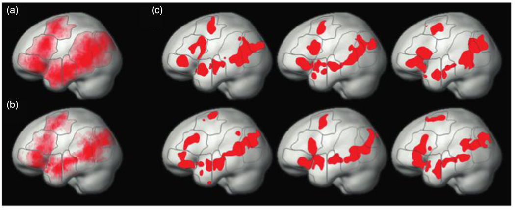
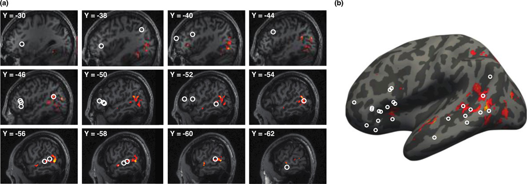
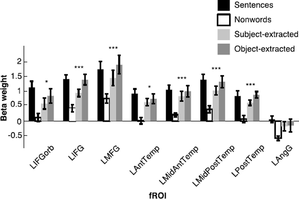
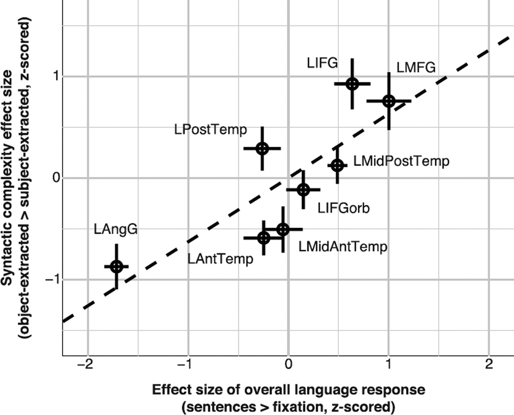
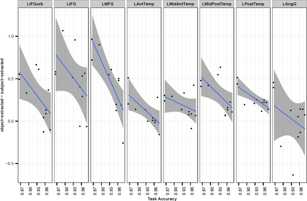
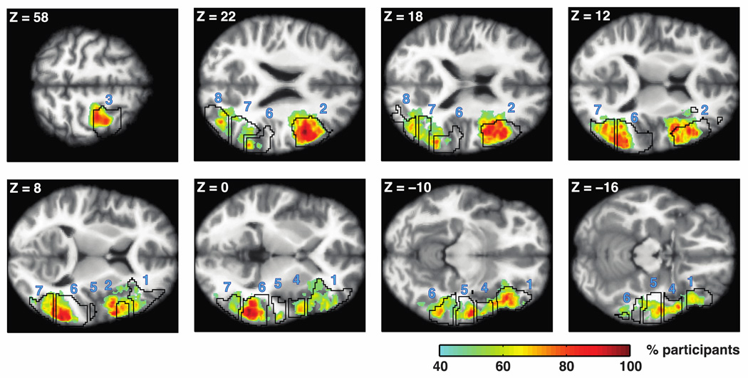
Language comprehension recruits an extended set of regions in the human brain. Is syntactic processing localized to a particular region or regions within this system, or is it distributed across the entire ensemble of brain regions that support high-level linguistic processing? Evidence from aphasic patients is more consistent with the latter possibility: damage to many different language regions and to white-matter tracts connecting them has been shown to lead to similar syntactic comprehension deficits. However, brain imaging investigations of syntactic processing continue to focus on particular regions within the language system, often parts of Broca's area and regions in the posterior temporal cortex. We hypothesized that, whereas the entire language system is in fact sensitive to syntactic complexity, the effects in some regions may be difficult to detect because of the overall lower response to language stimuli. Using an individual-subjects approach to localizing the language system, shown in prior work to be more sensitive than traditional group analyses, we indeed find responses to syntactic complexity throughout this system, consistent with the findings from the neuropsychological patient literature. We speculate that such distributed nature of syntactic processing could perhaps imply that syntax is inseparable from other aspects of language comprehension (e.g., lexico-semantic processing), in line with current linguistic and psycholinguistic theories and evidence. Neuroimaging investigations of syntactic processing thus need to expand their scope to include the entire system of high-level language processing regions in order to fully understand how syntax is instantiated in the human brain.

Keywords: Functional MRI; Language; Syntactic complexity; Syntactic processing.


Figures

Figure 1
Figure 1
Schematic illustration of sample trials in the object-extracted condition. In these instances, the picture matching the sentence is on the left.
Figure 2
Figure 2
Functional regions of interest (fROIs) in the language system. (a) The probabilistic overlap map for the contrast sentences > nonwords in a prior dataset of 25 subjects (Experiments 1 and 2 in Fedorenko et al., 2010). This map was used for generating group-based masks (outlined in gray) which were then used in the current experiment to constrain the selection of individual subjects’ fROIs. (b) The probabilistic overlap map of individual fROIs in the current experiment (shown in red), constrained to fall within the masks (outlined in gray) that were defined based on the prior data shown in (a). (c) Individual fROIs in six sample subjects in the current experiment.
Figure 3
Figure 3
Syntactic complexity effects in the left hemisphere identified with traditional group analysis. Both (a) and (b) show the activation map of our critical contrast, object-extraction > subject-extraction (p<0.001, uncorrected for whole-brain multiple comparisons) in hot colors. White circles show the locations of activations to similar syntactic complexity contrasts reported in prior studies as reviewed by Friederici (2011; referred to in that paper as “studies of movement”). Notice that the activations in the current study fall within the same general locations found previously, namely, the posterior middle temporal gyrus and the inferior frontal gyrus. (a) The effects are superimposed on sagittal slices of an anatomical scan from one of our participants. (b) The effects are projected onto an inflated cortical surface of an average brain in MNI space.
Figure 4
Figure 4
Responses of the language fROIs to the conditions of the language localizer and the critical experiment. Error bars represent standard errors of the mean by participants. The sentences > nonwords contrast is highly significant (p<10−4) in every region (this analysis was carried out using across-runs cross-validation, so that the data used to define the fROIs and estimate the responses are independent, as described in section 2.6). For the object-extracted > subject-extracted contrast: * significance at the p<0.05 level, and *** significance at the p<10−3 level or stronger. All effects remain significant after an FDR correction for the number of regions (n=8). (Note that it is difficult to directly compare the magnitudes of response to the sentences condition of the localizer task and the magnitudes of response to the two critical conditions, because of many differences in the design, materials and procedure across the two experiments.)
Figure 5
Figure 5
The syntactic complexity effect size co-varies with overall sensitivity to language. The mean size, across participants, of the syntactic complexity effect (object-extracted > subject-extracted) is plotted against the mean effect size of the overall response to language, as estimated in the localizer experiment (sentences > fixation). To control for inter-individual differences in the overall response strength, data for the eight fROIs were z-scored within each participant prior to averaging. Crosses show standard errors across participants for both effects. A dashed, black line depicts the linear regression line for predicting the syntactic complexity effect based on the overall language response, and was estimated for visualization purposes only (the linear mixed-effects regression reported in the Results section was carried out using individual data from all participants).
Figure 6
Figure 6
The relationship between task accuracy and the size of the syntactic complexity effect (object-extracted > subject-extracted) in fMRI. Data is shown for each of the 8 fROIs, which all show a downward trend. Blue lines are based on a simple linear regression for each region, with smoothed 95% confidence intervals shaded in gray. Most of the points fall above 0, which shows the main effect of increased fMRI response to the object-extracted condition relative to the subject-extracted condition.
Figure 7
Figure 7
Overlap across participants in the anatomical location of the syntactic complexity effect. Heat maps depict voxels in which more than 40% of participants have an “activation neighborhood” for the syntactic complexity contrast (object-extracted > subject-extracted). Neighborhoods were defined as maximal sets of contiguous voxels that surrounded an activation peak and had contrast estimates numerically greater than zero. Black contours depict our group-based masks (from Fedorenko et al., 2010) used to define fROIs. Numbers correspond to the order of fROIs in Figure 4: 1, LIFGorb; 2, LIFG; 3, LMFG; 4, LAntTemp; 5, LMidAntTemp; 6, LMidPostTemp; 7, LPostTemp; 8, LAngG. Data are superimposed on horizontal slices of Freesurfer’s average T1 scan in common MNI space. Slices were chosen to maximize visibility of the greatest overlap in each mask. Note the especially high overlap (shown in dark red) in the LIFG and LMidPostTemp.

References

    1. Amici S, Ogar J, Brambati SM, Miller BL, Neuhaus J, Dronkers NL, Gorno-Tempini ML. Performance in specific language tasks correlates with regional volume changes in progressive aphasia. Cognitive and Behavioral Neurology. 2007;20(4):203–211. - PubMed
    1. Amunts K, Schleicher A, Bürgel U, Mohlberg H, Uylings H, Zilles K. Broca's region revisited: Cytoarchitecture and intersubject variability. Journal of Comparative Neurology. 1999;412(2):319–341. - PubMed
    1. Ances BM, Leontiev O, Perthen JE, Liang C, Lansing AE, Buxton RB. Regional differences in the coupling of cerebral blood flow and oxygen metabolism changes in response to activation: Implications for BOLD-fMRI. Neuroimage. 2008;39(4):1510–1521. - PMC - PubMed
    1. Baggio G, Hagoort P. The balance between memory and unification in semantics: A dynamic account of the N400. Language and Cognitive Processes. 2011;26(9):1338–1367.
    1. Barde LH, Yeatman JD, Lee ES, Glover G, Feldman HM. Differences in neural activation between preterm and full term born adolescents on a sentence comprehension task: Implications for educational accommodations. Developmental Cognitive Neuroscience. 2012;2:S114–S128. - PMC - PubMed

Publication types