

Exosomes: Fundamental Biology and Roles in Cardiovascular Physiology
- PMID: 26667071
- PMCID: PMC5425157
- DOI: 10.1146/annurev-physiol-021115-104929
Exosomes: Fundamental Biology and Roles in Cardiovascular Physiology
Abstract
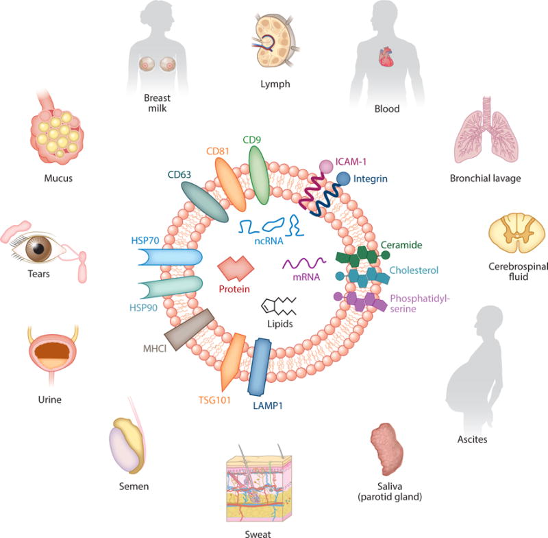
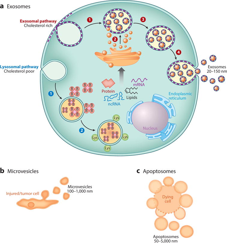
Exosomes are nanosized membrane particles that are secreted by cells that transmit information from cell to cell. The information within exosomes prominently includes their protein and RNA payloads. Exosomal microRNAs in particular can potently and fundamentally alter the transcriptome of recipient cells. Here we summarize what is known about exosome biogenesis, content, and transmission, with a focus on cardiovascular physiology and pathophysiology. We also highlight some of the questions currently under active investigation regarding these extracellular membrane vesicles and their potential in diagnostic and therapeutic applications.
Keywords: cardiovascular disease; extracellular vesicles; intracellular communication; microRNA; regenerative medicine; stem cells.
Figures
Similar articles
-
Characteristics and Roles of Exosomes in Cardiovascular Disease.DNA Cell Biol. 2017 Mar;36(3):202-211. doi: 10.1089/dna.2016.3496. Epub 2017 Jan 23. DNA Cell Biol. 2017. PMID: 28112546 Review.
-
Exosomes and exosomal miRNAs in cardiovascular protection and repair.Vascul Pharmacol. 2015 Aug;71:24-30. doi: 10.1016/j.vph.2015.02.008. Epub 2015 Apr 11. Vascul Pharmacol. 2015. PMID: 25869502 Free PMC article. Review.
-
Cardiac Myocyte Exosome Isolation.Methods Mol Biol. 2016;1448:237-48. doi: 10.1007/978-1-4939-3753-0_17. Methods Mol Biol. 2016. PMID: 27317185 Free PMC article.
-
Mesenchymal Stem Cell-Derived Exosomes: Applications in Regenerative Medicine.Cells. 2021 Aug 1;10(8):1959. doi: 10.3390/cells10081959. Cells. 2021. PMID: 34440728 Free PMC article. Review.
-
Exosome-like Extracellular Vesicles from MYCN-amplified Neuroblastoma Cells Contain Oncogenic miRNAs.Anticancer Res. 2015 May;35(5):2521-30. Anticancer Res. 2015. PMID: 25964525
Cited by
-
Cell-Based Therapy Approaches in Treatment of Non-obstructive Azoospermia.Reprod Sci. 2023 May;30(5):1482-1494. doi: 10.1007/s43032-022-01115-6. Epub 2022 Nov 15. Reprod Sci. 2023. PMID: 36380137 Free PMC article. Review.
-
Plasmon-Enhanced Biosensing for Multiplexed Profiling of Extracellular Vesicles.Adv Biosyst. 2020 Dec;4(12):e2000003. doi: 10.1002/adbi.202000003. Epub 2020 Aug 19. Adv Biosyst. 2020. PMID: 32815321 Free PMC article.
-
Drug Value of Drynariae Rhizoma Root-Derived Extracellular Vesicles for Neurodegenerative Diseases Based on Proteomics and Bioinformatics.Plant Signal Behav. 2022 Dec 31;17(1):2129290. doi: 10.1080/15592324.2022.2129290. Plant Signal Behav. 2022. PMID: 36196516 Free PMC article.
-
TMAO-Activated Hepatocyte-Derived Exosomes Are Widely Distributed in Mice with Different Patterns and Promote Vascular Inflammation.Cardiol Res Pract. 2022 Feb 14;2022:5166302. doi: 10.1155/2022/5166302. eCollection 2022. Cardiol Res Pract. 2022. PMID: 35198242 Free PMC article.
-
RNA profiling of sEV (small extracellular vesicles)/exosomes reveals biomarkers and vascular endothelial dysplasia with moyamoya disease.J Cereb Blood Flow Metab. 2023 Jul;43(7):1194-1205. doi: 10.1177/0271678X231162184. Epub 2023 Mar 8. J Cereb Blood Flow Metab. 2023. PMID: 36883376 Free PMC article.
References
-
- Chargaff E, West R. The biological significance of the thromboplastic protein of blood. J Biol Chem. 1946;166:189–97. - PubMed
-
- Wolf P. The nature and significance of platelet products in human plasma. Br J Haematol. 1967;13:269–88. - PubMed
-
- Johnstone RM. Exosomes biological significance: a concise review. Blood Cells Mol Dis. 2006;36:315–21. - PubMed
-
- Johnstone RM, Adam M, Hammond JR, Orr L, Turbide C. Vesicle formation during reticulocyte maturation. Association of plasma membrane activities with released vesicles (exosomes) J Biol Chem. 1987;262:9412–20. - PubMed
Publication types
MeSH terms
Substances
Grants and funding
LinkOut - more resources
Full Text Sources
Other Literature Sources
