Review
. 2016:78:243-76.
doi: 10.1146/annurev-physiol-021014-071720. Epub 2015 Nov 19.

ADAM Proteases and Gastrointestinal Function

Affiliations
Review

ADAM Proteases and Gastrointestinal Function

Jennifer C Jones et al. Annu Rev Physiol. 2016.

Abstract

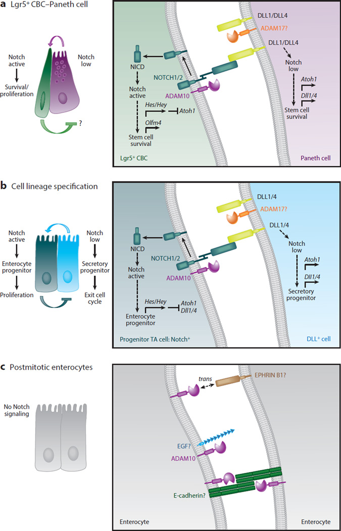
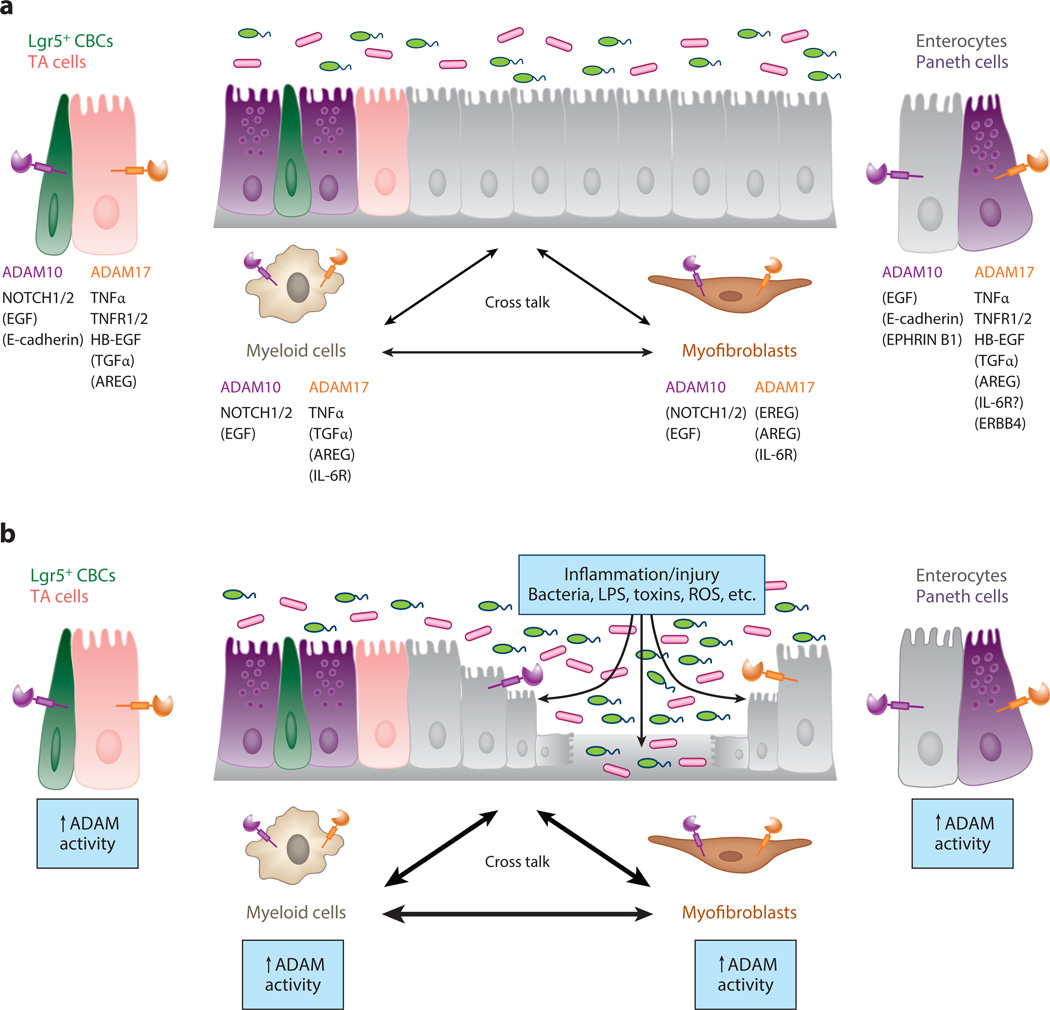
A disintegrin and metalloproteinases (ADAMs) are a family of cell surface proteases that regulate diverse cellular functions, including cell adhesion, migration, cellular signaling, and proteolysis. Proteolytically active ADAMs are responsible for ectodomain shedding of membrane-associated proteins. ADAMs rapidly modulate key cell signaling pathways in response to changes in the extracellular environment (e.g., inflammation) and play a central role in coordinating intercellular communication within the local microenvironment. ADAM10 and ADAM17 are the most studied members of the ADAM family in the gastrointestinal tract. ADAMs regulate many cellular processes associated with intestinal development, cell fate specification, and the maintenance of intestinal stem cell/progenitor populations. Several signaling pathway molecules that undergo ectodomain shedding by ADAMs [e.g., ligands and receptors from epidermal growth factor receptor (EGFR)/ErbB and tumor necrosis factor α (TNFα) receptor (TNFR) families] help drive and control intestinal inflammation and injury/repair responses. Dysregulation of these processes through aberrant ADAM expression or sustained ADAM activity is linked to chronic inflammation, inflammation-associated cancer, and tumorigenesis.

Keywords: ADAM10; ADAM17; EGFR; Notch; TNFα; intestinal stem cells.


Figures

Figure 1
Figure 1
Proteolytically active A disintegrin and metalloproteinases (ADAMs): structure and function. (a) ADAMs are members of the adamalysin subfamily of metzincin metalloproteinases, which also includes ADAMs containing thrombospondin motifs (ADAMTSs) and snake venom metalloproteinases (SVMPs). Different ADAMTSs contain variable numbers of thrombospondin-like motifs (represented by the black X) and other functional domains at the C terminus (not shown). Most ADAMs are synthesized as transmembrane (TM) proteins, whereas all ADAMTSs and SVMPs are secreted proteins. Another important difference between the proteins is that all SVMPs and ADAMTSs are predicted to be catalytically active, but only ~60% of ADAMs have intact metalloproteinase domains capable of proteolytic activity. (b) Phylogenetic tree built from aligned full-length amino acid sequences (left; tree adapted from References and 7) and the tissue distribution of all human proteolytically active ADAMs (right). (c) ADAM domain structure and function. (Left) Color representation of structural domains: prodomain, metalloproteinase domain, disintegrin domain, cysteine-rich domain, EGF-like/proximal membrane domain, transmembrane domain, and cytoplasmic domain. Putative furin-like cleavage sites between the prodomain and metalloproteinase domain are shown by an asterisk. (Right) Summary of domain functions. Many ADAMs display broad tissue distribution, but detailed analysis of their cellular expression in different tissues is lacking.
Figure 2
Figure 2
Diverse extracellular signaling pathways are regulated by A disintegrin and metalloproteinase (ADAM)-mediated shedding events. (a) TNFα/TNFR signaling pathway. ADAM17 is the principal protease responsible for the cleavage of the TNFαligand and both TNFR1 and TNFR2 receptors, all of which are type II transmembrane proteins. (i ) Membrane-anchored TNFα (mTNFα) precursors engage in juxtacrine signaling with cell surface TNFRs, particularly high-affinity TNFR2 receptors. (ii ) ADAM17 cleaves mTNFαto release soluble TNFαligand (sTNFα) that can bind to cell surface TNFRs in an autocrine or paracrine manner. (iii ) ADAM17 can also cleave TNFRs, reducing the level of functional TNFR signaling at the cell surface. Soluble TNFRs (sTNFRs) can act as decoy receptors by sequestering sTNFα. (b) ErbB ligand/ErbB receptor signaling pathway. Multiple ADAMs, particularly ADAM10 and ADAM17, are responsible for cleaving different ErbB ligands and ErbB receptors, which are all type I transmembrane proteins. Both ADAM10 and ADAM17 cleave several ErbB ligands, such as HB-EGF and neuregulin (NRG1), under different experimental conditions. Mouse genetic loss-of-function studies have implicated other ADAMs (e.g., ADAM9 and ADAM19) in the processing of ErbB ligands (not shown). (i ) Membrane-anchored ErbB ligand precursors, such as HB-EGF, engage in juxtacrine signaling with cell surface ErbB receptors. (ii ) ADAM10 and ADAM17 cleave different membrane-anchored ErbB ligands, releasing soluble ligands that function in an autocrine or paracrine manner. (iii ) ADAM10 and ADAM17 can cleave ERBB2 and ERBB4 receptors, respectively, reducing functional ErbB receptor signaling at the cell surface. Soluble ErbB receptors can act as decoy receptors, and soluble ERBB2 (sERBB2) can reduce the therapeutic efficacy of neutralizing antibodies against ERBB2 receptors. (c) IL-6 trans-signaling. The IL-6 receptor (IL-6R) can be cleaved by ADAM10 and ADAM17, but ADAM specificity is species dependent. In classical IL-6 signaling, IL-6 binds to cell surface IL-6R, which recruits two molecules of GP130 into a functional ligand/receptor signaling complex. However, in IL-6 trans-signaling, IL-6R is cleaved by either ADAM10 or 17 to release soluble IL-6R (sIL-6R) that can bind to soluble IL-6 ligand. The IL-6/sIL-6R complex has high affinity for cells expressing GP130 receptor and activates them. Classic IL-6 signaling mediates the activation of anti-inflammatory and regenerative pathways, whereas IL-6 trans-signaling is primarily observed in inflammatory and stress conditions. (d ) Cell adhesion. ADAM10 and ADAM17 cleave different cell adhesion molecules that alter cell-cell interactions. (i ) Homotypic E-cadherin protein interactions are involved in maintaining adherens junction formation between epithelial cells. ADAM10 cleaves E-cadherin, resulting in (ii ) decreased cell-cell interaction and altered epithelial cell migration. (iii ) L-selectin and VCAM1 are examples of cell adhesion molecules involved in leukocyte rolling and adhesion to endothelial cells (not shown). (e) Canonical Notch signaling. ADAM10 initiates the processing and activation of the Notch receptors. (i ) Notch ligand expressed on the signal-sending cell engages the Notch receptor on the signal-receiving cell. (ii ) Normally, the negative regulatory region (NRR) within the Notch receptor masks the α-secretase (S2) cleavage site close to the transmembrane domain. Notch ligand binding to its receptor is proposed to confer a conformational change in the NRR domain, allowing ADAM10 to access the Notch S2 cleavage site. ADAM10 is responsible for cleavage of the NOTCH1, -2, and -3 receptors. (iii ) The Notch remnant is subject to intramembrane proteolysis, in which the γ-secretase complex cleaves within the intramembrane domain at the S3 cleavage site to release the Notch intracellular domain (NICD) into the cytoplasm. After translocation into the nucleus, the NICD associates with other transcriptional cofactors and activates expression of Notch-responsive genes such as Hes1. (iv) Under certain experimental conditions, Notch ligands can be subject to extracellular cleavage by ADAM proteases, such as ADAM17. Notch ligand processing may limit active ligand availability, or it may be involved in ligand sequential processing or bidirectional signaling by the ligand intracellular domain. Alternatively, soluble Notch ligand may bind to and activate Notch receptors via a noncanonical pathway. ( f ) Exosome signaling. ADAM10 and substrates such as L1, CD44, and Notch are enriched in exosomes, providing a mechanism for short- and long-range cellular communication. ADAM10 and its substrates on the cell surface are trafficked through the endosomal compartment and then enriched in intraluminal vesicles (ILVs) produced within multivesicular bodies (MVBs). Upon MVB fusion with the plasma membrane (PM), IVLs are released as exosomes into the extracellular environment. (i ) L1 and CD44 can be shed from ADAM10-expressing exosomes into the extracellular space (not shown). (ii ) Exosomes can also interact with cells at distant cellular sites. In addition, ADAMs may be expressed on ectosomes generated by outward budding of the PM (not shown).
Figure 3
Figure 3
ADAM proteolytic activity can be regulated at multiple levels. (a) Constitutive and stimulus-induced ADAM-mediated substrate shedding can be regulated at multiple levels. Although constitutive shedding may require functional interactions with the ADAM cytoplasmic domain, it is generally accepted that rapid, stimulus-induced ADAM shedding events result from posttranslational modifications of the ADAM extracellular domain and act independently of the ADAM cytoplasmic domain. (i ) Extracellular stimuli such as ROS can directly interact with the ADAM extracellular domain to regulate proteolytic activity and substrate recognition (22). (ii ) Cell surface receptor activation via signaling intermediates may generate signals (e.g., ROS) that are released into the extracellular environment and act directly on the ADAM extracellular domain. (iii ) Alternatively, cell surface receptor signaling or other extracellular signals can regulate ADAM transcription, trafficking, and protein-protein interactions, in addition to posttranslational modifications of the ADAM intracellular domain. Many of these same signaling pathways are also likely to act on substrates directly, regulating their presentation to and recognition by ADAMs. (b) Schematic of signaling activity generated by a substrate shed under constitutive and stimulus-induced shedding conditions. The ability to rapidly stimulate substrate shedding (e.g., of ErbB ligands) provides a mechanism to reach a signaling threshold, or signaling pulse, that can produce a distinct stimulus-induced cellular response. However, stimulus-induced shedding can rapidly reduce the reservoir of substrate that is available to be shed from the cell surface. If substrate levels are not replenished at the cell surface, signaling activity is predicted to decrease (possibly below constitutive levels), and time may be required before sufficient substrate is available to repeat this process. Abbreviations: ADAM, A disintegrin and metalloproteinase; ECD, ADAM extracellular domain; ER, endoplasmic reticulum; GFs, growth factors; GPCRs, G-protein coupled receptors; ICD, ADAM intracellular domain; PDI, protein disulfide isomerase; LPS, lipopolysaccharide; RA, retinoic acid; ROS, reactive oxygen species; TIMP, tissue inhibitor of metalloproteinase.
Figure 4
Figure 4
Overview of intestinal homeostasis: regulation of the intestinal stem cell (ISC) niche and cell fate specification in the small intestine. (a) Crypt-villus architecture and signaling gradients involved in maintaining crypt homeostasis. The mouse small intestine is composed of repeating crypt-villus units. The replenishment of the entire epithelial lining of the intestine is a dynamic process that occurs every ~5–7 days. Complex interactions between multiple signaling pathways are precisely integrated to maintain epithelial cell renewal and crypt homeostasis. (Left) Hematoxylin and eosin staining of the mouse small intestine shows the repeating crypt-villus units (inset). (Right) Schematic showing the distribution of gradients for the Wnt, Notch, epidermal growth factor receptor (EGFR)/ErbB, BMP, and Eph/Ephrin signaling pathways along the crypt-villus axis. (b) Cell types of the intestine (left) and signaling within the stem cell niche (right). (Left) Schematic showing the cellular composition of the stem cell niche in the crypt compartment. Leu-rich repeat–containing G protein–coupled receptor 5–expressing (Lgr5+) crypt base columnar stem cells (CBCs) are positioned at the crypt base, intercalated between Paneth cells. Lgr5+ CBCs are an essential component of the ISC niche and give rise to rapidly proliferating transit-amplifying (TA) progenitor cells. +4 cells and DLL1+ cells are facultative reserve stem cells located at positions 4 and 5 from the crypt base. TA cells appear above the stem cell niche and rapidly migrate toward the crypt-villus junction. Before TA cells exit the crypts, they differentiate into distinct absorptive and secretory cell lineages. All differentiated postmitotic intestinal cells emerge from the crypts, migrate along the villus surface, and are eventually shed from the villus tips into the gut lumen. The one exception is Paneth cells, which first appear above the stem cell niche but are then retained at the crypt base. Paneth cells have a longer life span (>30 days) than other differentiated cell types (~5–7 days). (Right) Paneth cells provide signals required for regulating and maintaining Lgr5+ CBCs in the stem cell niche (inset). They produce Wnt ligands (e.g., Wnt3a), which bind to LRP5/6/Frizzled receptor complexes on Lgr5+ CBCs. The binding of R-spondin to LGR4/5 receptors enhances Wnt activity in Lgr5+ CBCs. Paneth cells release EGF (and other ErbB ligands) that bind to EGFR/ErbB receptors on Lgr5+ CBCs. LRIG1, a negative regulator of EGFR/ErbB signaling, can modulate stem cell/progenitor proliferation. DLL4 and DLL1 on the surface of Paneth cells bind to and activate Notch receptors on Lgr5+ CBCs. Wnt, EGFR/ErbB, and Notch signaling promotes stem cell survival, proliferation, and renewal. However, BMP ligands bind to BMPR receptors on Lgr5+ CBCs to limit cell proliferation and increase differentiation. BMPs are produced by myofibroblasts in the lamina propria, whereas the cellular sources of R-spondins are still under investigation. Other accessory cells within the lamina propria (e.g., pericryptal myofibroblasts, immune cells, and endothelial cells) contribute paracrine, and often redundant, signals (both agonistic and antagonistic) that can regulate the stem cell niche, particularly during tissue regeneration following injury and inflammation (not shown). (c) Notch regulates intestinal cell fate specification. Notch signaling is required for Lgr5+ CBC proliferation and survival. Notch also controls cell fate decisions of short-lived TA progenitors by regulating the key transcription factor Atoh1. NOTCH1 and NOTCH2 receptors and DLL1 and DLL4 control these events (see Figure 5 for more details). Notch+ (Dll-low) TA cells are fated toward the enterocyte lineage. Absorptive progenitors differentiate into enterocytes, the major cell type lining the villi. Dll+ (Notch-low) TA cells are fated toward the secretory lineage. Secretory progenitors undergo further specification and differentiate into goblet cells, enteroendocrine cells, tuft cells, and Paneth cells. Other specialized epithelial cell types (e.g., M cells associated with Peyer’s patches and cup cells) are found in the intestine, but their fate mapping is still poorly understood. Panels a and b adapted from Reference .
Figure 5
Figure 5
Cell-autonomous A disintegrin and metalloproteinase (ADAM)10 signaling acts iteratively to regulate Notch signaling in intestinal stem cells (ISCs) and transit-amplifying (TA) progenitors during crypt homeostasis. (a) ADAM10-mediated Notch signaling is required for the survival and maintenance of Leu-rich repeat–containing G protein–coupled receptor 5–expressing (Lgr5+) crypt base columnar stem cells (CBCs). Lgr5+ CBCs expressing NOTCH1/2 receptors are intercalated between Paneth cells expressing DLL1 and DLL4 ligands in the small intestinal stem cell niche. High Notch activity in Lgr5+ CBCs is required for the proliferation and survival of stem cells and maintenance of the stem cell pool. Notch signaling is activated in Lgr5+ CBCs when Dll ligand, found on the surface of Paneth cells, binds to Notch receptors expressed on the surface of Lgr5+ CBCs. Several studies have shown that the intestine tolerates Paneth cell depletion, suggesting that Notch ligand is likely provided by other cells as well. Notch is sequentially cleaved by ADAM10 and γ-secretase to generate the Notch intracellular domain (NICD), which translocates to the nucleus, where its forms an active transcriptional complex. In Notch-active Lgr5+ CBCs, Notch targets genes including those that encode the Hes/Hey transcription factors, which repress Atoh1 and Dll1/4 ligand transcription and enhance expression of the stem cell marker Olfm4. In DLL+ Paneth cells, low Notch activity, reinforced through Notch lateral inhibition, allows derepression of Atoh1 and Dll1/4 ligand expression. ADAM10 is not required in Paneth cells to maintain the Lgr5+ CBC stem cell pool, but it may be involved in other redundant signaling pathways (e.g., EGF) that contribute to the stem cell niche. (b) ADAM10-mediated Notch signaling is required for the fate specification of TA cells. Classical Notch lateral inhibition determines whether a TA cell becomes an absorptive or secretory progenitor. In Notch-active TA progenitors, Notch targets genes including those that encode the Hes/Hey transcription factors, which repress Atoh1 and Dll1/4 ligand transcription. These cells are fated to become absorptive progenitors, which undergo several rounds of proliferation before differentiating into postmitotic enterocytes. In DLL+ TA cells, low Notch activity allows derepression of Atoh1 and Dll1/4 ligand expression. These cells are fated to become secretory progenitors, which rapidly exit the cell cycle and differentiate into distinct secretory cell types. Under certain experimental conditions, Notch ligands can be subject to extracellular cleavage by ADAM proteases such as ADAM17 (as shown in panels a and b). (c) ADAM10 signaling in postmitotic differentiated intestinal epithelial cells (IECs). Although Notch signaling is restricted to the crypt compartment, ADAM10 is abundantly expressed on all differentiated IECs. This implies that ADAM10 is involved in other shedding events in these postmitotic IECs. Potential ADAM10 substrates include E-cadherin, EGF, and EPHRIN B1. The profound effects of ADAM10-deficiency in the ISC/progenitor compartment have hindered analysis of other ADAM10 substrates in vivo.
Figure 6
Figure 6
A disintegrin and metalloproteinases (ADAMs) play a central role in intercellular communication during intestinal homeostasis and upon injury/inflammation. (a) A schematic diagram showing the specific roles of ADAM10 and ADAM17 signaling in cross talk between different cell types during normal intestinal homeostasis. For simplicity, only ADAM-mediated signaling between intestinal epithelial cells (IECs), myofibroblasts, and macrophages/myeloid cells is shown. ADAM activity is required in many other cell types of the gastrointestinal tract, including other immune cell populations (e.g., dendritic cells, T cells, and B cells), endothelial cells, and enteric neurons (not shown). For each cell type, ADAM substrate specificity and hierarchy may be different. For ADAM substrates, direct evidence exists for cleavage events in the gastrointestinal tract. For ADAM substrates listed in parentheses, in vitro data strongly suggest that ADAM-mediated cleavage events occur in the gastrointestinal tract. TA denotes transit amplifying. (b) Under conditions of intestinal inflammation and/or IEC injury, ADAM activity is upregulated. Loss of barrier integrity allows gut luminal contents to directly interact with ADAMs expressed on the basolateral surface of IECs. Numerous proinflammatory stimuli [e.g., lipopolysaccharide (LPS), reactive oxygen species (ROS), cytokines] will stimulate ADAM expression and proteolytic activity. Normally, enhanced ADAM activity and signaling cross talk are carefully integrated to resolve inflammation/injury and promote epithelial restitution and regeneration. However, under conditions of chronic and relapsing inflammation, these same signals, if sustained, may increase the risk of inflammation-associated cancer.

Similar articles

Cited by

References

    1. Weber S, Saftig P. Ectodomain shedding and ADAMs in development. Development. 2012;139:3693–3709. - PubMed
    1. Edwards DR, Handsley MM, Pennington CJ. The ADAM metalloproteinases. Mol. Asp. Med. 2008;29:258–289. - PMC - PubMed
    1. Endres K, Fahrenholz F. Regulation of α-secretase ADAM10 expression and activity. Exp. Brain Res. 2012;217:343–352. - PubMed
    1. Reiss K, Saftig P. The “A disintegrin and metalloprotease” (ADAM) family of sheddases: physiological and cellular functions. Semin. Cell Dev. Biol. 2009;20:126–137. - PubMed
    1. Scheller J, Chalaris A, Garbers C, Rose-John S. ADAM17: a molecular switch to control inflammation and tissue regeneration. Trends Immunol. 2011;32:380–387. - PubMed

Publication types

LinkOut - more resources