

Neural and mesenchymal stem cells in animal models of Huntington's disease: past experiences and future challenges
- PMID: 26667114
- PMCID: PMC4678723
- DOI: 10.1186/s13287-015-0248-1
Neural and mesenchymal stem cells in animal models of Huntington's disease: past experiences and future challenges
Abstract
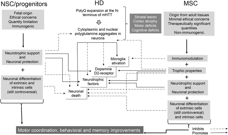
Huntington's disease (HD) is an inherited disease that causes progressive nerve cell degeneration. It is triggered by a mutation in the HTT gene that strongly influences functional abilities and usually results in movement, cognitive and psychiatric disorders. HD is incurable, although treatments are available to help manage symptoms and to delay the physical, mental and behavioral declines associated with the condition. Stem cells are the essential building blocks of life, and play a crucial role in the genesis and development of all higher organisms. Ablative surgical procedures and fetal tissue cell transplantation, which are still experimental, demonstrate low rates of recovery in HD patients. Due to neuronal cell death caused by accumulation of the mutated huntingtin (mHTT) protein, it is unlikely that such brain damage can be treated solely by drug-based therapies. Stem cell-based therapies are important in order to reconstruct damaged brain areas in HD patients. These therapies have a dual role: stem cell paracrine action, stimulating local cell survival, and brain tissue regeneration through the production of new neurons from the intrinsic and likely from donor stem cells. This review summarizes current knowledge on neural stem/progenitor cell and mesenchymal stem cell transplantation, which has been carried out in several animal models of HD, discussing cell distribution, survival and differentiation after transplantation, as well as functional recovery and anatomic improvements associated with these approaches. We also discuss the usefulness of this information for future preclinical and clinical studies in HD.
Figures
Similar articles
-
Transplants of adult mesenchymal and neural stem cells provide neuroprotection and behavioral sparing in a transgenic rat model of Huntington's disease.Stem Cells. 2014 Feb;32(2):500-9. doi: 10.1002/stem.1508. Stem Cells. 2014. PMID: 23939879
-
Stem cell transplantation for Huntington's diseases.Methods. 2018 Jan 15;133:104-112. doi: 10.1016/j.ymeth.2017.08.017. Epub 2017 Sep 1. Methods. 2018. PMID: 28867501 Review.
-
Stem cell therapy and cellular engineering for treatment of neuronal dysfunction in Huntington's disease.Biotechnol J. 2014 Jul;9(7):882-94. doi: 10.1002/biot.201300560. Epub 2014 May 15. Biotechnol J. 2014. PMID: 24827816 Review.
-
Stem cell-based therapy for Huntington's disease.J Cell Biochem. 2013 Apr;114(4):754-63. doi: 10.1002/jcb.24432. J Cell Biochem. 2013. PMID: 23097329 Review.
-
Reductions in behavioral deficits and neuropathology in the R6/2 mouse model of Huntington's disease following transplantation of bone-marrow-derived mesenchymal stem cells is dependent on passage number.Stem Cell Res Ther. 2015 Feb 19;6(1):9. doi: 10.1186/scrt545. Stem Cell Res Ther. 2015. PMID: 25971780 Free PMC article.
Cited by
-
Mouse Degenerating Optic Axons Survived by Human Embryonic Stem Cell-Derived Neural Progenitor Cells.Cell J. 2022 Mar;24(3):120-126. doi: 10.22074/cellj.2022.7873. Cell J. 2022. PMID: 35451581 Free PMC article.
-
Rotating and Neurochemical Activity of Rats Lesioned with Quinolinic Acid and Transplanted with Bone Marrow Mononuclear Cells.Behav Sci (Basel). 2018 Sep 20;8(10):87. doi: 10.3390/bs8100087. Behav Sci (Basel). 2018. PMID: 30241338 Free PMC article.
-
Safety and tracking of intrathecal allogeneic mesenchymal stem cell transplantation in healthy and diseased horses.Stem Cell Res Ther. 2018 Apr 10;9(1):96. doi: 10.1186/s13287-018-0849-6. Stem Cell Res Ther. 2018. PMID: 29631634 Free PMC article.
-
Function and mechanism of mesenchymal stem cells in the healing of diabetic foot wounds.Front Endocrinol (Lausanne). 2023 Mar 16;14:1099310. doi: 10.3389/fendo.2023.1099310. eCollection 2023. Front Endocrinol (Lausanne). 2023. PMID: 37008908 Free PMC article. Review.
-
Conditioned medium derived from bovine umbilical mesenchymal stem cells as an alternative source of cell-free therapy.Vet World. 2021 Oct;14(10):2588-2595. doi: 10.14202/vetworld.2021.2588-2595. Epub 2021 Oct 5. Vet World. 2021. PMID: 34903913 Free PMC article. Review.
References
-
- Barker RA, Mason SL, Harrower TP, Swain RA, Ho AK, Sahakian BJ, et al. The long-term safety and efficacy of bilateral transplantation of human fetal striatal tissue in patients with mild to moderate Huntington’s disease. J Neurol Neurosurg Psychiatry. 2013;84:657–65. doi: 10.1136/jnnp-2012-302441. - DOI - PMC - PubMed
Publication types
MeSH terms
LinkOut - more resources
Full Text Sources
Other Literature Sources
Medical
Miscellaneous
