. 2016 Jan 15;196(2):691-702.
doi: 10.4049/jimmunol.1500710. Epub 2015 Dec 14.

Mannose-Capped Lipoarabinomannan from Mycobacterium tuberculosis Induces CD4+ T Cell Anergy via GRAIL

Affiliations

Mannose-Capped Lipoarabinomannan from Mycobacterium tuberculosis Induces CD4+ T Cell Anergy via GRAIL

Obondo J Sande et al. J Immunol. .

Abstract

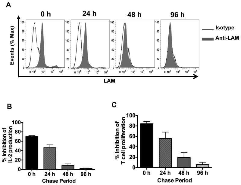
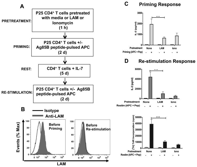
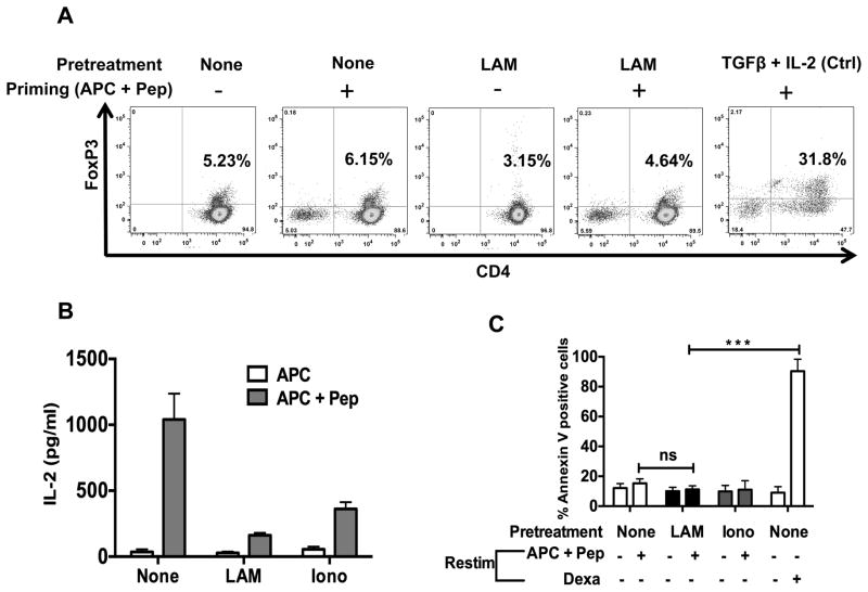
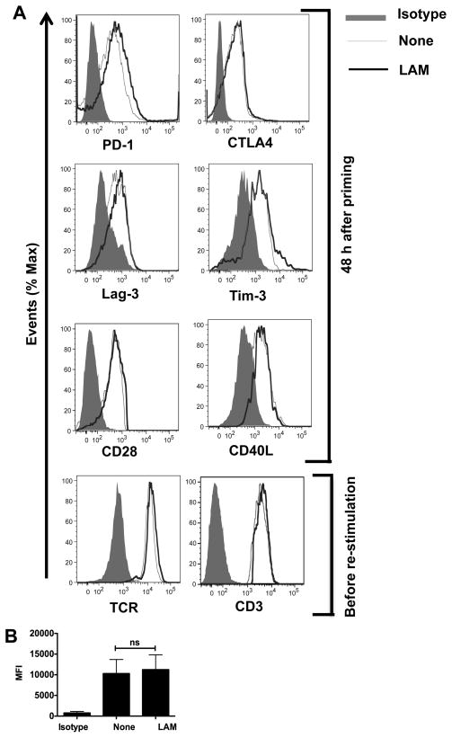
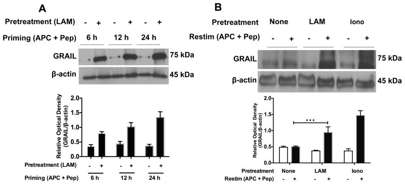
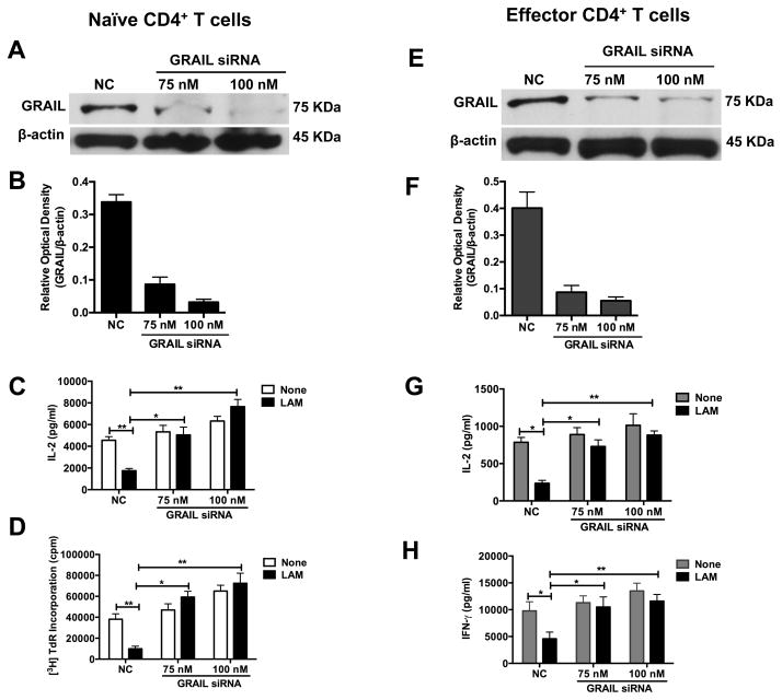
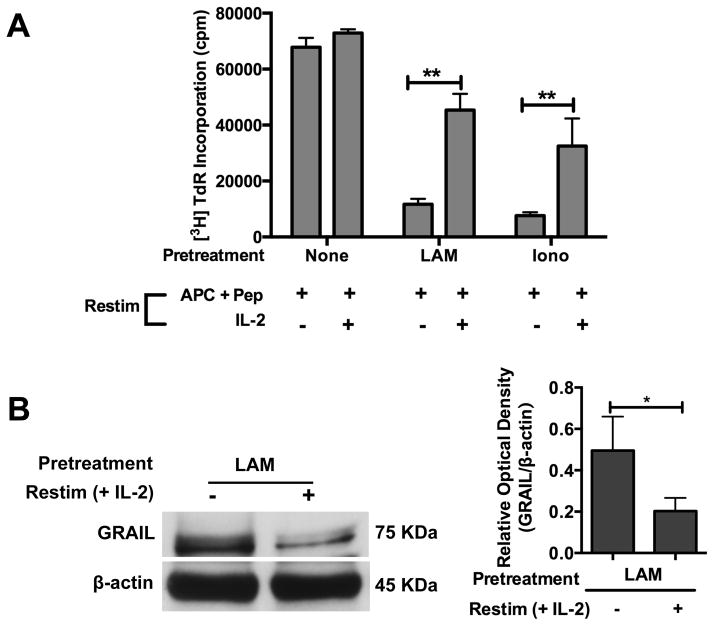
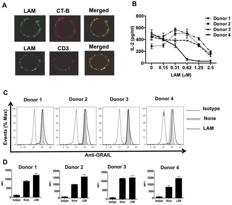
Mycobacterium tuberculosis cell wall glycolipid, lipoarabinomannan, can inhibit CD4(+) T cell activation by downregulating the phosphorylation of key proximal TCR signaling molecules: Lck, CD3ζ, ZAP70, and LAT. Inhibition of proximal TCR signaling can result in T cell anergy, in which T cells are inactivated following an Ag encounter, yet remain viable and hyporesponsive. We tested whether mannose-capped lipoarabinomannan (LAM)-induced inhibition of CD4(+) T cell activation resulted in CD4(+) T cell anergy. The presence of LAM during primary stimulation of P25 TCR-transgenic murine CD4(+) T cells with M. tuberculosis Ag85B peptide resulted in decreased proliferation and IL-2 production. P25 TCR-transgenic CD4(+) T cells primed in the presence of LAM also exhibited decreased response upon restimulation with Ag85B. The T cell anergic state persisted after the removal of LAM. Hyporesponsiveness to restimulation was not due to apoptosis, generation of Foxp3-positive regulatory T cells, or inhibitory cytokines. Acquisition of the anergic phenotype correlated with upregulation of gene related to anergy in lymphocytes (GRAIL) protein in CD4(+) T cells. Inhibition of human CD4(+) T cell activation by LAM also was associated with increased GRAIL expression. Small interfering RNA-mediated knockdown of GRAIL before LAM treatment abrogated LAM-induced hyporesponsiveness. In addition, exogenous IL-2 reversed defective proliferation by downregulating GRAIL expression. These results demonstrate that LAM upregulates GRAIL to induce anergy in Ag-reactive CD4(+) T cells. Induction of CD4(+) T cell anergy by LAM may represent one mechanism by which M. tuberculosis evades T cell recognition.


Figures

FIGURE 1
FIGURE 1
Presence of LAM on CD4+ T cell membranes is required for inhibition of CD4+ T cell activation after primary stimulation. (A) CD4+ T cells (1×10⁶) were pre-treated with LAM (1 μM) or media (untreated) for 1 h at 37°C, washed and incubated with IL-7 (20 ng/ml). Cells were chased for 0, 24, 48 and 96 h. At the end of each chase period, cell aliquots were removed and washed with media, prior to LAM staining. LAM-treated T cells were stained with anti-LAM mAb or isotype control and analyzed by flow cytometry. (B, C) LAM-treated and untreated CD4+ T cells at the end of each chase period were activated with plate-bound anti-CD3 (1 μg/ml) and soluble anti-CD28 (1 μg/ml). IL-2 was measured in 24 h culture supernatants by ELISA (B) and T cell proliferation was measured after 48 h by [3H] thymidine incorporation (C). Percent inhibition reflects the ratio of IL-2 (B) or proliferation (C) between LAM-treated and untreated T cells x 100. Error bars indicate mean +/− SD of triplicate wells of one representative experiment (n=3).
FIGURE 2
FIGURE 2
LAM induces anergy in P25 CD4+ T cells. (A) Experimental design. (B) P25 TCR-Tg CD4+ T cells (1×10⁶) pretreated with LAM for 1 h were stained with anti-LAM mAb or with an isotype control mAb before priming (left histogram) or 5 d after priming and before re-stimulation (right histogram), and analyzed by flow cytometry. (C) 1 h after pretreatment, untreated (none), LAM-treated or ionomycin-treated T cells were primed with Ag 85B peptide-pulsed APC for 48 h. IL-2 was measured in 24-h culture supernatants by ELISA. (D) Primed P25 TCR-Tg CD4+ T cells (in C) were cultured for 5 d in IL-7. Cells were washed and re-stimulated with Ag85B peptide-pulsed APC. IL-2 was measured in 24 h-culture supernatants by ELISA (upper panel) and cell proliferation was determined after 48 h by [3H] thymidine incorporation (lower panel). Data in C through D are means +/− SEM of three independent experiments conducted in triplicate. ***P<0.001 (paired t test)
FIGURE 3
FIGURE 3
LAM does not induce FoxP3-positive regulatory T cells and/or increase activation-Induced cell death (apoptosis). (A) 1×10⁶ P25 TCR-Tg CD4+ T cells left untreated (none) or treated with LAM (as described in Fig. 2) were primed with APC-Ag85B peptide for 48 h. Cells were washed and cultured in IL-7 for 5 d. Cells were washed, and Foxp3-positive T cells were determined by intracellular staining and analyzed by flow cytometry. (B) 1×10⁶ T reg depleted P25 TCR-Tg CD4+ T cells were left untreated (none) or pre-treated with LAM (1 μM) for 1 h. Cells were washed and primed with Ag 85B peptide-pulsed (APC + Pep) or mock-pulsed APC (APC). After 48 h cells were washed and rested in media containing IL-7 for 5 days. CD4+ T cells were then washed and re-stimulated for 24 h. IL-2 was measured in 24-h culture supernatants by ELISA. (C) P25 TCR-Tg CD4+ T cells suppressed through treatment with LAM or ionomycin (as described in Fig. 2) were re-stimulated with APC-Ag85B peptide. After 24 h, T cells were stained with Annexin V eFluor 450 and apoptosis was measured by flow cytometry analysis of annexin V-positive T cells. At re-stimulation, T cells treated with 1 μM of dexamethasone (Dexa) were used as a positive control for apoptosis. Error bars indicate means +/− SD of triplicate wells of one representative experiment (n=3). ***P<0.001 (paired t test).
FIGURE 4
FIGURE 4
LAM does not affect receptor expression on LAM-anergized P25 TCR-Tg CD4+ T cells. (A) 1×10⁶ CD4+ T cells were pre-treated with either LAM or left untreated (none) and co-cultured with Ag85B-pulsed APCs. After 48 h, cells were collected, washed and labeled with anti-PD1, anti-Lag3 or anti-Tim 3, anti-CD28, anti-CD40L mAbs. Intracellular staining was performed for CTLA4. Gating was performed on live CD4+ T cells. Alternatively, after 48 h cells were collected, washed and cultured in IL-7 for 5 d. Cells were then stained for surface expression of TCR and CD3. Gating was performed on live CD4+ T cells. Representative histograms of at least three independent experiments are shown. (B) The mean fluorescence intensity (MFI) of PD-1 staining in LAM-treated (LAM) and untreated (none) T cells was quantified after 48 h of primary stimulation. Data are means +/− SEM of three independent experiments.
FIGURE 5
FIGURE 5
LAM induces increased GRAIL protein expression both during priming and upon re-stimulation. (A) Untreated and LAM-pre-treated P25 TCR-Tg CD4+ T cells were stimulated with Ag85B peptide-pulsed APC for the indicated times. GRAIL protein expression was measured in cell lysates from re-purified CD4+ T cells by Western blot. Blots were analyzed with ImageJ software (NIH) and the ratios of band intensities of GRAIL/beta-actin expressed as relative densities (bar graph). (B) P25 TCR-Tg CD4+ T cells were left untreated (none) or pretreated with LAM or ionomycin and primed with Ag85B peptide-pulsed APC. Cells were washed and cultured in IL-7 for 5 d. Cells were re-stimulated with Ag85B peptide-pulsed APC for 24 h. T cell lysates were obtained from re-stimulated CD4+ T cells, and Western blot performed for GRAIL protein. Data presented are the means +/− SEM of three independent experiments conducted in triplicate. ***P<0.001 (paired t test).
FIGURE 6
FIGURE 6. Knockdown of GRAIL expression by siRNA prevents inhibition of CD4+ T cell activation by LAM
(A, B) A total of 3×10⁶ pre-activated naïve CD4+ T cells plated at 3×10⁵ cells per well in a 24-well plate were transfected with 75 nM or 100 nM of siRNAs targeting GRAIL (GRAIL siRNA) or non-targeting control siRNA (NC) using 6 μl HiPerfect dissolved in serum-free Opti-MEM medium. After 6 h of transfection, an additional 400 μl DMEM medium with 10% FBS was added and cells allowed to incubate at 37°C for 24 h. After 24 h, GRAIL was measured by Western blot with β-actin as loading control. (C, D) GRAIL knocked-down or control naïve CD4+ T cells were left untreated (None) or treated with LAM for 1 h at 37°C. Cells were washed and re-stimulated with plate-bound anti-CD3ε and soluble anti-CD28 for 48 h. IL-2 was measured in 24 h culture supernatants by ELISA (C) and T cell proliferation after 48 h by [3H] thymidine incorporation (D). For A-D, representative blots and densitometry data are shown. (E, F) A total of 3×10⁶ Th1 effector CD4+ T cells plated at 3×10⁵ cells per well in a 24-well plate were transfected and cultured as in (A, B) above. After 24 h, GRAIL was measured by Western blot. (G, H) GRAIL knocked down or control Th1 effector CD4+ T cells were left untreated (None) or treated with LAM for 1 h at 37°C. Cells were washed and re-stimulated as above for 48 h. IL-2 (G) and IFN-γ (H) were measured in 24 h and 72 h culture supernatants respectively by ELISA. IL-2 and proliferation data shown are means +/− SEM of three independent experiments. *P< 0.05, **P< 0.01 (paired t test)
FIGURE 7
FIGURE 7. Exogenous IL-2 down-regulates GRAIL expression and restores T cell proliferation in LAM-anergized CD4+ T cells
(A) P25 TCR-Tg CD4+ T cells (1×10⁶) were left untreated (none) or treated with LAM (1 μM) or ionomycin (1 μM) for 1 h. Cells were washed and primed with Ag85B peptide-pulsed APC. After 48 h, cells were washed and rested in media containing IL-7 for 5 days. CD4+ T cells were washed and re-stimulated with Ag85B peptide-pulsed APC with or without addition of exogenous rIL-2 (20 ng/ml). Cell proliferation was measured after 48 h by [3H] thymidine incorporation. Data are means +/− SEM of three independent experiments conducted in triplicates. *P< 0.05, **P< 0.01 (paired t test). (B) P25 TCR-Tg CD4+ T cells were pretreated with LAM and primed with Ag85B peptide-pulsed APC. Cells were washed and cultured in IL-7 for 5 d. Cells were re-stimulated with Ag85B peptide-pulsed APC with or without addition of exogenous rIL-2 (20 ng/ml) for 48 h. T cell lysates were obtained and GRAIL protein measured by Western blot. A representative blot and densitometry for 3 experiments is shown.
FIGURE 8
FIGURE 8
LAM associates with lipid rafts and CD3 on human CD4+ T cells, and up-regulates GRAIL upon activation with anti-CD3/CD28. (A) Human CD4+ T cells were incubated with LAM for 1 h at 37°C. Top panels, after incubation with LAM, cells were incubated with Alexa Fluor 647 conjugated-cholera toxin subunit B (CT-B, 1 μg/ml) for 20 min on ice, washed extensively, labeled with anti-LAM mAb (clone Cs35) followed by Alexa Fluor 488 conjugated anti-mouse IgG and fixed with 1% paraformaldehyde. Lower panels, after incubation with LAM, cells were labeled on ice with anti-LAM mAb (clone Cs35) followed by Alexa Fluor 488 conjugated-anti-mouse IgG. Then, Alexa Fluor 647 congugated-anti-CD3 mAb was used to label the CD3-TCR complex. Cells were visualized and images merged in a Leica DM16000B confocal microscope (100X oil immersion lens). (B) Human CD4+ T cells isolated from four different Mtb uninfected adult donors were stimulated with plate-bound anti-CD3 and soluble anti-CD28 in the presence of indicated LAM concentrations for 24 h, followed by measurement of IL-2 levels by ELISA. (C) Human CD4+ T cells (1×10⁶) were left untreated (none) or pretreated with LAM (1 μM), followed by stimulation as above. Cells were stained for surface CD4 and intracellular GRAIL using APC-CD4 mAb and rabbit anti-GRAIL primary antibody, respectively. FITC-labeled anti-rabbit secondary antibody was used, followed by FACS analysis. (D) The mean fluorescence intensity (MFI) of GRAIL staining in LAM-treated (LAM) and untreated (none) T cells was quantified after 48 h of primary stimulation. Error bars indicate mean +/− SD of triplicate wells of one representative experiment.

Similar articles

Cited by

References

    1. Commandeur S, van Meijgaarden KE, Prins C, Pichugin AV, Dijkman K, van den Eeden SJ, Friggen AH, Franken KL, Dolganov G, Kramnik I, Schoolnik GK, Oftung F, Korsvold GE, Geluk A, Ottenhoff TH. An unbiased genome-wide Mycobacterium tuberculosis gene expression approach to discover antigens targeted by human T cells expressed during pulmonary infection. J immunol. 2013;190:1659–1671. - PubMed
    1. Lindestam Arlehamn CS, Gerasimova A, Mele F, Henderson R, Swann J, Greenbaum JA, Kim Y, Sidney J, James EA, Taplitz R, McKinney DM, Kwok WW, Grey H, Sallusto F, Peters B, Sette A. Memory T cells in latent Mycobacterium tuberculosis infection are directed against three antigenic islands and largely contained in a CXCR3+CCR6+ Th1 subset. PLoS Pathogens. 2013;9:e1003130. - PMC - PubMed
    1. Lindestam Arlehamn CS, Paul S, Mele F, Huang C, Greenbaum JA, Vita R, Sidney J, Peters B, Sallusto F, Sette A. Immunological consequences of intragenus conservation of Mycobacterium tuberculosis T-cell epitopes. Proc Natl Acad Sci USA. 2015;112:E147–155. - PMC - PubMed
    1. Sutherland JS, Lalor MK, Black GF, Ambrose LR, Loxton AG, Chegou NN, Kassa D. Analysis of Host Responses to Mycobacterium tuberculosis Antigens in a Multi-Site Study of Subjects with Different TB and HIV Infection States in Sub-Saharan Africa. PLOS ONE. 2013;8:e74080. - PMC - PubMed
    1. Comas I, Chakravartti J, Small PM, Galagan J, Niemann S, Kremer K, Ernst JD, Gagneux S. Human T cell epitopes of Mycobacterium tuberculosis are evolutionarily hyperconserved. Nat Genet. 2010;42:498–503. - PMC - PubMed

Publication types

MeSH terms