. 2016 Jun 1;22(11):2734-43.
doi: 10.1158/1078-0432.CCR-15-2361. Epub 2015 Dec 14.

Eradication of Large Solid Tumors by Gene Therapy with a T-Cell Receptor Targeting a Single Cancer-Specific Point Mutation

Affiliations

Eradication of Large Solid Tumors by Gene Therapy with a T-Cell Receptor Targeting a Single Cancer-Specific Point Mutation

Matthias Leisegang et al. Clin Cancer Res. .

Abstract

Purpose: Cancers usually contain multiple unique tumor-specific antigens produced by single amino acid substitutions (AAS) and encoded by somatic nonsynonymous single nucleotide substitutions. We determined whether adoptively transferred T cells can reject large, well-established solid tumors when engineered to express a single type of T-cell receptor (TCR) that is specific for a single AAS.

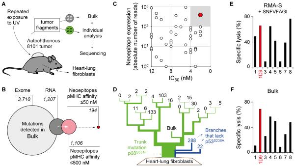
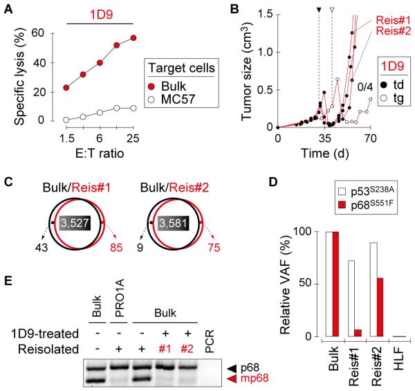
Experimental design: By exome and RNA sequencing of an UV-induced tumor, we identified an AAS in p68 (mp68), a co-activator of p53. This AAS seemed to be an ideal tumor-specific neoepitope because it is encoded by a trunk mutation in the primary autochthonous cancer and binds with highest affinity to the MHC. A high-avidity mp68-specific TCR was used to genetically engineer T cells as well as to generate TCR-transgenic mice for adoptive therapy.

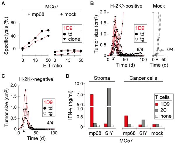
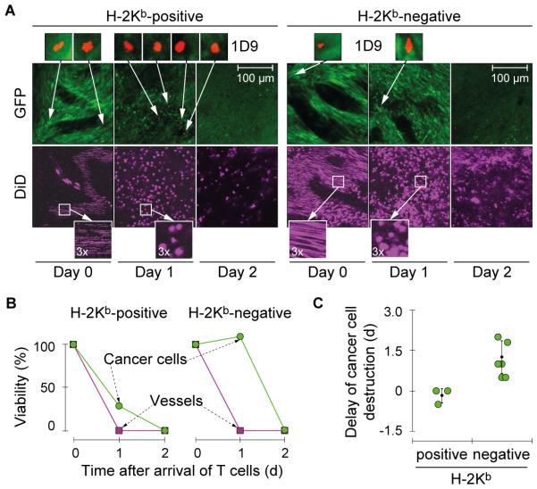
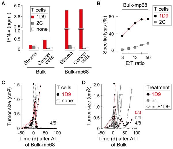
Results: When the neoepitope was expressed at high levels and by all cancer cells, their direct recognition sufficed to destroy intratumor vessels and eradicate large, long-established solid tumors. When the neoepitope was targeted as autochthonous antigen, T cells caused cancer regression followed by escape of antigen-negative variants. Escape could be thwarted by expressing the antigen at increased levels in all cancer cells or by combining T-cell therapy with local irradiation. Therapeutic efficacies of TCR-transduced and TCR-transgenic T cells were similar.

Conclusions: Gene therapy with a single TCR targeting a single AAS can eradicate large established cancer, but a uniform expression and/or sufficient levels of the targeted neoepitope or additional therapy are required to overcome tumor escape. Clin Cancer Res; 22(11); 2734-43. ©2015 AACRSee related commentary by Liu, p. 2602.


Figures

Fig. 1
Fig. 1. The neoepitope mp68, predicted by ‘reverse immunology’, is found throughout the genetically diverse tumor 8101 and is recognized by high-avidity T cell clones
A, UV-irradiation caused the development of an autochthonous tumor (8101). The tumor was excised and 20 individual tumor fragments were adapted to culture (Bulk) or analyzed separately using whole exome sequencing. Heart-lung fibroblasts (HLF) were generated as autologous tissue control. B, Identification of suitable neoepitopes as therapeutic targets by ‘reverse immunology’. Venn diagram (from left to right): Number of mutations detected in Bulk after whole exome and RNA sequencing. Number of neoepitopes (8-, 9-, or 10-mer peptides) found to be expressed and predicted to bind to H-2Kb or -Db with affinities of ≤500 nM or ≤50 nM (NetMHC 3.4). C, Binding affinity to MHC-I (IC50) and RNA expression level of neoepitopes identifies mp68 (SNFVFAGI, red) as highly expressed antigen with highest MHC affinity. D, Phylogenetic representation of somatic mutational frequency in the 8101 tumor identifies mp68 as trunk mutation. Green represents the trunk mutation p68S551F that was found in all fragments. Branches shown in blue lack the mutation p53S238A. Numbers on the top of each branching indicate unique mutations in 20 individual fragments and the Bulk tumor cell culture of 8101. E, F, The T cell clone 1D9 efficiently lyses Bulk tumor cells and is specific for the mp68 neoepitope. Specific lysis of (E) RMA-S cells loaded with 7.8 pM SNFVFAGI peptide or (F) Bulk tumor cells by mp68-specific T cell clones. The T cell clone 1D9 is highlighted in red and was used for subsequent TCR isolation.
Fig. 2
Fig. 2. Mutant-specific TCR gene therapy causes regression of primary 8101 tumors with the subsequent escape of antigen-negative variants
A, 1D9-engineered T cells efficiently lyse Bulk tumor cells. Specific lysis of Bulk and control MC57 tumor cells was analyzed in vitro using 1D9td T cells One representative experiment of three is shown. B, T cell therapy targeting mp68 causes regression of Bulk tumors that eventually escape. Mice with established Bulk tumors were treated with 1D9tg or 1D9td T cells. T cells were injected on day 29 (td) or 39 (tg), as indicated. Data are compiled from 3 independent experiments. Bulk tumor reisolates are indicated (Reis#1, Reis#2). C, Bulk tumor reisolates have similar mutational profiles compared to the parental tumor. DNA of Reis#1 and Reis#2 was analyzed by whole exome sequencing and compared to original Bulk tumor cells. The total number of mutations in each population is indicated. The numbers of shared mutations have colored background. D, Bulk tumor reisolates show diminished variant allele frequency (VAF) of mp68. The VAF of p53S238A and mp68 in Reis#1 and Reis#2 is shown in relation to the VAF detected in Bulk. E, Bulk tumor reisolates do not express the mp68 gene. RT-PCR was used to identify expression of the mutant allele of p68 in Reis#1 and Reis#2. Bulk tumor cells and a reisolated progressor variant of 8101 that escaped an immunocompetent normal host and lost the mp68 gene (PRO1A (33)) were used as controls.
Fig. 3
Fig. 3. Mutant-specific TCR gene therapy eradicates MC57 tumors expressing the mp68 neoepitope
A, T cells expressing the 1D9 TCR lyse MC57 tumor cells stably transfected with the mp68 neoepitope. Specific lysis of indicated target cells was analyzed in vitro using T cells expressing the 1D9 TCR (T cells from OT-IxRag−/− mice transduced with the 1D9 TCR (1D9td) or the original anti-mp68 1D9 T cell clone). One representative experiment of two is shown. B, TCR gene therapy targeting mp68 causes rejection of MC57-mp68 tumors. H-2Kb-positive Rag−/− mice with established MC57-mp68 tumors were treated with 1D9td or 1D9tg T cells (left panel) or with T cells transduced with an irrelevant TCR (Mock, right panel). T cells were injected between day 15 and 19 as indicated by the arrow heads. C, TCR gene therapy can reject MC57-mp68 tumors in absence of stromal cross-presentation. H-2Kb-negative Rag−/− mice bearing established MC57-mp68 were treated as in (B). Data in (B) and (C) were compiled from 7 independent experiments. D, Stromal cross-presentation of mp68 induces high levels of cytokine release by 1D9td T cells. CD11b+ stromal cells were isolated from untreated MC57-mp68 and MC57-SIY tumors. Enriched stromal cells and cancer cells of the respective lines were co-cultured with 1D9td or 2Ctd T cells. IFN-γ content of supernatants was determined by ELISA. One representative experiment of two is shown.
Fig. 4
Fig. 4. Stromal cross-presentation of mp68 accelerates elimination of cancer cells in established tumors whereas direct presentation by cancer cells suffices for tumor vessel destruction
A, Longitudinal confocal microscopy imaging of cancer cell and tumor vessel destruction following adoptive T cell transfer. Cross-presentation of mp68 by the tumor stroma cause rapid destruction of cancer cells by 1D9 T cells entering the tumor. The left panel shows the longitudinal imaging of MC57-mp68 tumors in a H-2Kb-positive and the right panel shows a H-2Kb-negative Rag−/− mouse following adoptive transfer of 1D9 T cells of YFP×1D9×Rag−/− mice. Day 0 is the time when the first 1D9 T cell was detected in the skinfold window (see magnification, red). Viability of tumor tissue was analyzed by monitoring GFP expression (cancer cells, green) and blood flow (see bottom magnification, DiD-stained erythrocytes, purple). Data are representative for 3 independent experiments. B, Quantification of the timing of cancer cell and vascular viability in tumors with or without cross-presentation of mp68 by the tumor stroma shown in (A). Areas on day 0 were defined as 100%. C, Quantification of cancer cell destruction in tumors with or without cross-presentation of mp68 by the tumor stroma. GFP and DiD signals were compared to calculate the delay of cancer cell destruction after collapse of blood flow. Mean values (± SD) obtained from individual mice are shown (p=0.036, Wilcoxon Rank Sum Test). Data were pooled from 3 independent experiments.
Fig. 5
Fig. 5. Escape of primary cancers from mp68-specific T cell therapy is thwarted by uniform and high expression of antigen or when T cell therapy follows local irradiation
A, Bulk-mp68 cancer cells or stromal cells isolated from Bulk-mp68 tumors induce release of high levels of IFN-γ by 1D9td T cells. CD11b+ stromal cells were isolated from Bulk tumors either unmodified or over-expressing mp68. Enriched stromal cells and cancer cells of the respective lines were co-cultured with 1D9td or 2Ctd T cells. IFN-γ content of supernatants was determined by ELISA. One representative experiment of two is shown. (B) Bulk tumor cells modified to express high levels of mp68 are recognized by 1D9-transduced T cells. Specific lysis of Bulk tumor cells over-expressing mp68 (Bulk-mp68) was analyzed in vitro using 1D9td T cells. T cells transduced with the 2C TCR were used as control. One representative experiment of two is shown. C, TCR gene therapy causes rejection of Bulk tumors overexpressing mp68. Mice with established Bulk-mp68 tumors were treated with 1D9tg T cells. T cells were injected between day 41 and 81 when tumors were established; timescale indicates time post T cell transfer. Data are compiled from 3 independent experiments. D, Irradiation prevents escape of parental Bulk tumors after TCR gene therapy. Growth of established Bulk tumors in Rag−/− mice after 1D9 T cell therapy combined with local radiation (1D9tg (n=5), 1D9td (n=3)). Control mice received only 1D9 T cells (1D9tg (n=2), 1D9td (n=1)) or radiation (n=3). Mice were treated between day 28 and 40 when tumors were established. Data are compiled from 4 independent experiments (p=0.01, Log-rank-test for progression-free survival of animals receiving either 1D9 T cells alone or in combination with local irradiation).

Comment in

References

    1. Bates GJ, Nicol SM, Wilson BJ, Jacobs A-MF, Bourdon J-C, Wardrop J, et al. The DEAD box protein p68: a novel transcriptional coactivator of the p53 tumour suppressor. EMBO J. 2005;24:543–53. - PMC - PubMed
    1. Nowell PC. The clonal evolution of tumor cell populations. Science. 1976;194:23–8. - PubMed
    1. Monach PA, Meredith SC, Siegel CT, Schreiber H. A unique tumor antigen produced by a single amino acid substitution. Immunity. 1995;2:45–59. - PubMed
    1. Wölfel T, Hauer M, Schneider J, Serrano M, Wölfel C, Klehmann-Hieb E, et al. A p16INK4a-insensitive CDK4 mutant targeted by cytolytic T lymphocytes in a human melanoma. Science. 1995;269:1281–4. - PubMed
    1. Coulie PG, Lehmann F, Lethé B, Herman J, Lurquin C, Andrawiss M, et al. A mutated intron sequence codes for an antigenic peptide recognized by cytolytic T lymphocytes on a human melanoma. Proc Natl Acad Sci USA. 1995;92:7976–80. - PMC - PubMed

Publication types

MeSH terms