. 2017 Jan;27(1):3-12.
doi: 10.1111/bpa.12344. Epub 2016 Jan 18.

Lewy Bodies and the Mechanisms of Neuronal Cell Death in Parkinson's Disease and Dementia with Lewy Bodies

Affiliations

Lewy Bodies and the Mechanisms of Neuronal Cell Death in Parkinson's Disease and Dementia with Lewy Bodies

John H T Power et al. Brain Pathol. 2017 Jan.

Abstract

Neuronal loss in specific brain regions and neurons with intracellular inclusions termed Lewy bodies are the pathologic hallmark in both Parkinson's disease (PD) and dementia with Lewy bodies (DLB). Lewy bodies comprise of aggregated intracellular vesicles and proteins and α-synuclein is reported to be a major protein component. Using human brain tissue from control, PD and DLB and light and confocal immunohistochemistry with antibodies to superoxide dismutase 2 as a marker for mitochondria, α-synuclein for Lewy bodies and βIII Tubulin for microtubules we have examined the relationship between Lewy bodies and mitochondrial loss. We have shown microtubule regression and mitochondrial and nuclear degradation in neurons with developing Lewy bodies. In PD, multiple Lewy bodies were often observed with α-synuclein interacting with DNA to cause marked nuclear degradation. In DLB, the mitochondria are drawn into the Lewy body and the mitochondrial integrity is lost. This work suggests that Lewy bodies are cytotoxic. In DLB, we suggest that microtubule regression and mitochondrial loss results in decreased cellular energy and axonal transport that leads to cell death. In PD, α-synuclein aggregations are associated with intact mitochondria but interacts with and causes nuclear degradation which may be the major cause of cell death.

Keywords: alpha synuclein; confocal microscopy; immunohistochemistry; microtubules; mitochondria; neurodegeneration.


Figures

Figure 1
Figure 1
Light immunohistochemistry (DAB substrate) showing SOD2 (1/1500) staining of cells in control tissue gray matter (A), control tissue white matter (C), DLB tissue gray matter (B), DLB tissue white matter (D). Bar = 20 μm.
Figure 2
Figure 2
Light immunohistochemistry using SOD2 antibodies (1/1500) (DAB substrate) at oil immersion (1000×) showing punctate staining of mitochondria within the axons in control tissue in mid frontal gyrus white matter (black arrows) (A). Reduction in the levels of SOD2 staining in the axons in DLB mid frontal gyrus white matter (red arrow) (B). Bar = 20 μm.
Figure 3
Figure 3
Confocal localization of the nuclear dye Dapi, SOD2 and MTCO2 in control tissue, [(A), SOD2 (1/200), DyLight™ 488‐green; (B) MTCO2 (1/100), Cy3‐red; (C) merged image; bar = 10 μm.
Figure 4
Figure 4
Confocal localization of the nuclear dye Dapi, SOD2 (1/200) (Cy3 red) in neurons in DLB tissue.
Figure 5
Figure 5
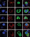
Confocal localization of the nuclear dye Dapi, SOD2 (1/200) (Cy3 red) and α‐synuclein (1/250) (DyLight 488‐green) in neurons in DLB tissue. Panel A,E,I, Dapi; B,F,J, SOD2; C,G,K, α‐synuclein; and D,H,L, merged image. White arrows indicate accumulating mitochondria with the early stage Lewy body in a DLB neuron. Bar = 10 μm.
Figure 6
Figure 6
Confocal localization of the nuclear dye Dapi, SOD2 (1/200) (Cy3 red) and α‐synuclein (1/250) (DyLight 488‐green) in neurons in PD tissue. Panel (A,E,I,M,Q) Dapi; (B,F,J,N,R) SOD2; (C,G,K,O,S) α‐synuclein; and (D,H,L,P,T) merged image. White arrow show unraveling DNA (enlarged in Figure 7). Yellow arrow indicates accumulating mitochondria with the early stage Lewy body in a PD neuron. Green arrow show accumulating mitochondria within α‐synuclein aggregation in a complex PD Lewy body. Bar = 5 μm.
Figure 7
Figure 7
High power confocal localization of the nuclear dye Dapi [panel (A), converted to white] and α‐synuclein panel (B) (white arrow in figure 6) and merged image panel (C). Bar = 5 μm.
Figure 8
Figure 8
Confocal localization of the nuclear dye Dapi, SOD2 (mitochondria, 1/200, Cy3 red), βIII tubulin (microtubules, 1/100, DyLight 488‐green) and α‐synuclein (Lewy bodies, 1/250, Cy5 blue) in neurons in control and DLB tissue. Panels (A, B)—two merged images in control cells. Panels (C, D) and (E, F)—merged image of a neurons with a Lewy body (C, E) and respective microtubules (D, F). White arrows show mitochondria associated with Lewy body. Yellow arrows shows wound up microtubules. Bar = 10 μm.

Similar articles

Cited by

References

    1. Alvarez‐Erviti L, Seow Y, Schapira AHV, Rodriguez‐Oroz MC, Obeso JA, Cooper JM (2013) Influence of microRNA deregulation on chaperone‐mediated autophagy and a‐synuclein pathology in Parkinson's disease. Cell Death Dis 4;e545; doi:10.1038. - PMC - PubMed
    1. Bartels T, Choi JG, Selkoe DJ (2011). α‐Synuclein occurs physiologically as a helically folded tetramer that resists aggregation. Nature 477:107–110. - PMC - PubMed
    1. Braak H, Sandmann‐Keil D, Gai WP, Braak E (1999) Extensive axonal Lewy neurites in Parkinson's disease: a novel pathological feature revealed by α‐synuclein immunocytochemistry. Neurosci Lett 265:67–69. - PubMed
    1. Choi BK, Choi MG, Kim JY, Yang Y, Lai Y, Kweon DH et al (2013) Large α‐synuclein oligomers inhibit neuronal SNARE‐mediated vesicle docking. Proc Natl Acad Sci USA 110:4087–4092. - PMC - PubMed
    1. Colla E, Jensen PH, Pletnikova O, Troncoso JC, Glabe C, Lee MK (2012) Accumulation of toxic α‐synuclein oligomer within endoplasmic reticulum occurs in α‐synucleinopathy in vivo. J Neurosci 32:3301–3305. - PMC - PubMed

MeSH terms