. 2016 Feb 19;291(8):3682-92.
doi: 10.1074/jbc.M115.678052. Epub 2015 Dec 14.

Gain-of-Function Mutation W493R in the Epithelial Sodium Channel Allosterically Reconfigures Intersubunit Coupling

Affiliations

Gain-of-Function Mutation W493R in the Epithelial Sodium Channel Allosterically Reconfigures Intersubunit Coupling

Mahmoud Shobair et al. J Biol Chem. .

Abstract

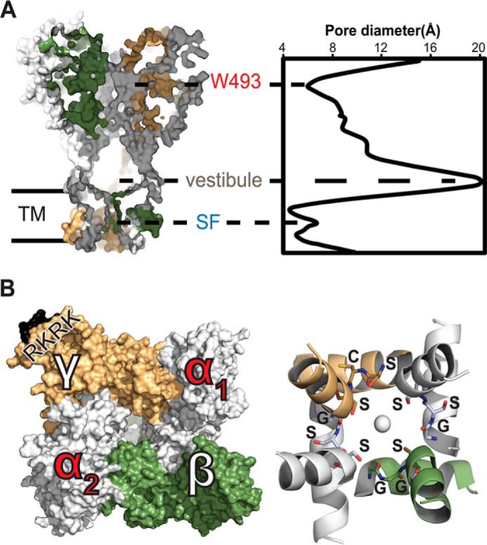
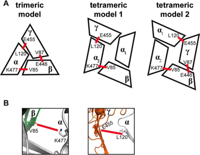
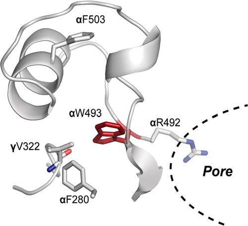
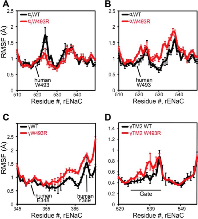
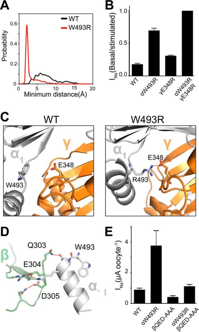
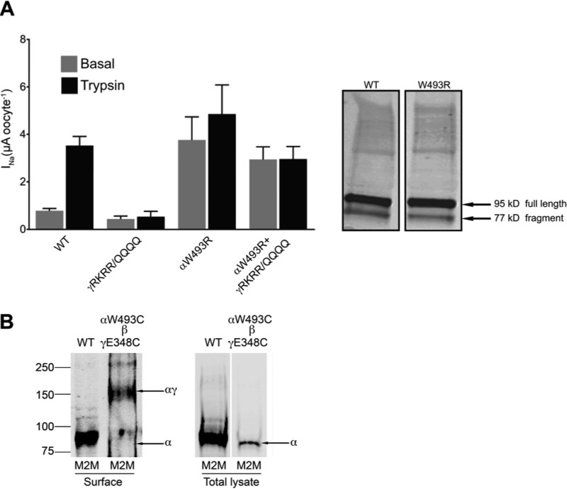
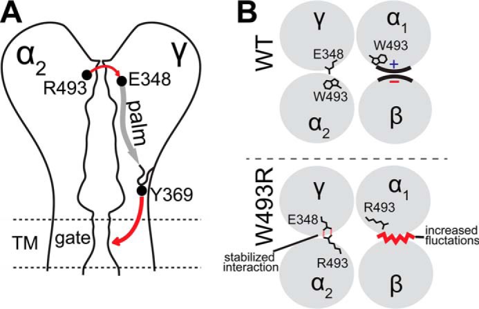
Sodium absorption in epithelial cells is rate-limited by the epithelial sodium channel (ENaC) activity in lung, kidney, and the distal colon. Pathophysiological conditions, such as cystic fibrosis and Liddle syndrome, result from water-electrolyte imbalance partly due to malfunction of ENaC regulation. Because the quaternary structure of ENaC is yet undetermined, the bases of pathologically linked mutations in ENaC subunits α, β, and γ are largely unknown. Here, we present a structural model of heterotetrameric ENaC α1βα2γ that is consistent with previous cross-linking results and site-directed mutagenesis experiments. By using this model, we show that the disease-causing mutation αW493R rewires structural dynamics of the intersubunit interfaces α1β and α2γ. Changes in dynamics can allosterically propagate to the channel gate. We demonstrate that cleavage of the γ-subunit, which is critical for full channel activation, does not mediate activation of ENaC by αW493R. Our molecular dynamics simulations led us to identify a channel-activating electrostatic interaction between α2Arg-493 and γGlu-348 at the α2γ interface. By neutralizing a sodium-binding acidic patch at the α1β interface, we reduced ENaC activation of αW493R by more than 2-fold. By combining homology modeling, molecular dynamics, cysteine cross-linking, and voltage clamp experiments, we propose a dynamics-driven model for the gain-of-function in ENaC by αW493R. Our integrated computational and experimental approach advances our understanding of structure, dynamics, and function of ENaC in its disease-causing state.

Keywords: allosteric activation; channelopathies; electrophysiology; ion channel; molecular docking; molecular dynamics; protein-protein interaction; tetramer model.


Figures

FIGURE 1.
FIGURE 1.
Heterotetramer α1βα2γ model of ENaC. A, left, side view, surface-rendering representation of intersubunit topology. Right, pore geometry of the channel calculated by HOLE (60). Constrictions along the pore axis are labeled at Trp-493, an extracellular site of gain-of-function, and the selectivity filter (SF) in the transmembrane helical bundle. Extracellular vestibule along the pore spans the widest region in the channel. B, left, top view, extracellular surface of intersubunit interfaces. The trypsin cleavage site is colored black and labeled RKRK on the γ-subunit. Right, bottom view, schematic representation of the helical bundle formed by the transmembrane domain TM2 based on the open state of cASIC (Protein Data Bank code 4NTY). The selectivity filter residues αGSS, βGGS, and γSCS are labeled on the structure and shown as sticks. A serine quartet surrounds a hypothetical sodium ion on the channel pore axis.
FIGURE 2.
FIGURE 2.
Sequence alignment of interfacial residues in the palm and β-ball domains. Alignment of sequences from the palm and β-ball domains of cASIC subunit and the α-, β-, and γ-subunits of ENaC indicates variable positions in loops connecting secondary structures. Highly conserved sequences are in boldface and colored in black and variable positions are colored in blue. Highlighted sequences are superimposed on cASIC crystal structure (Protein Data Bank code 2QTS). Experimentally tested Glu-348 is marked with an asterisk.
FIGURE 3.
FIGURE 3.
Comparison of tetrameric interfaces with a previously validated trimeric interface. A, schematic representations of a trimeric interface previously validated by cross-linking (35) and two previously proposed tetrameric configurations from functional studies of covalently linked concatemers (16). Cross-linking data from Collier et al. (41) suggest a trimeric clockwise topology of αγβ (35). These data also agree with two coexisting functional tetrameric configurations. Intersubunit angles vary slightly between trimer and tetramer models, resulting in slightly different interfaces as follows: in clockwise order, αβ, βγ, and γα, in addition to two unique interfaces to tetramers α1α2 and α1γ. B, schematic representation of α2β and α2γ interfaces in the tetramer model α1βα2γ. Shown as sticks, βV85 can cross-link with αLys-477, and γGlu-455 can cross-link with αLeu-120. Cross-linkers are represented as red bars.
FIGURE 4.
FIGURE 4.
Trp-493 binding pocket in a homology model of αENaC knuckle domain in α1βα2γ tetramer. Indole ring of Trp-493 is embedded inside the knuckle domain's cavity. Stabilizing phenylalanines Phe-280 and Phe-503 geometrically confine the pocket. Additional polar interactions stabilize the Trp-493's binding pose like γVal-322 from the neighboring γ-subunit. Trp-493 is connected to the pore-lining residue Arg-492 through the peptide chain.
FIGURE 5.
FIGURE 5.
Structural dynamics modulation in α1βα2γ by W493R gain-of-function. RMSF of Cα atoms in the α- and γ-subunits are from nine different 50-ns DMD trajectories; standard error is plotted for each residue. RMSF analysis is applied on equilibrated trajectories after discarding the first 5 ns. A and B, W493R destabilizes the knuckle domain of the α1-subunit near the Trp-493 site. However, in the α2-subunit, dynamics are slightly stabilized due to electrostatic interactions between α2Arg-493 and γGlu-348 at the α2γ interface. C, changes in dynamics of the γ-subunit propagate along a β-strand and enhance fluctuations of a loop, containing the signaling tyrosine Tyr-369, at the extracellular-transmembrane interface that communicates the activation signal (D); thus, fluctuations around the gate region containing the selectivity filter increase slightly.
FIGURE 6.
FIGURE 6.
W493R rewires intersubunit interactions. A, DMD simulations indicate an electrostatic interaction between α2Arg-493 (red) and γGlu-348 that is absent in the WT α2-subunit (black). Inter-residue interactions are quantified by a histogram of minimum distance between α2Arg-493 and γGlu-348. B, ENaC currents measured in oocytes injected with cRNA of WT and mutant ENaC subunits; basal current is normalized to trypsin-stimulated current to reflect basal activation. Perturbing the electrostatic interaction by the double mutant γE348R/αW493R channels shows no measurable population of near-silent channels, shown by lack of activation by trypsinization (n = 18, p < 0.0001). C, schematic representation of snapshots from DMD simulations at the α2γ interface. Trp-493/Arg-493 in the α2-subunit (light gray) and Glu-348 in the γ-subunit (orange) are shown as sticks, and hydrogen bonding between γGlu-348 and α2Arg-493 is shown by red dashed lines. D, hydrogen bond network at the α1β intersubunit interface by the acidic patch 303QED305 in the β-subunit with the knuckle domain in the α1-subunit. E, amiloride-sensitive current measured in oocytes. Neutralizing the acidic patch by alanine substitutions decreases W493R activation by ∼2-fold (n = 12, p = 0.002). β303AAA305 slightly inhibits WT ENaC (n = 12, p = 0.008).
FIGURE 7.
FIGURE 7.
A, cleavage of the γ-subunit does not contribute to the gain-of-function in ENaC by αW493R. Left, amiloride-sensitive current measured in oocytes. Protease-insensitive γ135QQQQ138 substitution prevents proteolytic activation by trypsin (n = 12, p < 0.00001). γ135QQQQ138/αW493R double mutant channels recapitulate the gain-of-function effect indicated by >3-fold increase in basal current and insensitivity to trypsinization (n = 12, p = 0.0003). Right, Western blots of the full-length and cleaved γ-subunit. Lysates from oocytes expressing the α-, β-, and HA-tagged γ-subunits were immunoblotted with anti-HA antibody. Marked by arrows, bands of full-length γ-subunit (95 kDa) and its cleaved form (77 kDa) are shown. Intensities of cleaved WT γENaC are comparable with the γENaC fragment in αW493R channels. B, cross-linking αW493C and γE348C using the cross-linker M2M at 200 μm. Cross-linking intact oocytes produced an ∼175-kDa band that corresponds to the αγ dimer.
FIGURE 8.
FIGURE 8.
Superposition of coevolving residues in the ENaC/Degenerin family on cASIC structure. A, highly coevolving residues in the ENaC/Degenerin family computed from Mutual Information Server to Confer Coevolution (MISTIC) (42) superimposed on cASIC crystal structure. A spatially connected allosteric path of coevolving residues lines the thumb, knuckle, and the transmembrane spanning domain TM2. B, zooming in on a specific coevolving interaction between Lys-43 and Asp-454, which may stabilize the transmembrane structure by polar interactions. Charged residues are colored in blue (positive) and red (negative). Cysteines in disulfide bridges are colored in black. Hydrophobic residues are colored in yellow.
FIGURE 9.
FIGURE 9.
Allosteric model of activation of ENaC by αW493R. A, path of allosteric signaling mapped on a schematic diagram of the α2γ interface. Initiation of activation signal is mediated by an electrostatic stabilizing interaction between α2Arg-493 and γE348; the signal is then transmitted through a β-strand in the palm domain reaching a signaling loop. Loop fluctuations are predicted to enhance fluctuations in the gate region by γTyr-369 through π-π stacking interactions at the palm-transmembrane interface. B, top view of intersubunit interfaces. Fluctuations in hydrogen bonding network at WT α1β interface (top) are increased at the mutant interface (bottom). Structural dynamics at the mutant α2γ interface (bottom) are stabilized by electrostatic interactions between γGlu-348 and α2Arg-493.

References

    1. Bhalla V., and Hallows K. R. (2008) Mechanisms of ENaC regulation and clinical implications. J. Am. Soc. Nephrol. 19, 1845–1854 - PubMed
    1. Shehata M. F. (2009) Regulation of the epithelial sodium channel [ENaC] in kidneys of salt-sensitive Dahl rats: insights on alternative splicing. Int. Arch. Med. 2, 28. - PMC - PubMed
    1. Baker S. E., Wheatley C. M., Cassuto N. A., Foxx-Lupo W. T., Sprissler R., and Snyder E. M. (2011) Genetic variation of αENaC influences lung diffusion during exercise in humans. Respir. Physiol. Neurobiol. 179, 212–218 - PMC - PubMed
    1. Azad A. K., Rauh R., Vermeulen F., Jaspers M., Korbmacher J., Boissier B., Bassinet L., Fichou Y., des Georges M., Stanke F., De Boeck K., Dupont L., Balascáková M., Hjelte L., Lebecque P., et al. (2009) Mutations in the amiloride-sensitive epithelial sodium channel in patients with cystic fibrosis-like disease. Hum. Mutat 30, 1093–1103 - PubMed
    1. Howsham C., and Danahay H. (2014) in Ion Channel Drug Discovery (Cox B., and Gosling M., eds) pp. 135–155, Royal Society of Chemistry, London

Publication types

LinkOut - more resources