. 2016 Mar 18;291(12):6272-80.
doi: 10.1074/jbc.M115.694372. Epub 2015 Dec 14.

Perturbation of Critical Prolines in Gloeobacter violaceus Ligand-gated Ion Channel (GLIC) Supports Conserved Gating Motions among Cys-loop Receptors

Affiliations

Perturbation of Critical Prolines in Gloeobacter violaceus Ligand-gated Ion Channel (GLIC) Supports Conserved Gating Motions among Cys-loop Receptors

Matthew Rienzo et al. J Biol Chem. .

Abstract

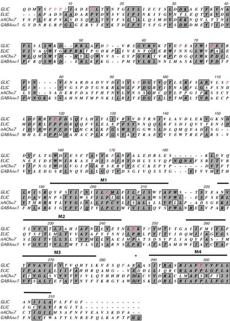
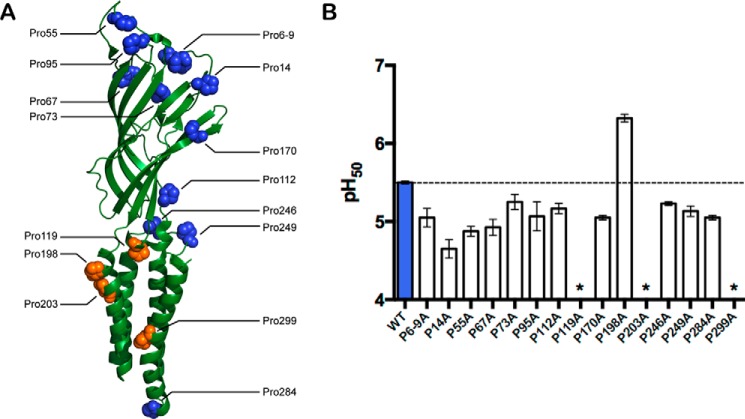
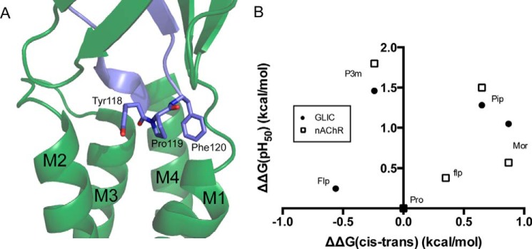
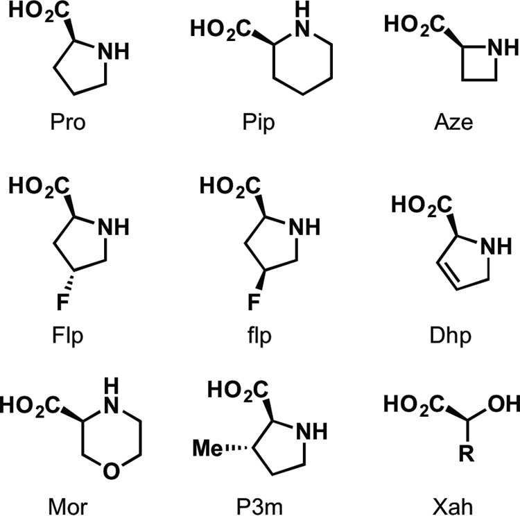
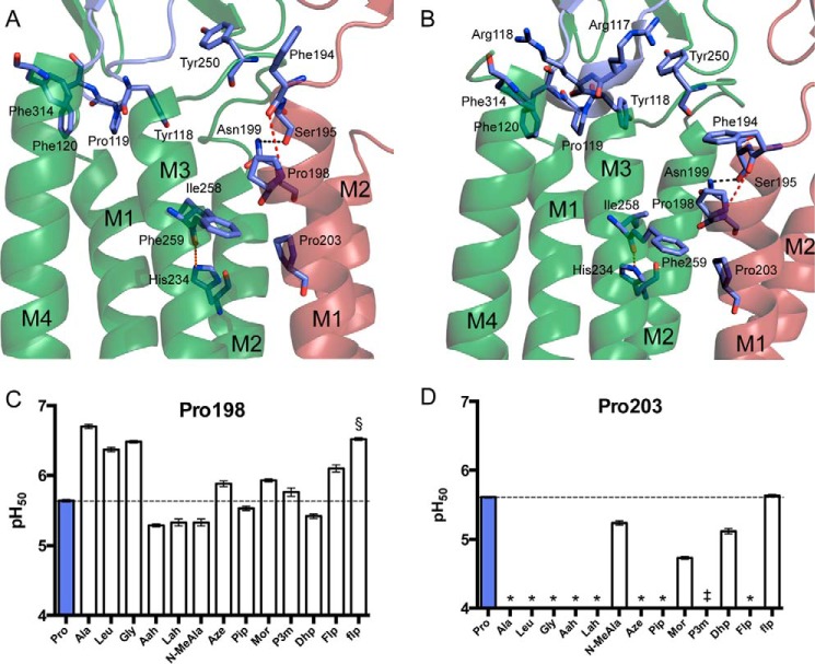
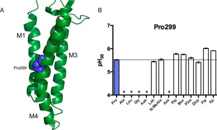
Gloeobacter violaceus ligand-gated ion channel (GLIC) has served as a valuable structural and functional model for the eukaryotic Cys-loop receptor superfamily. In Cys-loop and other receptors, we have previously demonstrated the crucial roles played by several conserved prolines. Here we explore the role of prolines in the gating transitions of GLIC. As conventional substitutions at some positions resulted in nonfunctional proteins, we used in vivo non-canonical amino acid mutagenesis to determine the specific structural requirements at these sites. Receptors were expressed heterologously in Xenopus laevis oocytes, and whole-cell electrophysiology was used to monitor channel activity. Pro-119 in the Cys-loop, Pro-198 and Pro-203 in the M1 helix, and Pro-299 in the M4 helix were sensitive to substitution, and distinct roles in receptor activity were revealed for each. In the context of the available structural data for GLIC, the behaviors of Pro-119, Pro-203, and Pro-299 mutants are consistent with earlier proline mutagenesis work. However, the Pro-198 site displays a unique phenotype that gives evidence of the importance of the region surrounding this residue for the correct functioning of GLIC.

Keywords: Cys-loop receptor; Gloeobacter violaceus; electrophysiology; mutagenesis; non-canonical amino acid; non-standard mutagenesis; nonsense suppression; proline analogs; protein conformation; structure-function.


Figures

FIGURE 1.
FIGURE 1.
Sequence alignment of GLIC, ELIC, and two example eukaryotic pLGIC subunits: nAChR α7 and GABAA R α1. Prolines examined in this study are shown in red. Shaded residues are conserved or similar in side-chain character among sequences. The M3/M4 loop of the eukaryotic receptors has been removed at *.
FIGURE 2.
FIGURE 2.
Proline sites examined in this study. A, GLIC subunit structure, indicating proline residues (PDB: 3EHZ). B, conventional mutagenesis of GLIC Pro residues. Substitution with Ala mostly had little or no effect on pH50 values, although P119A and P203A were nonfunctional, and P198A had increased sensitivity. P6-9A indicates simultaneous substitution of prolines at sites 6, 7, 8, and 9 with Ala; individual Ala substitutions at these sites yielded similar pH50 values (not shown). Data shown are mean ± S.E., n = 3–4. *, pH-induced responses were comparable with uninjected cells. Typical maximal responses for WT and all Ala-containing mutants were 20–40 μA, except for P14A, where maximal responses were 2–5 μA.
FIGURE 3.
FIGURE 3.
The GLIC YPF motif. A, the ECD/TM interface in GLIC, highlighting the YPF motif in the cis conformation (PDB: 3EAM). B, relationship between mean pH50 values and cis-trans preferences for Pro analogs at Pro-119 in GLIC (filled circles), when compared with the analogous position (Pro-136) in the muscle-type nAChR (open squares) (17). Typical maximal currents for functional mutants generated by nonsense suppression were 0.5–2 μA, with P119Pip giving responses as high as 10 μA.
FIGURE 4.
FIGURE 4.
Structures of amino acid analogs used in this study.
FIGURE 5.
FIGURE 5.
The GLIC M1 prolines. A and B, Pro-198 and Pro-203 in the top of the M1 helix at pH 7 (PDB: 4NPQ) (A) and pH 4 (PDB: 3EHZ) (B). Shown are a hydrogen bond between Asn-199 and the main-chain carbonyl of Ser-195 (black), the disrupted hydrogen bond hypothesized to be restored by conventional mutagenesis at Pro-198 (red), and the interhelix hydrogen bond between His-234 and the Ile-258 carbonyl (green). C and D, pH50 values for GLIC Pro-198 (C) and Pro-203 (D) mutants. Data shown are mean ± S.E., with n = 8–20. Conventional mutants at Pro-198 gave currents comparable with wild type GLIC. Typical maximal currents from functional mutants generated by nonsense suppression were 0.5–3 μA, with P198Lah giving responses as high as 20 μA. *, pH-induced responses comparable with uninjected cells; §, a biphasic curve was observed with a low-sensitivity component having pH50 < 4.5; ‡, currents were observed, but pH50 was too low to measure (< 4.5) due to nonspecific acid-induced currents.
FIGURE 6.
FIGURE 6.
GLIC Pro-299. A, The GLIC TM domain, highlighting Pro-299 (PDB: 3EHZ). B, pH50 values for GLIC Pro-299 mutants. Data shown are mean ± S.E., with n = 7–20. Typical maximal currents from functional mutants generated by nonsense suppression were 0.5–5 μA. *, pH-induced responses comparable with uninjected cells.

References

    1. dacosta C. J. B., and Baenziger J. E. (2013) Gating of pentameric ligand-gated ion channels: structural insights and ambiguities. Structure 21, 1271–1283 - PubMed
    1. Nys M., Kesters D., and Ulens C. (2013) Structural insights into Cys-loop receptor function and ligand recognition. Biochem. Pharmacol. 86, 1042–1053 - PubMed
    1. Miller P. S., and Aricescu A. R. (2014) Crystal structure of a human GABAA receptor. Nature 512, 270–275 - PMC - PubMed
    1. Hassaine G., Deluz C., Grasso L., Wyss R., Tol M. B., Hovius R., Graff A., Stahlberg H., Tomizaki T., Desmyter A., Moreau C., Li X.-D., Poitevin F., Vogel H., and Nury H. (2014) X-ray structure of the mouse serotonin 5-HT3 receptor. Nature 512, 276–281 - PubMed
    1. Du J., Lü W., Wu S., Cheng Y., and Gouaux E. (2015) Glycine receptor mechanism elucidated by electron cryo-microscopy. Nature 526, 224–229 - PMC - PubMed

Publication types