

Ultrastructure of the Epidermal Cell Wall and Cuticle of Tomato Fruit (Solanum lycopersicum L.) during Development
- PMID: 26668335
- PMCID: PMC4734585
- DOI: 10.1104/pp.15.01725
Ultrastructure of the Epidermal Cell Wall and Cuticle of Tomato Fruit (Solanum lycopersicum L.) during Development
Abstract
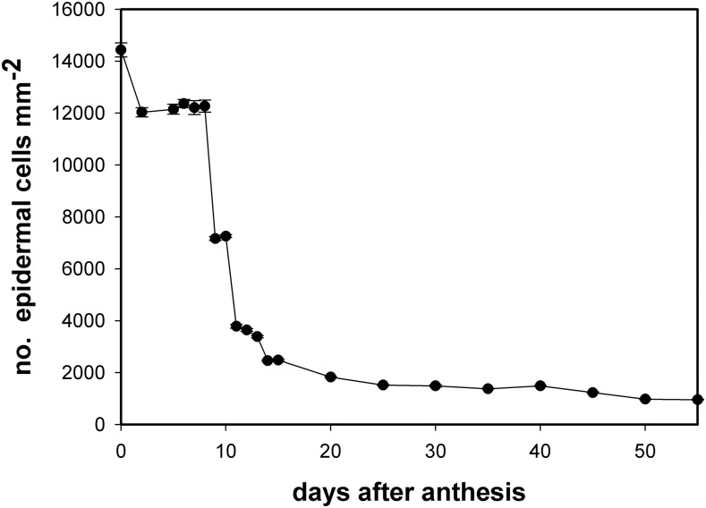
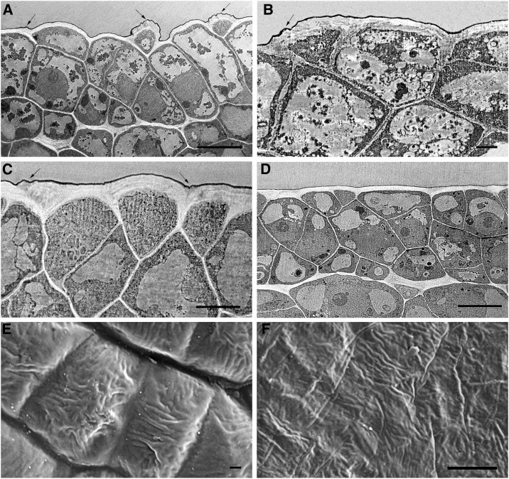
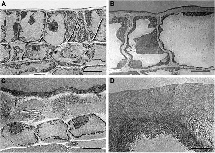
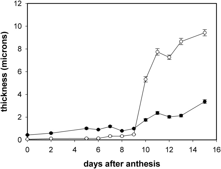
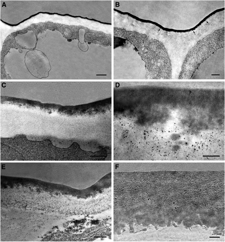
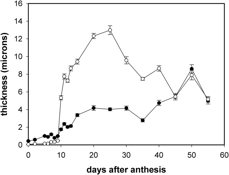
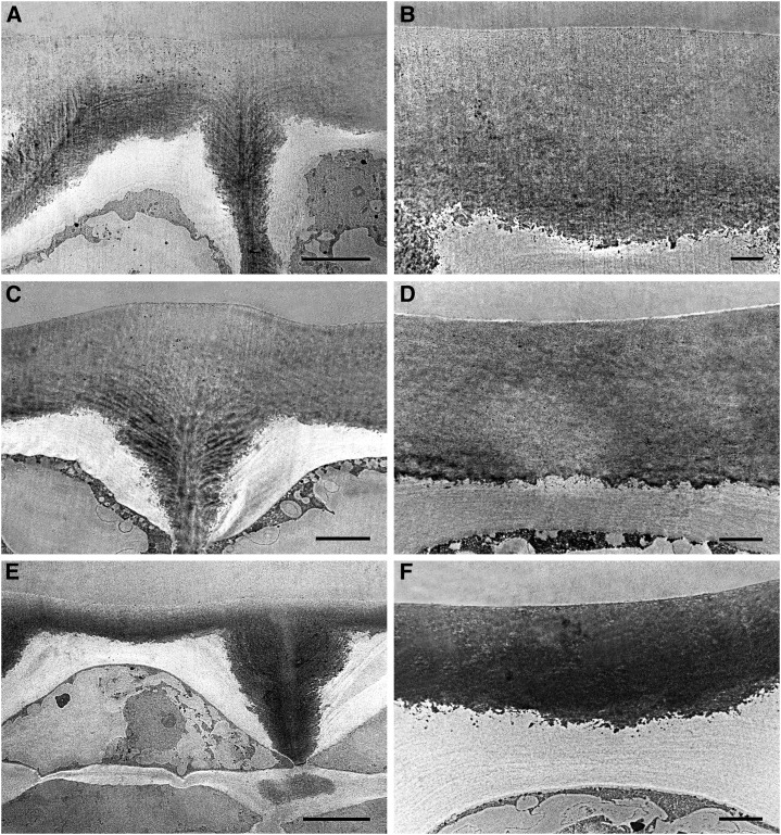
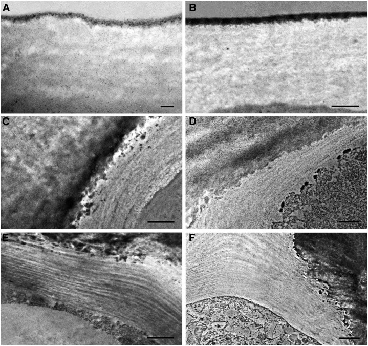
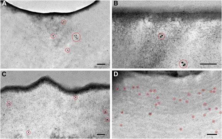
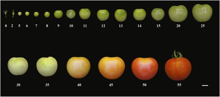
The epidermis plays a pivotal role in plant development and interaction with the environment. However, it is still poorly understood, especially its outer epidermal wall: a singular wall covered by a cuticle. Changes in the cuticle and cell wall structures are important to fully understand their functions. In this work, an ultrastructure and immunocytochemical approach was taken to identify changes in the cuticle and the main components of the epidermal cell wall during tomato fruit development. A thin and uniform procuticle was already present before fruit set. During cell division, the inner side of the procuticle showed a globular structure with vesicle-like particles in the cell wall close to the cuticle. Transition between cell division and elongation was accompanied by a dramatic increase in cuticle thickness, which represented more than half of the outer epidermal wall, and the lamellate arrangement of the non-cutinized cell wall. Changes in this non-cutinized outer wall during development showed specific features not shared with other cell walls. The coordinated nature of the changes observed in the cuticle and the epidermal cell wall indicate a deep interaction between these two supramolecular structures. Hence, the cuticle should be interpreted within the context of the outer epidermal wall.
© 2016 American Society of Plant Biologists. All Rights Reserved.
Figures
References
-
- Anastasiou E, Lenhard M (2007) Growing up to one’s standard. Curr Opin Plant Biol 10: 63–69 - PubMed
-
- Baker EA, Bukovac MJ, Hunt GM (1982) Composition of tomato fruit cuticle as related to fruit growth and development. In Cutler DF, Alvin KL, Price CE, eds, The Plant Cuticle. Academic Press, London, UK, pp 33–44
-
- Bargel H, Koch K, Cerman Z, Neinhuis C (2006) Structure–function relationships of the plant cuticle and cuticular waxes—a smart material? Funct Plant Biol 33: 893–910 - PubMed
-
- Becker T, Knoche M (2012) Deposition, strain, and microcracking of the cuticle in the developing ‘Riesling’ grape berries. Vitis 51: 1–6
-
- Beisson F, Li-Beisson Y, Pollard M (2012) Solving the puzzles of cutin and suberin polymer biosynthesis. Curr Opin Plant Biol 15: 329–337 - PubMed
Publication types
MeSH terms
Substances
LinkOut - more resources
Full Text Sources
Other Literature Sources
