. 2015 Dec 14:5:42.
doi: 10.1186/s13395-015-0067-1. eCollection 2015.

Pervasive satellite cell contribution to uninjured adult muscle fibers

Affiliations

Pervasive satellite cell contribution to uninjured adult muscle fibers

Bradley Pawlikowski et al. Skelet Muscle. .

Abstract

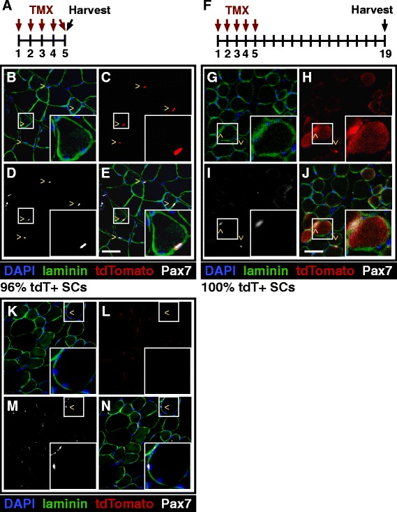
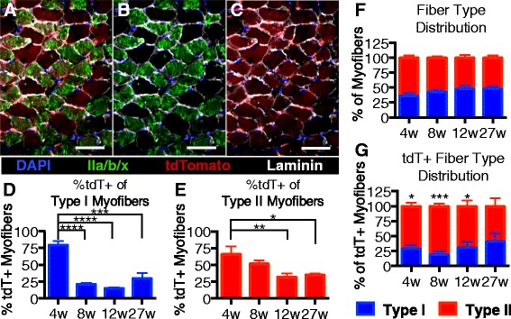
Background: Adult skeletal muscle adapts to functional needs, maintaining consistent numbers of myonuclei and stem cells. Although resident muscle stem cells or satellite cells are required for muscle growth and repair, in uninjured muscle, these cells appear quiescent and metabolically inactive. To investigate the satellite cell contribution to myofibers in adult uninjured skeletal muscle, we labeled satellite cells by inducing a recombination of LSL-tdTomato in Pax7(CreER) mice and scoring tdTomato+ myofibers as an indicator of satellite cell fusion.

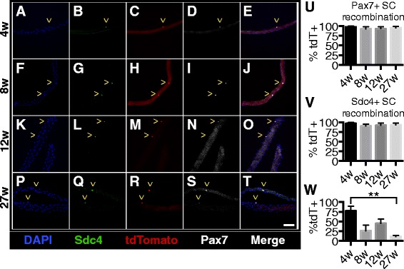
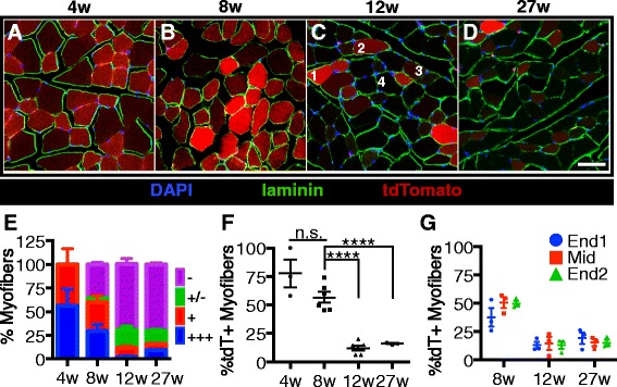
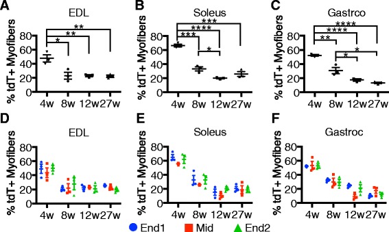
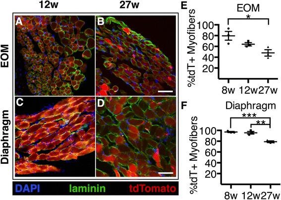
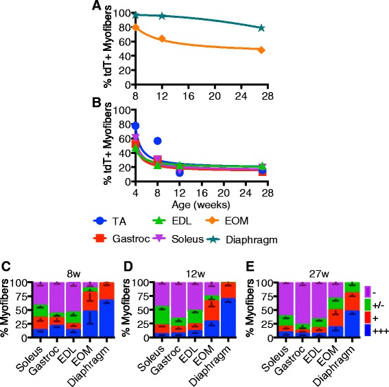
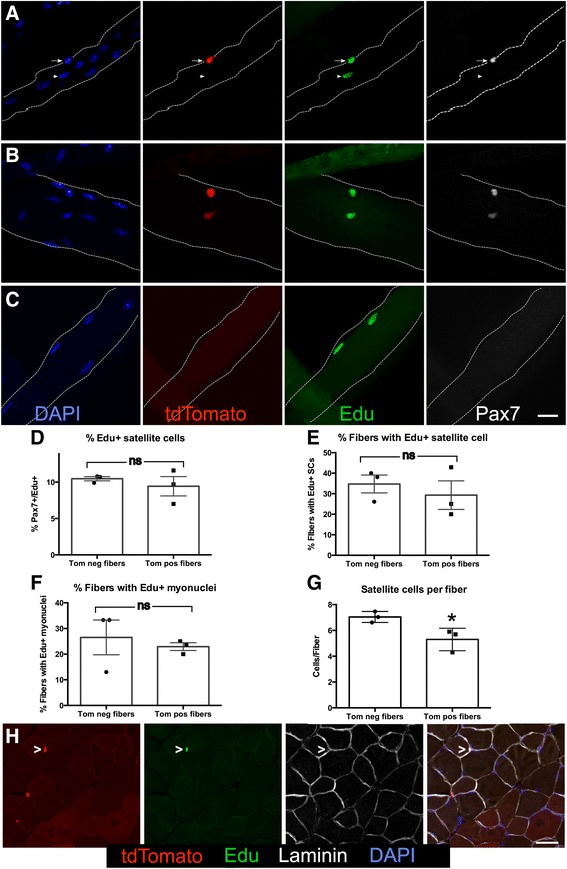
Results: Satellite cell fusion into myofibers plateaus postnatally between 8 and 12 weeks of age, reaching a steady state in hindlimb muscles, but in extra ocular or diaphragm muscles, satellite cell fusion is maintained at postnatal levels irrespective of the age assayed. Upon recombination and following a 2-week chase in 6-month-old mice, tdTomato-labeled satellite cells fused into myofibers as 20, 50, and 80 % of hindlimb, extra ocular, and diaphragm myofibers, respectively, were tdTomato+. Satellite cells contribute to uninjured myofibers either following a cell division or directly without an intervening cell division.

Conclusions: The frequency of satellite cell fusion into the skeletal muscle fibers is greater than previously estimated, suggesting an important functional role for satellite cell fusion into adult myofibers and a requirement for active maintenance of satellite cell numbers in uninjured skeletal muscle.

Keywords: Homeostasis; Pax7; Satellite cell; Skeletal muscle.


Figures

Fig. 1
Fig. 1
Induction of tdTomato expression in satellite cells labels myofibers by satellite cell fusion. a Pax7Cre;tdTomato mice were injected with tamoxifen once a day for five consecutive days to induce tdTomato expression in satellite cells. Images shown in this figure are from 6-month-old mice. be Representative images of TA sections show that 96 % of Pax7-expressing satellite cells are also expressing tdTomato. Insert highlights a Pax7+, tdTomato+ sub-laminar satellite cell. f Pax7Cre;tdTomato mice were injected with tamoxifen once a day for five consecutive days and then tissue was collected 2 weeks following the last injection. gj Representative images of TA sections show Pax7-expressing satellite cells as tdTomato+ but also unexpectedly show tdTomato+ myofibers. Insert highlights a tdTomato-expressing myofiber with an associated Pax7+, tdTomato+ satellite cell. kn Representative images of TA sections from Pax7Cre;tdTomato mice that were injected with only with corn oil showing no basal tdTomato expression prior to tamoxifen injection. Images display DAPI (blue), Sdc4 (green), tdTomato (red), Pax7 (white). ^ marks indicate satellite cells. Scale bars are 50 μm
Fig. 2
Fig. 2
Induction of tdTomato expression does not vary with age of tamoxifen injection. at Images of EDL myofibers that were harvested 2 weeks after the final tamoxifen injection show that almost all satellite cells are expressing tdTomato. Images show DAPI (blue), Syndecan-4 (green), tdTomato (red), and Pax7 (white). ^ marks within micrographs indicate satellite cells. uv Quantification showing the percentages of satellite cells on myofibers that are tdTomato+. w Quantification showing the percentages of myofibers that are tdTomato+. Scale bar is 50 μm. Three biological replicates were analyzed per time point
Fig. 3
Fig. 3
Satellite cell contribution to tibialis anterior muscle plateaus between 8 and 12 weeks of age. ad Images of TA muscle sections from mice that were harvested 2 weeks after the final tamoxifen injection show the percentages of tdTomato expressing myofibers decrease with age. Images display DAPI (blue), laminin (green), and tdTomato (red). Numbers in the 12-week image c illustrate tdTomato scoring metric. e Quantification of tdTomato-expressing myofibers in TA muscle sections by tdTomato expression level. +++ myofibers express high levels of tdTomato (1 in a), + myofibers clearly express tdTomato (2 in a), +/− myofibers have very low levels of tdTomato and are scored as negative (3 in a), and – myofibers do not express tdTomato at observable levels (4 in a). f Quantification showing the percentages of myofibers that express tdTomato (+ or +++) in TA muscle sections. g Quantification showing the percentages of tdTomato-expressing TA myofibers by location within the muscle. Scale bar is 50 μm. ****P < 0.0001. Mean ± standard error of the mean is plotted. Significance was determined by Student’s t test. Three to six biological replicates were analyzed per time point
Fig. 4
Fig. 4
Satellite cell contribution is similar in all hindlimb muscles as a function of age. ac Quantification of tdTomato-expressing myofibers among the EDL (a), soleus (b), and gastroc (c) muscles 2 weeks after the final tamoxifen injection. df Quantification of tdTomato-expressing myofibers by location within the EDL (d), soleus (e), and gastroc (f). *P < 0.05, **P < 0.01, ***P < 0.001, ****P < 0.0001. For all graphs, mean ± standard error of the mean is plotted. Significance was determined by Student’s t test. Three biological replicates were analyzed per time point
Fig. 5
Fig. 5
Differential contribution of satellite cells to fast and slow twitch myofibers. ac Images shows soleus muscle sections from mice given tamoxifen injections at 27 weeks old and collected 2 weeks post injection. tdTomato-expressing myofibers are shown in red, and myofibers labeled with an antibody to identify fast twitch myofibers are shown in green. d Quantification of tdTomato expression in type I myofibers. e Quantification of tdTomato expression in type II myofibers. f Soleus fiber type distribution by age. g Fiber type distribution of tdTomato-expressing myofibers by age. Pseudocolored images display DAPI (blue), laminin (white), tdTomato (red), and fast myosin heavy chain (green, IIa, IIb, and IIx). Scale bars are 50 μm. *P < 0.05, ***P < 0.001. Mean ± standard error of the mean is plotted. Significance was determined by Student’s t test. Three biological replicates were analyzed per time point
Fig. 6
Fig. 6
Satellite cell contribution to EOM and diaphragm muscles does not plateau with age. ad Images show tdTomato expressing myofibers in the EOM and diaphragm 2 weeks after the final tamoxifen injection. ef Quantification showing the percentages of tdTomato-expressing myofibers in sections of the e EOM and f diaphragm. Images display DAPI (blue), laminin (green), and tdTomato (red). Scale bar is 50 μm. *P < 0.05, **P < 0.01. For all graphs, mean ± standard error of the mean is plotted. Significance was determined by Student’s t test. Three biological replicates were analyzed per time point
Fig. 7
Fig. 7
Satellite cell contribution to myofibers is age dependent and varies in different muscles. ab Non-linear regression trend lines for percentages of tdTomato-expressing myofibers through experimental time points from the EOM and diaphragm (a) and EDL, soleus, and gastrocnemius (b). ce Quantification of myofiber tdTomato-expression of hindlimb, EOM, and diaphragm muscles with recombination induced at 4 (c), 12 (d), or 27 weeks (e). Quantification of tdTomato was evaluated at four relative levels of expression: +++ very high tdTomato intensity (see example myofiber “1” in Fig. 3c, 12 weeks), + intermediate tdTomato intensity (see example myofiber “2” in Fig. 3c, 12 weeks), +/− very low tdTomato intensity myofibers and scored as negative (see example myofiber “3” in Fig. 3c, 12 weeks), and – fibers do tdTomato intensity indistinguishable from background (see example myofiber “4” in Fig. 3c, 12 weeks)
Fig. 8
Fig. 8
Satellite cells fuse into uninjured myofibers either with our without an intervening cell division. ac Representative images show EdU+ satellite cells and EdU-labeled nuclei within myofibers. a Myofiber showing an EdU+ satellite cell (arrow) and an EdU+ differentiated myonuclei (arrowhead). b Images show a myofiber with two satellite cells that are EdU+. c A myofiber with two EdU+ myonuclei within the myofiber. For ac, DAPI (blue), tdTomato (red), EdU (green), and Pax7 (white). Scale bar is 20 μm. d Quantification showing the percentages of satellite cells on dtTomato+ (Tom pos) myofibers versus dtTomato− myofibers (Tom neg) that were labeled with EdU. e Bar graph showing the percentages of myofibers that have at least one EdU+ associated satellite cell. f Quantification showing the percentages of myofibers that have at least one EdU+ nuclei within the myofiber. g Quantification of the satellite cell number present on dtTomato+ myofibers versus dtTomato− myofibers (*p ≤ 0.01). h Images of TA muscle sections show an EdU+, tomato+ SC. tdTomato (red), EdU (green), laminin (white), and DAPI (blue). Scale bar is 40 μm. For bar graphs, ns denotes not significant. Significance was determined by Student’s t test. N = 3. For all graphs, the mean with the standard error of the mean is plotted

References

    1. Fuchs E. The tortoise and the hair: slow-cycling cells in the stem cell race. Cell. 2009;137(5):811–9. doi: 10.1016/j.cell.2009.05.002. - DOI - PMC - PubMed
    1. Bentzinger CF, von Maltzahn J, Rudnicki MA. Extrinsic regulation of satellite cell specification. Stem Cell Res Ther. 2010;1(3):27. doi: 10.1186/scrt27. - DOI - PMC - PubMed
    1. Murphy MM, Lawson JA, Mathew SJ, Hutcheson DA, Kardon G. Satellite cells, connective tissue fibroblasts and their interactions are crucial for muscle regeneration. Development. 2011;138(17):3625–37. doi: 10.1242/dev.064162. - DOI - PMC - PubMed
    1. Sousa-Victor P, García-Prat L, Serrano AL, Perdiguero E, Muñoz-Cánoves P. Muscle stem cell aging: regulation and rejuvenation. Trends Endocrinol Metab. 2015. doi:10.1016/j.tem.2015.03.006. - PubMed
    1. Keefe AC, Lawson JA, Flygare SD, Fox ZD, Colasanto MP, Mathew SJ, et al. Muscle stem cells contribute to myofibres in sedentary adult mice. Nat Commun. 2015;6:7087. doi: 10.1038/ncomms8087. - DOI - PMC - PubMed