Review
. 2015 Nov;48(6):466-75.
doi: 10.5946/ce.2015.48.6.466. Epub 2015 Nov 30.

The Past, Present, and Future of Image-Enhanced Endoscopy

Affiliations
Review

The Past, Present, and Future of Image-Enhanced Endoscopy

Jae-Young Jang. Clin Endosc. 2015 Nov.

Abstract

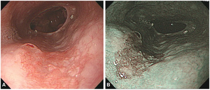
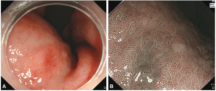
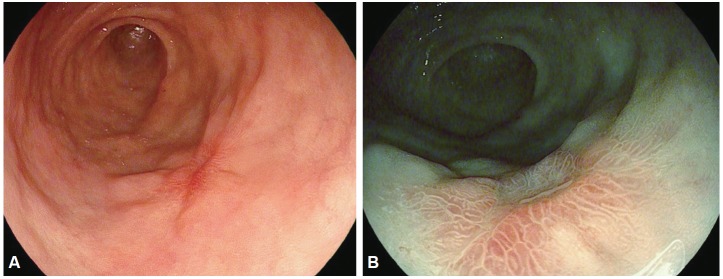
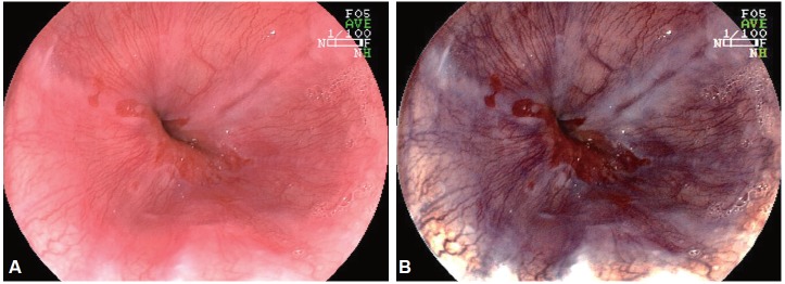
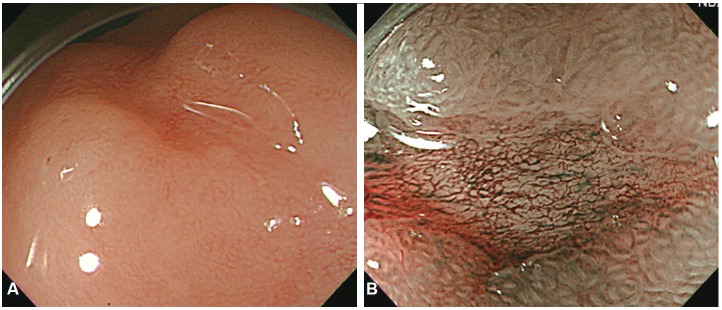
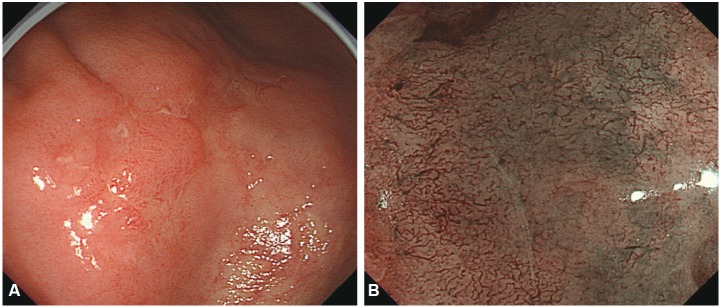
Despite the remarkable progress recently made to enhance the resolution of white-light endoscopy, detection, and diagnosis of premalignant lesions, such as adenomas and subtle early-stage cancers, remains a great challenge. As for example, although chromoendoscopy, such as endoscopy using indigo carmine, is useful for the early diagnosis of subtle lesions, the technique presents various disadvantages ranging from the time required for spray application of the dye and suctioning of excess dye to the increased difficulty in identifying lesions in the presence of severe inflammation and obstruction of visual field due to the pooling of solution in depressed-type lesions. To overcome these diagnostic problems associated with chromoendoscopy, research has focused on the development of endoscopes based on new optical technologies. Several types of image-enhanced endoscopy methods have recently been presented. In particular, image-enhanced endoscopy has emerged as a new paradigm for the diagnosis of gastrointestinal disorders. Image-enhanced endoscopes provide high-contrast images of lesions by means of optical or electronic technologies, including the contrast enhancement of the mucosal surface and of blood vessels. Chromoendoscopy, narrow-band imaging, i-SCAN, and flexible spectral imaging color enhancement are representative examples of image-enhanced endoscopy discussed in this paper.

Keywords: Chromoendoscopy; Flexible spectral imaging color enhancement; Image-enhanced endoscopy; Narrow band imaging; i-SCAN.


Conflict of interest statement

Conflicts of Interest: The author have no financial conflicts of interest.

Figures

Fig. 1.
Fig. 1.
The resolution of endoscopic image. (A) Standard definition (SD) endoscopic image of gastroesophageal (GE) junction, (B) high definition endoscopic image of GE junction can be observed more clearly compared to the SD endoscopic image.
Fig. 2.
Fig. 2.
Magnifying endoscopic finding of normal gastric mucosa. (A) In the gastric antrum, microvascular architecture is a coil-shaped. The pits demonstrate a linear or reticular patterns. (B) In the gastric body, microvascular architecture is a honeycomb shaped, and the pits demonstrate a round or oval shape.
Fig. 3.
Fig. 3.
Esophageal squamous cell carcinoma. (A) A depressed lesion with irregular nodularity and redness is noted at the mid esophagus. (B) With iodine staining, it is shown as an iodine-void area with a well-defined boundary.
Fig. 4.
Fig. 4.
Early gastric cancer. (A) Indistinct margined gastric cancer due to marked atrophy and intestinal metaplasia around the lesions are often observed. (B) Indigo carmine chromoendoscopy delineated the lesion more clearly.
Fig. 5.
Fig. 5.
Esophageal squamous cell carcinoma. (A) A depressed lesion with irregular nodularity and redness is noted at the lower esophagus. (B) In narrow-band imaging mode, is seen as dark brown lesion due to its copious vascularity.
Fig. 6.
Fig. 6.
Early gastric cancer. (A) About 0.6-cm sized slightly depressed lesion was noted at the lesser curvature side of mid body in white light imaging. (B) Well delineated margin, loss of pit, and irregular microvessels were noted in near focus.
Fig. 7.
Fig. 7.
i-SCAN image of depressed typed early gastric cancer. (A) Small depressed lesion is noted at antrum. (B) i-SCAN TE-g makes it more clear delineation of the tumor.
Fig. 8.
Fig. 8.
Reflux esophagitis. (A) Gastroesophageal junction and a mucosal break are noted in white light imaging. (B) Flexible spectral imaging color enhancement imaging enhanced the visualization of mucosal structure and vessels.
Fig. 9.
Fig. 9.
Early gastric cancer. (A) A depressed lesion was noted at the antrum. (B) Magnifying narrow-band imaging demonstrated demarcation line, loss of fine mucosal structure, and presence of an irregular microvascular pattern. The lesion was suspected to mucosal well differentiated gastric adenocarcinoma. Endoscopic submucosal dissection was performed.
Fig. 10.
Fig. 10.
Early gastric cancer. (A) Ill defined, pale mucosal lesion with central nodule is noted on the antrum. (B) Magnifying narrow-band imaging findings of that pale mucosa showed loss of the regular microvascular pattern and corkscrew pattern. The lesion was suspected to poorly differentiated gastric adenocarcinoma. Gastrectomy with lymph node dissection was performed.

References

    1. Subramanian V, Ragunath K. Advanced endoscopic imaging: a review of commercially available technologies. Clin Gastroenterol Hepatol. 2014;12:368.e1–376.e1. - PubMed
    1. ASGE Technology Committee. Kwon RS, Adler DG, et al. High-resolution and high-magnification endoscopes. Gastrointest Endosc. 2009;69(3 Pt 1):399–407. - PubMed
    1. Bruno MJ. Magnification endoscopy, high resolution endoscopy, and chromoscopy; towards a better optical diagnosis. Gut. 2003;52 Suppl 4:iv7–iv11. - PMC - PubMed
    1. Peitz U, Malfertheiner P. Chromoendoscopy: from a research tool to clinical progress. Dig Dis. 2002;20:111–119. - PubMed
    1. Woolf GM, Riddell RH, Irvine EJ, Hunt RH. A study to examine agreement between endoscopy and histology for the diagnosis of columnar lined (Barrett’s) esophagus. Gastrointest Endosc. 1989;35:541–544. - PubMed

LinkOut - more resources