

Endoscopic Extraction of Biliary Fascioliasis Diagnosed Using Intraductal Ultrasonography in a Patient with Acute Cholangitis
- PMID: 26668810
- PMCID: PMC4676668
- DOI: 10.5946/ce.2015.48.6.579
Endoscopic Extraction of Biliary Fascioliasis Diagnosed Using Intraductal Ultrasonography in a Patient with Acute Cholangitis
Abstract
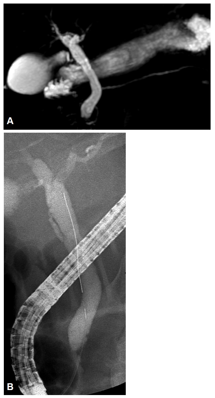
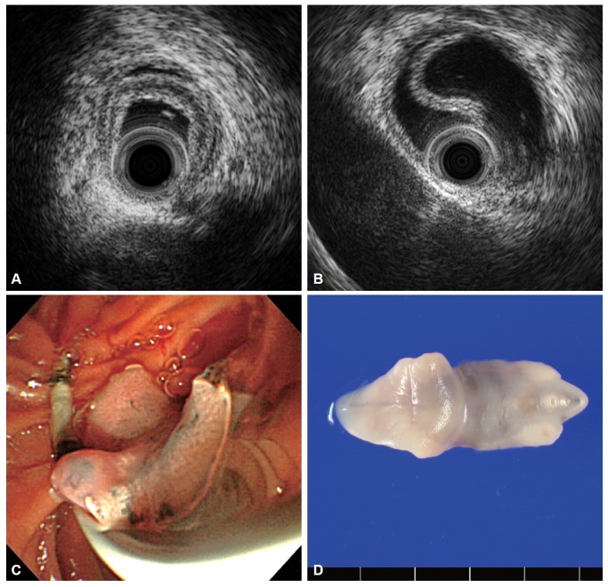
Fasciola hepatica infection may result in biliary obstruction with or without cholangitis in the chronic biliary phase. Because clinical symptoms and signs of F. hepatica are similar to other biliary diseases that cause bile duct obstruction, such as stones or bile duct malignancies, that are, in fact, more common, this condition may not be suspected and diagnosis may be overlooked and delayed. Patients undergoing endoscopic retrograde cholangiopancreatography or endoscopic ultrasonography for the evaluation of bile duct obstruction may be incidentally detected with the worm, and diagnosis can be confirmed by extraction of the leaf-like trematode from the bile duct. Intraductal ultrasonography (IDUS) can provide high-resolution cross-sectional images of the bile duct, and is useful in evaluating indeterminate biliary diseases. We present a case of biliary fascioliasis that was diagnosed using IDUS and managed endoscopically in a patient with acute cholangitis.
Keywords: Acute cholangitis; Fascioliasis; Intraductal ultrasonography.
Conflict of interest statement
Figures
Similar articles
-
Endoscopic extraction of living fasciola hepatica: case report and literature review.Turk J Gastroenterol. 2003 Mar;14(1):74-7. Turk J Gastroenterol. 2003. PMID: 14593544 Review.
-
A case of Fasciola hepatica infection mimicking cholangiocarcinoma and ITS-1 sequencing of the worm.Korean J Parasitol. 2014 Apr;52(2):193-6. doi: 10.3347/kjp.2014.52.2.193. Epub 2014 Apr 18. Korean J Parasitol. 2014. PMID: 24850964 Free PMC article.
-
Endoscopic diagnosis and treatment of biliary obstruction due to acute cholangitis and acute pancreatitis secondary to Fasciola hepatica infection.Ulus Travma Acil Cerrahi Derg. 2018 Jan;24(1):71-73. doi: 10.5505/tjtes.2017.89490. Ulus Travma Acil Cerrahi Derg. 2018. PMID: 29350372
-
Biliary fascioliasis--an uncommon cause of recurrent biliary colics: report of a case and brief review.Ger Med Sci. 2012;10:Doc10. doi: 10.3205/000161. Epub 2012 May 2. Ger Med Sci. 2012. PMID: 22566787 Free PMC article. Review.
-
Endoscopic management of biliary fascioliasis: a case report.J Med Case Rep. 2010 Mar 6;4:83. doi: 10.1186/1752-1947-4-83. J Med Case Rep. 2010. PMID: 20205932 Free PMC article.
Cited by
-
A Curious Case of Walking Cholangitis in a Nonendemic Region: Hepatobiliary Fascioliasis.ACG Case Rep J. 2025 May 5;12(5):e01700. doi: 10.14309/crj.0000000000001700. eCollection 2025 May. ACG Case Rep J. 2025. PMID: 40330503 Free PMC article. No abstract available.
-
Biliary Fascioliasis in Chronic Calcific Pancreatitis Presenting with Ascending Cholangitis and Biliary Stricture.Case Rep Gastroenterol. 2019 Oct 9;13(3):438-444. doi: 10.1159/000503277. eCollection 2019 Sep-Dec. Case Rep Gastroenterol. 2019. PMID: 31762732 Free PMC article.
-
A 53-Year-Old Man with Intermittent Colicky Abdominal Pain due to Fasciola Incarceration in Common Bile Duct: A Case Report.Iran J Parasitol. 2018 Oct-Dec;13(4):664-668. Iran J Parasitol. 2018. PMID: 30697324 Free PMC article.
-
An Update on the Pathogenesis of Fascioliasis: What Do We Know?Res Rep Trop Med. 2024 Feb 13;15:13-24. doi: 10.2147/RRTM.S397138. eCollection 2024. Res Rep Trop Med. 2024. PMID: 38371362 Free PMC article. Review.
-
Endoscopic Diagnosis of Biliary Fascioliasis in Non-endemic Region.Dig Dis Sci. 2023 Sep;68(9):3476-3478. doi: 10.1007/s10620-023-08045-6. Epub 2023 Jul 27. Dig Dis Sci. 2023. PMID: 37498416 No abstract available.
References
-
- Rana SS, Bhasin DK, Nanda M, Singh K. Parasitic infestations of the biliary tract. Curr Gastroenterol Rep. 2007;9:156–164. - PubMed
-
- el-Newihi HM, Waked IA, Mihas AA. Biliary complications of Fasciola hepatica: the role of endoscopic retrograde cholangiography in management. J Clin Gastroenterol. 1995;21:309–311. - PubMed
-
- Dias LM, Silva R, Viana HL, Palhinhas M, Viana RL. Biliary fascioliasis: diagnosis, treatment and follow-up by ERCP. Gastrointest Endosc. 1996;43:616–620. - PubMed
-
- Tamada K, Inui K, Menzel J. Intraductal ultrasonography of the bile duct system. Endoscopy. 2001;33:878–885. - PubMed
LinkOut - more resources
Full Text Sources
Other Literature Sources
