. 2015 Dec 29;112(52):15790-7.
doi: 10.1073/pnas.1521919112. Epub 2015 Dec 15.

mTOR inhibition activates overall protein degradation by the ubiquitin proteasome system as well as by autophagy

Affiliations

mTOR inhibition activates overall protein degradation by the ubiquitin proteasome system as well as by autophagy

Jinghui Zhao et al. Proc Natl Acad Sci U S A. .

Abstract

Growth factors and nutrients enhance protein synthesis and suppress overall protein degradation by activating the protein kinase mammalian target of rapamycin (mTOR). Conversely, nutrient or serum deprivation inhibits mTOR and stimulates protein breakdown by inducing autophagy, which provides the starved cells with amino acids for protein synthesis and energy production. However, it is unclear whether proteolysis by the ubiquitin proteasome system (UPS), which catalyzes most protein degradation in mammalian cells, also increases when mTOR activity decreases. Here we show that inhibiting mTOR with rapamycin or Torin1 rapidly increases the degradation of long-lived cell proteins, but not short-lived ones, by stimulating proteolysis by proteasomes, in addition to autophagy. This enhanced proteasomal degradation required protein ubiquitination, and within 30 min after mTOR inhibition, the cellular content of K48-linked ubiquitinated proteins increased without any change in proteasome content or activity. This rapid increase in UPS-mediated proteolysis continued for many hours and resulted primarily from inhibition of mTORC1 (not mTORC2), but did not require new protein synthesis or key mTOR targets: S6Ks, 4E-BPs, or Ulks. These findings do not support the recent report that mTORC1 inhibition reduces proteolysis by suppressing proteasome expression [Zhang Y, et al. (2014) Nature 513(7518):440-443]. Several growth-related proteins were identified that were ubiquitinated and degraded more rapidly after mTOR inhibition, including HMG-CoA synthase, whose enhanced degradation probably limits cholesterol biosynthesis upon insulin deficiency. Thus, mTOR inhibition coordinately activates the UPS and autophagy, which provide essential amino acids and, together with the enhanced ubiquitination of anabolic proteins, help slow growth.

Keywords: autophagy; mTOR; proteasome; ubiquitination.


Conflict of interest statement

The authors declare no conflict of interest.

Figures

Fig. 1.
Fig. 1.
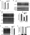
mTOR inhibitors enhance the degradation of long-lived, but not short-lived, cell proteins by activating proteasomal proteolysis, as well as autophagy. (A) Rapamycin and Torin1 did not affect the degradation of short-lived cell proteins in HEK293 cells. Newly synthesized proteins were radiolabeled by incubating cells in complete medium supplemented with 3H-Phe for 20 min. After two quick washes of the cells with medium containing 2 mM nonradioactive Phe and cycloheximide to prevent reuptake and reincorporation, control (♦, vehicle), rapamycin (□, 300 nM), or Torin1 (▲, 100 nM) were added at time 0, and the release of 3H-Phe from cell proteins into the media was measured especially during the first hour when short-lived proteins are selectively degraded. (B) Rapamycin and Torin1 increased the degradation of long-lived proteins in HEK293 cells. To label long-lived proteins selectively, cells were incubated with 3H-Phe for 20 h. After switching to chase medium for 2 h to allow the degradation of short-lived components, fresh chase medium containing mTOR inhibitors was added, and proteolysis assayed. The fraction of radioactive proteins degraded to TCA-soluble material at different times was expressed as a percentage of total radioactivity incorporated into cell proteins at t = 0. Significant increases upon mTOR inhibition were observed at 90 min and thereafter. (C) mTOR inhibition with rapamycin or Torin1 increased degradation by proteasomes, which was measured as CCA-resistant proteolysis in HEK293 cells and MEFs (Left), or by BTZ-sensitive proteolysis in Atg5−/− MEFs (Right). (D) mTOR inhibition increased lysosomal (CCA-sensitive) proteolysis in HEK293 cells and wild type MEFs, but not in Atg5−/− MEFs. Error bars (SEM) were provided in C and D and for every time point in A and B (but were hidden by labels in B). *P < 0.05.
Fig. 2.
Fig. 2.
mTOR inhibition rapidly increases the levels of K48-linked Ub conjugates in cells and mouse liver without affecting proteasomal activity. (A) The increase in proteasomal degradation by Torin1 treatment in HEK293 cells did not require protein synthesis. Proteasomal proteolysis was determined in the presence or absence of 20 μg/mL cycloheximide. (B) mTOR inhibition increased the cellular content of Ub conjugates but not SUMO2/3 conjugates in MEFs, which were pretreated with cycloheximide for 1 h and then with rapamycin or Torin1 for 1 h. Ub conjugates, free Ub, and SUMO2/3 conjugates were determined by Western blot, and their intensities quantitated and normalized to that of β-actin. Phospho-S6K and -Akt were measured to confirm the efficacy of rapamycin and Torin1 treatments. (C) mTOR inhibition raised K48-linked, but not K63-linked, Ub conjugates in MEFs. MEFs were pretreated with cycloheximide for 1 h and then treated with rapamycin or Torin1 for 1 h. K48 or K63-linked conjugates were determined by Western blot with linkage-specific antibodies, and their intensities quantitated and plotted in SI Appendix, Fig. S11. (D) Rapamycin raised the level of Ub conjugates in mouse liver. CD1 mice (body weights ∼35 g) were injected intraperitoneally with 2.5 mg/kg rapamycin or PBS. 2 h later, mice were killed, livers homogenized, and 100 µg total proteins were analyzed by Western blot. (E) Torin1 treatment did not increase peptidase activity of 26S proteasomes. HEK293 cells were first treated with Torin1 or vehicle for 1 h before lysis, and activity of 26S proteasomes in lysates was measured by using the fluorogenic substrate Suc-LLVY-amc. Second, we treated a mouse myoblast cell line that expresses a FLAG-tagged PSMB2 with Torin1 or vehicle for 1 h before lysis. 26S proteasomes were then isolated with a FLAG-antibody resin, and peptidase activity of the purified 26S proteasomes measured. Western blots of α-subunits of the proteasome are included to show equal loading. *P < 0.05.
Fig. 3.
Fig. 3.
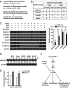
The increased proteasomal degradation by Torin1 requires ubiquitination, but not Akts, S6Ks, and Ulks, and results primarily from mTORC1 inhibition. (A) The increase in proteasomal degradation induced by mTOR inhibition requires ubiquitination. Proteasomal and lysosomal proteolysis were determined in the presence or absence of 10 μM Ube1 inhibitor in MEFs. (B) Blocking ubiquitination markedly reduced the cellular content of Ub conjugates. MEFs were treated with the Ube1 inhibitor or vehicle for 1 h before lysis, and the levels of Ub conjugates was measured by Western blot with anti-Ub (FK2). (C) The increase in proteasomal proteolysis by Torin1 was independent of S6Ks or 4E-BPs. Proteasomal proteolysis was determined in wild-type MEFs and MEFs lacking 4E-BP1 and 2 or S6K1 and 2. (D) Overexpression of Ulk1 or Ulk2 enhanced lysosomal but not proteasomal degradation. Proteolysis was determined in HEK293 cells after 20 h of transfection with vectors expressing Ulk1, Ulk2, or an inactive mutant of Ulk1 (Ulk1-K46N). (E) Unlike Torin1, the Akt inhibitor, Akti-1/2, (2 μM) did not increase proteasomal proteolysis in MEFs. (F) Akt inhibition did not induce the degradation of HMGCS1 and SUPT6H. The contents of these proteins were measured by Western blot after treatments for 6 h in the presence of cycloheximide. Phospho-Pras40 was measured to confirm the efficacy of Akti-1/2. (G) Torin1 increased proteasomal degradation in mTORC2-deficient (Rictor-null) MEFs. (H) Rictor-null MEFs showed no mTORC2 activity, reduced mTORC1 activity, and similar levels of HMGCS1, SUPT6H, and α-taxilin proteins compared with wild-type MEFs. (I) Selective activation or inhibition of mTORC2 did not affect proteasomal degradation in HEK293 cells. Proteasomal proteolysis was measured as described in Methods. For EBSS treatment, cells were washed once with EBSS and incubated with EBSS plus or minus insulin for 30 min, and then Torin1 was added for 1 h before measuring proteolysis. *P < 0.05; n.s., not significant.
Fig. 4.
Fig. 4.
Identification of proteins that are ubiquitinated and degraded more rapidly after Torin1 treatment. (A) Protocol for identifying proteins with increased ubiquitination after Torin1 treatment of HEK293 cells. (B) Examples of the 55 proteins with increased ubiquitination after Torin1 treatment, as shown by increased number of peptides identified by mass spectrometry of ubiquitinated proteins. (C) Torin1 treatment increased the proteasomal degradation of 4 of the 12 proteins for which antibodies were obtained. Western blot of cell extracts after treatments of MEFs for 6 h in the presence of cycloheximide, and their levels were quantified. (D) The marked reduction in HMGCS1 protein levels after treatment with Torin1 or EBSS buffer for 3–6 h was prevented by BTZ treatment. (E) Torin1-caused increase in proteasomal proteolysis did not require a cullin-based Ub ligase. MLN4924 (2 μM) was used to inactivate all cullin-based Ub ligases. (F) Summary of the regulation of proteasomal and autophagic proteolysis by mTOR, downstream of nutrient and growth factor signaling. *P < 0.05; #P < 0.01.

References

    1. Goldberg AL, St John AC. Intracellular protein degradation in mammalian and bacterial cells: Part 2. Annu Rev Biochem. 1976;45:747–803. - PubMed
    1. Ma XM, Blenis J. Molecular mechanisms of mTOR-mediated translational control. Nat Rev Mol Cell Biol. 2009;10(5):307–318. - PubMed
    1. Feng Y, Yao Z, Klionsky DJ. How to control self-digestion: Transcriptional, post-transcriptional, and post-translational regulation of autophagy. Trends Cell Biol. 2015;25(6):354–363. - PMC - PubMed
    1. Chantranupong L, Wolfson RL, Sabatini DM. Nutrient-sensing mechanisms across evolution. Cell. 2015;161(1):67–83. - PMC - PubMed
    1. Shimobayashi M, Hall MN. Making new contacts: The mTOR network in metabolism and signalling crosstalk. Nat Rev Mol Cell Biol. 2014;15(3):155–162. - PubMed

Publication types

Substances

LinkOut - more resources