

Early Detection and Quantification of Cerebral Venous Thrombosis by Magnetic Resonance Black-Blood Thrombus Imaging
- PMID: 26670082
- PMCID: PMC4729606
- DOI: 10.1161/STROKEAHA.115.011369
Early Detection and Quantification of Cerebral Venous Thrombosis by Magnetic Resonance Black-Blood Thrombus Imaging
Erratum in
-
Correction. Early detection and quantification of cerebral venous thrombosis by magnetic resonance blackblood thrombus imaging.Stroke. 2016 Feb;47(2):e39. doi: 10.1161/STR.0000000000000096. Stroke. 2016. PMID: 26811334 No abstract available.
Abstract
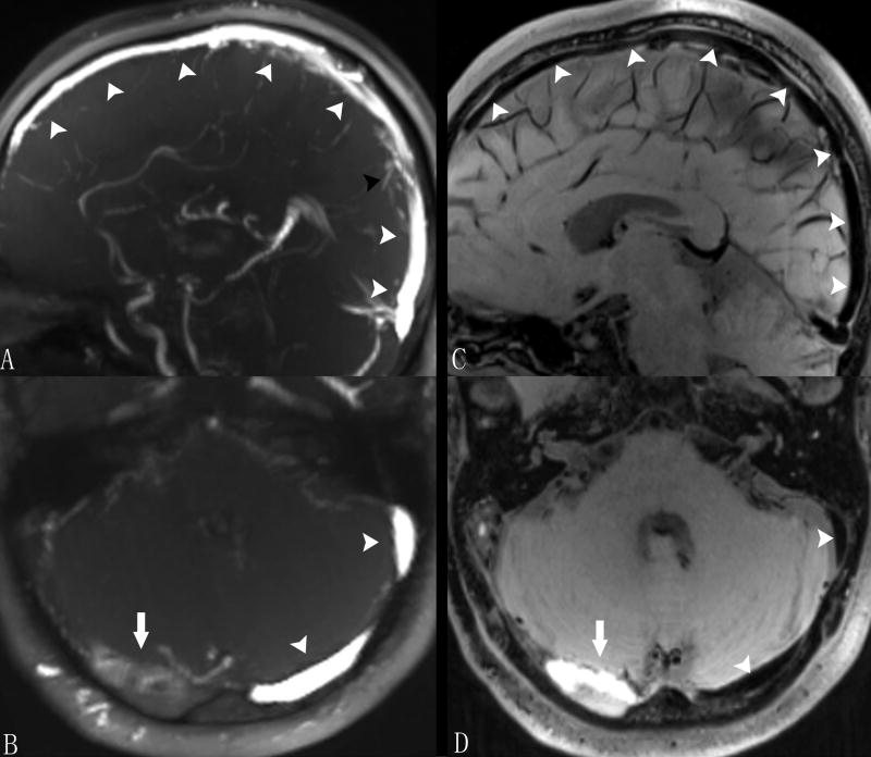
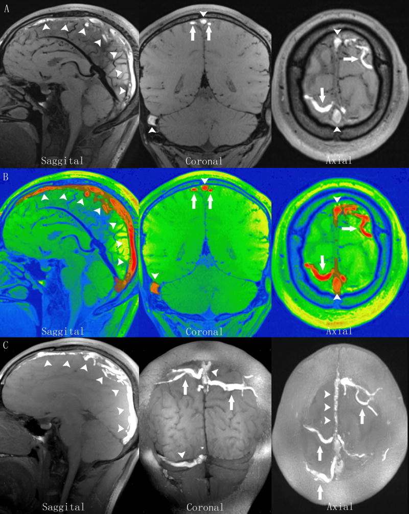
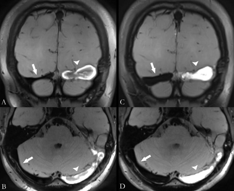
Background and purpose: Early diagnosis of cerebral venous thrombosis (CVT) is currently a major clinical challenge. We proposed a novel magnetic resonance black-blood thrombus imaging technique (MRBTI) for detection and quantification of CVT.
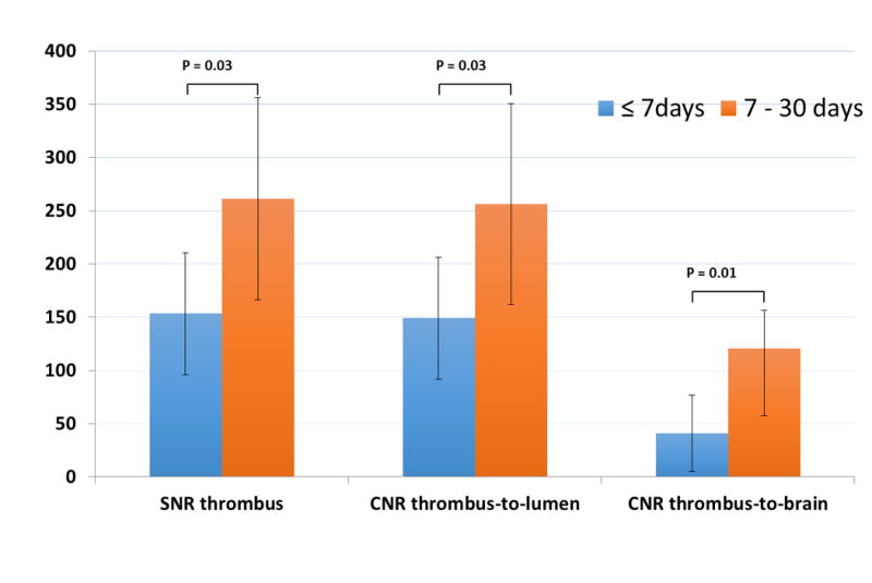
Methods: MRBTI was performed on 23 patients with proven CVT and 24 patients with negative CVT confirmed by conventional imaging techniques. Patients were divided into 2 groups based on the duration of clinical onset: ≤7 days (group 1) and between 7 and 30 days (group 2). Signal/noise ratio was calculated for the detected thrombus, and contrast/noise ratio was measured between thrombus and lumen and also between thrombus and brain tissue. The feasibility of using MRBTI for thrombus volume measurement was explored, and total thrombus volume was calculated for each patient.
Results: In 23 patients with proven CVT, MRBTI correctly identified 113 of 116 segments with a sensitivity of 97.4%. Thrombus signal/noise ratio was 153±57 and 261±95 for group 1 (n=10) and group 2 (n=13), respectively (P<0.01). Thrombus to lumen contrast/noise ratio was 149±57 and 256±94 for group 1 and group 2, respectively. Thrombus to brain tissue contrast/noise ratio was 41±36 and 120±63 (P<0.01), respectively. Quantification of thrombus volume was successfully conducted in all patients with CVT, and mean volume of thrombus was 10.5±6.9 mL.
Conclusions: The current findings support that with effectively suppressed blood signal, MRBTI allows selective visualization of thrombus as opposed to indirect detection of venous flow perturbation and can be used as a promising first-line diagnostic imaging tool.
Keywords: magnetic resonance imaging; sinus thrombosis, intracranial; venous thrombosis.
© 2015 American Heart Association, Inc.
Conflict of interest statement
Figures
References
-
- Stam J. Thrombosis of the Cerebral Veins and Sinuses. N Engl J Med. 2005;352:1791–1798. - PubMed
-
- Bousser MG, Ferro JM. Cerebral venous thrombosis: an update. The Lancet Neurology. 2007;6:162–170. - PubMed
-
- Coutinho JM, Zuurbier SM, Stam J. Declining Mortality in Cerebral Venous Thrombosis A Systematic Review. Stroke. 2014;45:1338–1341. - PubMed
-
- Ferro JM. Prognosis of Cerebral Vein and Dural Sinus Thrombosis: Results of the International Study on Cerebral Vein and Dural Sinus Thrombosis (ISCVT) Stroke. 2004;35:664–670. - PubMed
-
- Saposnik G, Barinagarrementeria F, Brown RD, Bushnell CD, Cucchiara B, Cushman M, et al. American Heart Association Stroke Council and the Council on Epidemiology and Prevention. Diagnosis and management of cerebral venous thrombosis: a statement for healthcare professionals from the American Heart Association/American Stroke Association. Stroke. 2011;42:1158–1192. - PubMed
Publication types
MeSH terms
Grants and funding
LinkOut - more resources
Full Text Sources
Other Literature Sources
Medical
