. 2015 Dec 16:5:18300.
doi: 10.1038/srep18300.

Attrition of Hepatic Damage Inflicted by Angiotensin II with α-Tocopherol and β-Carotene in Experimental Apolipoprotein E Knock-out Mice

Affiliations

Attrition of Hepatic Damage Inflicted by Angiotensin II with α-Tocopherol and β-Carotene in Experimental Apolipoprotein E Knock-out Mice

Kaliappan Gopal et al. Sci Rep. .

Abstract

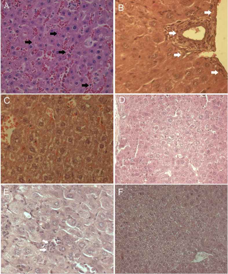
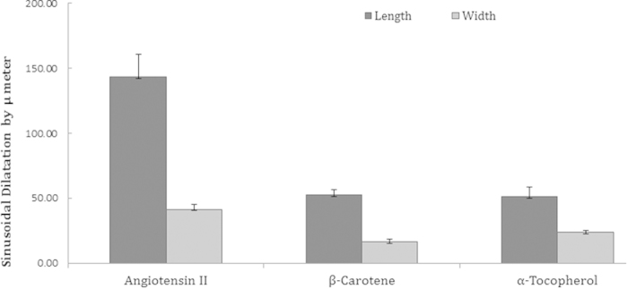
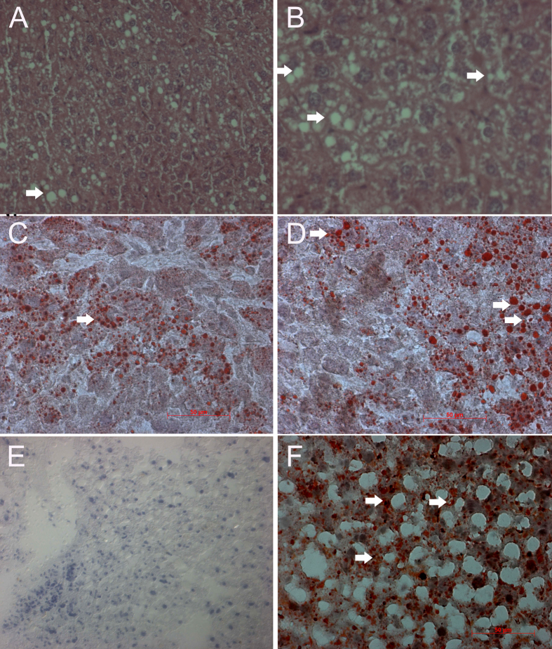
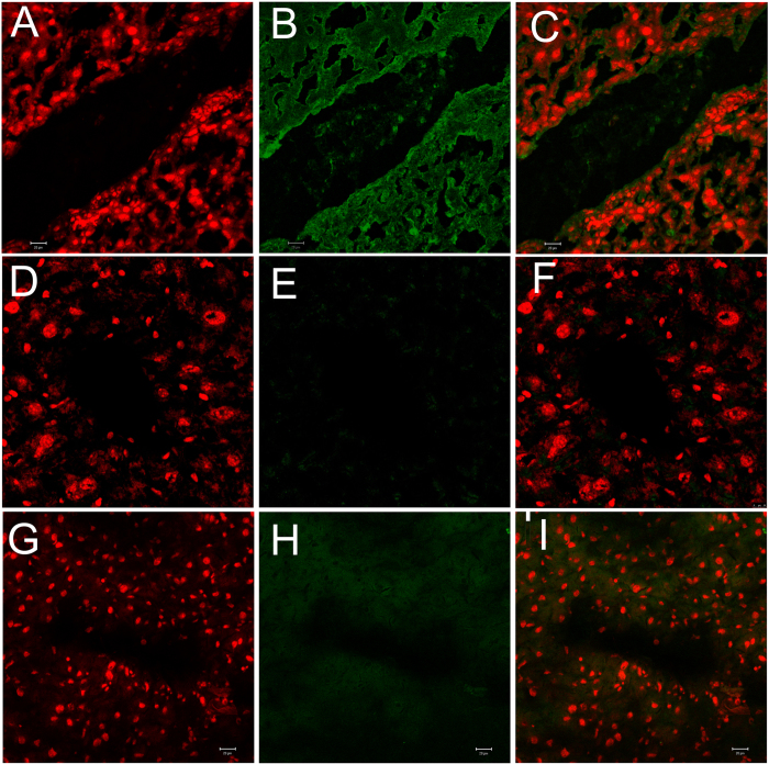
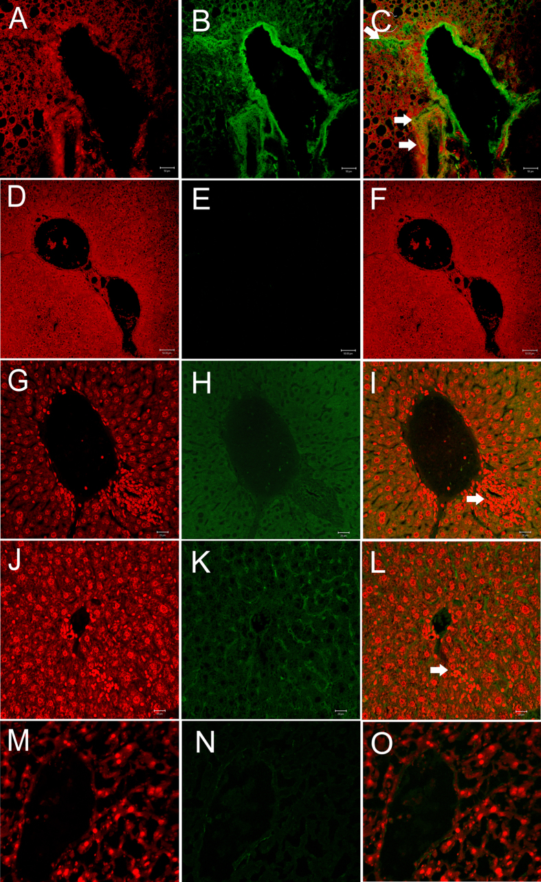
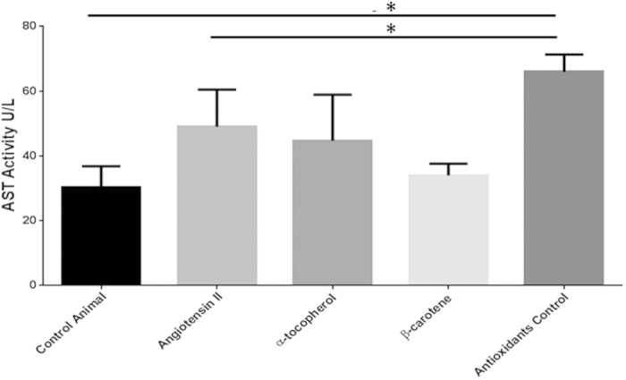
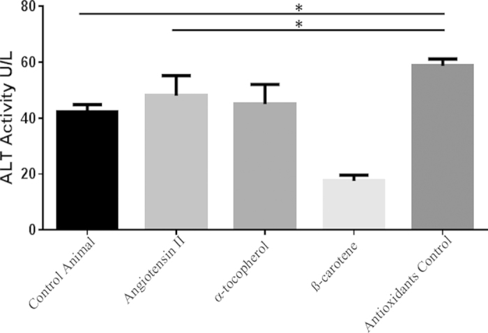
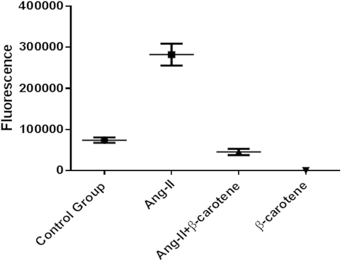
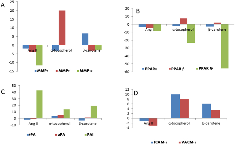
Angiotensin II is one of the key regulatory peptides implicated in the pathogenesis of liver disease. The mechanisms underlying the salubrious role of α-tocopherol and β-carotene on liver pathology have not been comprehensively assessed. Here, we investigated the mechanisms underlying the role of Angiotensin II on hepatic damage and if α-tocopherol and β-carotene supplementation attenuates hepatic damage. Hepatic damage was induced in Apoe(-/-)mice by infusion of Angiotensin II followed by oral administration with α-tocopherol and β-carotene-enriched diet for 60 days. Investigations showed fibrosis, kupffer cell hyperplasia, hepatocyte degeneration and hepatic cell apoptosis; sinusoidal dilatation along with haemorrhages; evidence of fluid accumulation; increased ROS level and increased AST and ALT activities. In addition, tPA and uPA were down-regulated due to 42-fold up-regulation of PAI-1. MMP-2, MMP-9, MMP-12, and M-CSF were down-regulated in Angiotensin II-treated animals. Notably, α-tocopherol and β-carotene treatment controlled ROS, fibrosis, hepatocyte degeneration, kupffer cell hyperplasia, hepatocyte apoptosis, sinusoidal dilatation and fluid accumulation in the liver sinusoids, and liver enzyme levels. In addition, PAI-1, tPA and uPA expressions were markedly controlled by β-carotene treatment. Thus, Angiotensin II markedly influenced hepatic damage possibly by restraining fibrinolytic system. We concluded that α-tocopherol and β-carotene treatment has salubrious role in repairing hepatic pathology.


Figures

Figure 1
Figure 1. Histopathological analysis of pathological changes in the liver of Angiotensin-II- treated Apoe−/− mice using Hematoxylin & Eosin and Masson trichrome staining methods.
(A,B) Individual hepatocyte degeneration (white arrow), mild bile duct hyperplasia, periductular/peri billary fibrosis (black arrow) and few foci of individual hepatocyte apoptosis (red arrow) seen in the liver of Angiotensin-II-treated Apoe−/− mice. (C,D) Hepatic necrosis with infiltration of inflammatory cells (white arrow); (E,F) Peribiliary and/or periportal fibrosis shown by Masson trichrome staining in Angiotensin-II- treated Apoe−/− mice (arrow); (G) Liver tissue stained with Masson trichrome of β-carotene-treated Apoe−/− mice lacking evidence of peribiliary or periportal fibrosis; (H) Control liver not showing any fibrosis.
Figure 2
Figure 2. Histological analysis of vascular changes in the liver of Angiotensin-II-treated, α-tocopherol and β-carotene-treated Apoe−/− mice.
(A,B) Moderate to severe sinusoidal dilatation along with haemorrhages (black arrows) and mild to moderate inflammatory reactions where periportal/ peribillary fibrosis (white arrows) seen in Angiotensin-II-treated Apoe−/− mice. (C,D) Vascular and inflammatory changes were not observed in β-carotene-treated Apoe−/− mice. (E,F) Lack of fibrotic and vascular changes, with mild Kupffer cell proliferation in α-tocopherol-treated Apoe−/− mice.
Figure 3
Figure 3. Sinusoidal dilatation in the angiotensin II-treated experimental animals and compared to β-carotene and α-tocopherol treated Apoe−/− mice.
Figure 4
Figure 4. Oil-red ‘O’ staining for pathological examination of liver of α-tocopherol- and β-carotene-treated Apoe−/− mice.
(A,B) Moderate vacoular degeneration (arrow) in the hepatocytes of β-carotene-treated Apoe−/− mice. (C,D) β-carotene-treated liver show moderate accumulation of red colour lipid droplets in the cytoplasm of hepatocytes (arrow). (E) α-tocopherol-treated liver lacking accumulation of lipid droplets as compared to positive control high fat diet-treated liver of Apoe−/− mice. (F) Accumulation of red colour lipid droplets in the cytoplasm of hepatocytes in high fat diet-treated liver of Apoe−/− mice (arrow) (positive control).
Figure 5
Figure 5. Confocal microscopy analysis of Mac3 expression in Angiotensin-II and β-carotene-treated Apoe−/− mice.
(A–C) Mac3 over expressed marginally in the liver tissues of Ang II- treated Apoe−/− mice; (D–F) Mac3 show no expression in β-carotene-treated Apoe−/− mice; (G–I) No expression of Mac3 is noticed in control liver.
Figure 6
Figure 6. Confocal microscopy analysis of K18, p53 and caspase 3 expressions in Angiotensin-II and β-carotene-treated Apoe−/− mice.
(A–C) Cytokeratin 18 over-expression (arrow) in Angiotensin-II-treated as compared to (D–F) β-carotene-treated Apoe−/− mice; (G–I) Caspase 3 over-expressed moderately in the inflammatory regions of Angiotensin-II-treated Apoe−/− mice (arrow); (J–L) p53 show mild over-expression in the inflammatory regions (white arrow) of Angiotensin-II-treated Apoe−/− mice as compared to β-carotene-treated Apoe−/− mice (M-O).
Figure 7
Figure 7. AST activity in different treatment groups of Apoe−/− mice.
Determined AST activity in the different treatment groups were compared with control Apoe−/− mice. AST activity levels of Angiotensin-II-treated group were remarkably elevated as compared to controls. AST activity levels in the antioxidant control group (Angiotensin-II infused and no antioxidant supplements) were significantly elevated as compared to the Angiotensin-II and control groups. Footnotes: *Indicates significant difference between the individual treatment groups marked by arrow line.
Figure 8
Figure 8. ALT activity in different treatment groups of Apoe−/− mice.
Examined ALT activity in the different treatment groups were compared with control Apoe−/− mice. ALT activity levels of Angiotensin-II-treated group were remarkably elevated as compared to control (p~0.007). AST levels of antioxidants control group (Angiotensin-II infused and no antioxidant supplements) were significantly further elevated as compared to the Angiotensin-II and control groups. Footnotes: *Indicates significant difference between the individual treatment groups marked by arrow line.
Figure 9
Figure 9. Angiotensin-II inflicted ROS levels controlled by β-carotene in Apoe−/− mice.
Determined ROS release in the liver tissues of different treatment groups were compared with control Apoe−/− mice, and found significant difference between the treatment groups (p~0.0001). ROS releases were significantly increased in the livers of Ang-II infused Apoe−/− mice as compared to control Apoe−/− mice (p~0.002). ROS releases were significantly controlled in the β-carotene supplement treated Apoe−/− mice as compared to Ang-II infused (p~0.002) and control Apoe−/− mice (p~0.001). Footnotes: *Indicates significant difference between the individual treatment groups marked by arrow line.
Figure 10
Figure 10. Fold changes of selective candidate genes mRNA level in Angiotensin-II-infused, and α-tocopherol and β-carotene-treated Apoe−/− mice.
(A) MMP-3, MMP-9 and MMP-12 fold gene expression. MMP-12 down-regulated in the liver of Angiotensin-II treated group; (B) PPAR-α, PPAR-β and PPAR-γ down-regulated in Angiotensin-II- and both α-tocopherol- and β-carotene-treated animals; (C) PAI-1 up-regulated, and both tPA and uPA down-regulated in the Angiotensin-II-treated mice. α-tocopherol- and β-carotene-treated animals controlled PAI-1 expression and improved the levels of both tPA and uPA mRNA. (D) ICAM-1 and VCAM-1 down-regulated in the Angiotensin-II-treated group and up-regulated in both α-tocopherol- and β-carotene-treated Apoe−/− mice.

References

    1. Torok N. J. Recent advances in the pathogenesis and diagnosis of liver fibrosis. J Gastroenterol. 43, 315–321 (2008). - PubMed
    1. Hirose A. et al. Angiotensin II type 1 receptor blocker inhibits fibrosis in rat nonalcoholic steatohepatitis. Hepatology. 45, 1375–1381 (2007). - PubMed
    1. Browatzki M. et al. Angiotensin II stimulates matrix metalloproteinase secretion in human vascular smooth muscle cells via nuclear factor- kappaB and activator protein 1 in a redox-sensitive manner. J Vasc Res. 42, 415–423 (2005). - PubMed
    1. Gopal K. et al. β-Carotene Attenuates Angiotensin II-Induced Aortic Aneurysm by Alleviating Macrophage Recruitment in Apoe −/− Mice. PLoS One. 8, e67098 (2013). - PMC - PubMed
    1. Yoshiji H. & Fukui H. Renin-angiotensin system and progression of chronic liver diseases. J Gastroenterol. 41, 1020–1022 (2006). - PubMed

Publication types

MeSH terms