. 2015;18(5):696-704.
doi: 10.18433/j3cc9k.

In vitro Treatment with cis-[Ru(H-dcbpy-)2(Cl)(NO)] Improves the Endothelial Function in Aortic Rings with Endothelial Dysfunction

Affiliations

In vitro Treatment with cis-[Ru(H-dcbpy-)2(Cl)(NO)] Improves the Endothelial Function in Aortic Rings with Endothelial Dysfunction

Jorge Camargo Oishi et al. J Pharm Pharm Sci. 2015.

Abstract

Purpose: The ruthenium complex cis-[Ru(H-dcbpy-)2(Cl)(NO)] (DCBPY) is a nitric oxide (NO) donor and studies suggested that the ruthenium compounds can inactivate O2-. The aim of this study is to test if DCBPY can revert and/or prevent the endothelial dysfunction.

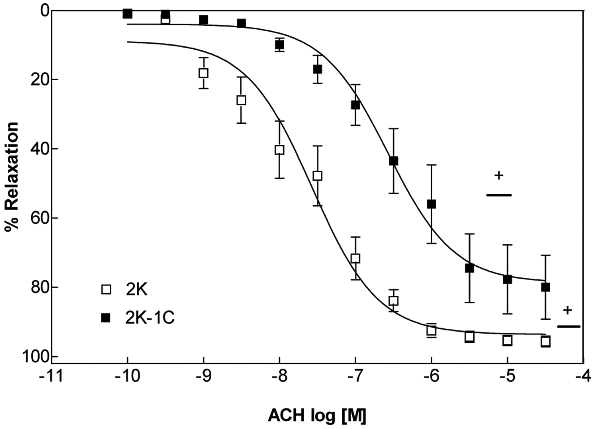
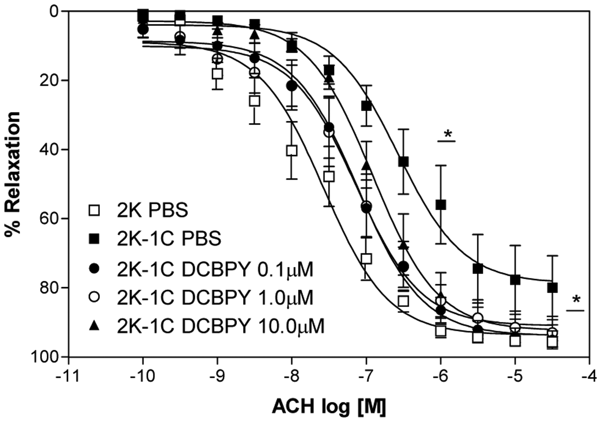
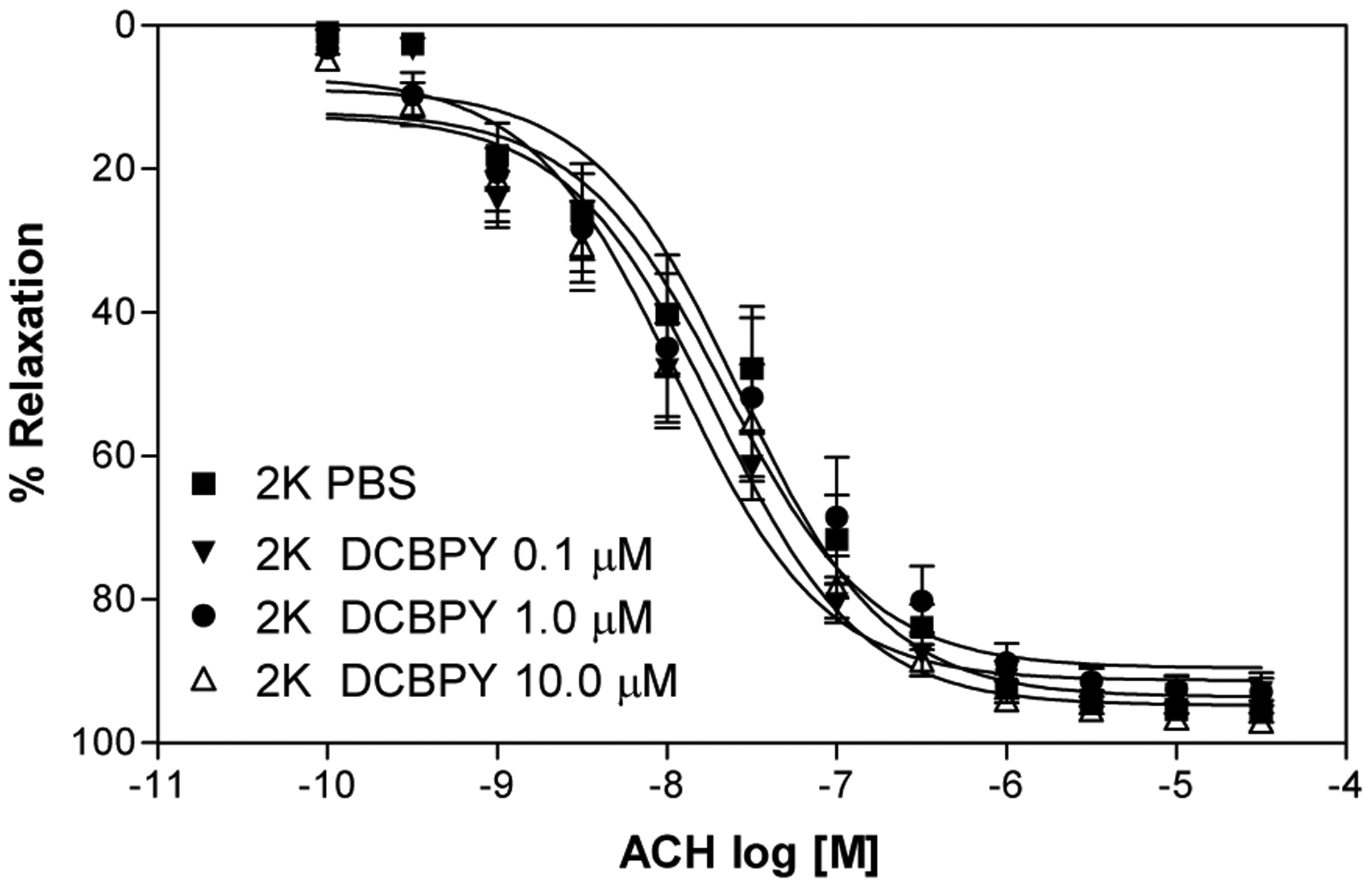
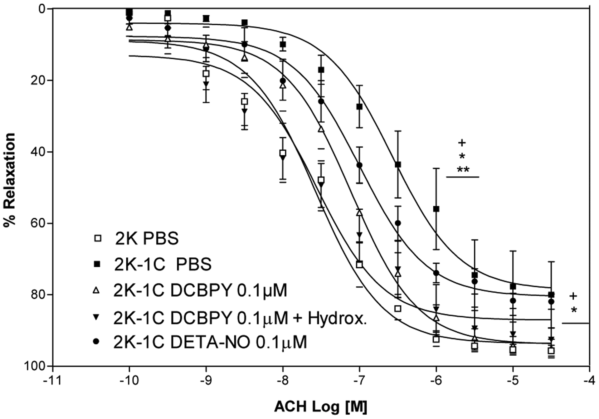
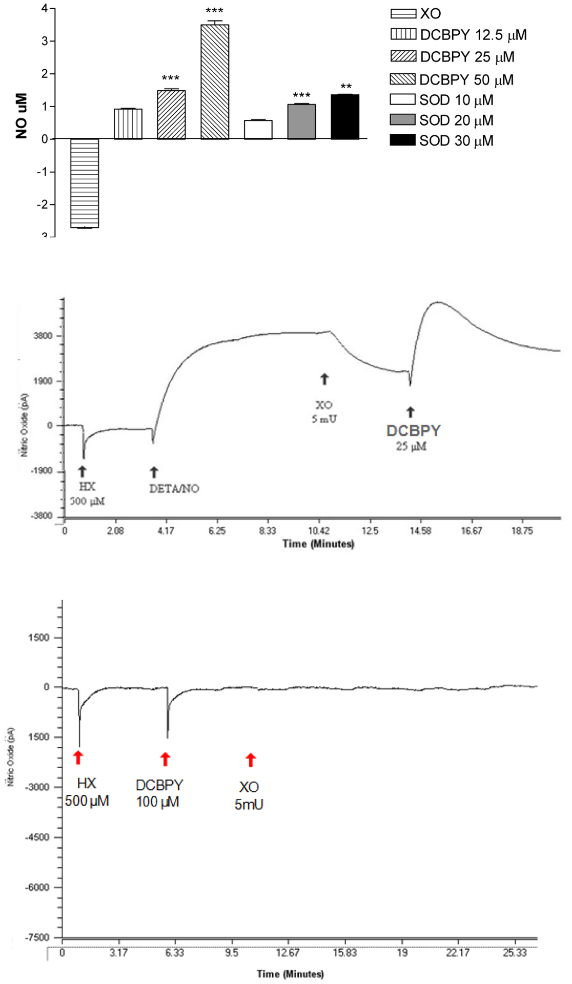
Methods: Normotensive (2K) and hypertensive (2K-1C) wistar rats were used. To vascular reactivity study, thoracic aortas were isolated, rings with intact endothelium were incubated with: DCBPY: 0.1; 1 and 10μM, DCBPY plus hydroxocobalin (NO scavenger) or tempol during 30 minutes, and concentration effect curves to acetylcholine were performed. The potency values (pD2) and maximum effect (ME) were analyzed. The O2- was generated using hypoxantine xantine oxidase and the reduction of cytochrome c, NO consumption by O2- and the effect in avoid NO consumption was measured.

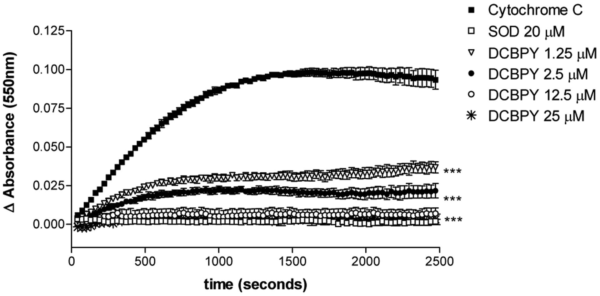
Results: In 2K-1C DCBPY at 0.1; 1 or 10μM improved the relaxation endothelium dependent induced by acetylcholine in aortic rings compared to control 2K-1C, and also improved ME. In rings from 2K incubation with DCBPY (0.1; 1.0 and 10 μM) did not change pD2 or ME. Incubation with 0.1 μM of DCBPY plus hydroxocobalamin did not modify the potency and ME in 2K-1C compared to DCBPY (0.1 μM). DCBPY and SOD inhibits the reduction of cytochrome c and inhibited the NO consumption by O2-, showing that O2- has been removed from the solution.

Conclusion: Our results suggest that DCBPY at a lower concentration (0.1 µM) is not an NO generator, but can inactivate superoxide and improves the endothelial function.


Figures

Figure 1.
Figure 1.
Concentration–response curves (n=8) for acetylcholine in intact endothelium- aortic rings contracted with phenylephrine. Values are mean ± S.E.M. of experiments performed on preparations obtained from different animals. + indicates significant difference (p< 0.05) in pD2 and ME values for 2K vs. 2K-1C
Figure 2.
Figure 2.
Concentration–response curves for acetylcholine in aortic rings with intact endothelium and incubated with different concentrations of DCBPY and contracted with phenylephrine. Values are mean ± S.E.M. of experiments performed on preparations obtained from different animals. * indicates significant difference (p< 0.05) in pD2 and ME values for 2K-1C PBS (n=8) vs. 2K-1C DCBPY 0.1/1.0/10.0 μM (n=8).
Figure 3.
Figure 3.
Concentration–response curves for acetylcholine in 2K aortic rings intact endothelium and incubated with different concentrations of DCBPY and contracted with phenylephrine. Values are mean ± S.E.M. of experiments performed on preparations obtained from different animals (n=8).
Figure 4.
Figure 4.
Concentration–response curves for acetylcholine after incubation with DCBPY 0,1 μM in the absence or presence of Hydroxocobalamin (100 μM) or DETA-NO, in aortic rings intact endothelium- contracted with phenylephrine. Values are mean ± S.E.M. of experiments performed on preparations obtained from different animals. + indicates significant difference (p< 0.05) in pD2 and ME values for 2K vs. 2K-1C; * indicates significant difference (p< 0.05) in pD2 and ME values for 2K-1C PBS (n=8) vs. 2K-1C DCBPY 0.1 μM (n=8); ** indicates significant difference (p< 0.05) in pD2 and ME values for 2K-1C PBS (n=8) vs DCBPY + DETA-NO (n=8).
Figure 5.
Figure 5.
Concentration–response curves for acetylcholine after incubation with DCBPY 0.1 μM, and TEMPOL 10 μM in aortic rings intact endothelium- contracted with phenylephrine. Values are mean ± S.E.M. of experiments performed on preparations obtained from different animals. *** indicates significant difference (p< 0.001) in pD2 and ME values for 2K-1C PBS (n=8) vs 2K-1C TEMPOL (n=8).
Figure 6.
Figure 6.
Measurement of intracellular NO production in HUVEC. Cells were treated with 0.1 μM of DCBPY and DETA-NO for 30 min. The NO production measured by fluorescence intensity of DAF-2T from stained cells. Bars (top panel) represent data of NO production are presented as means ± SEM. *p< 0.05 versus control, in 3 independent experiments of each protocol. The bottom panel represents images of NO detection by fluorescent images of DAF stained cells.
Figure 7.
Figure 7.
Measurement of the reduction of cytochrome c by superoxide (O2), at 550nm. Difference (Δ) in the absorbance (AU) was obtained, and the superoxide-scavenging effect of the compound DCBPY and superoxide dismutase (SOD) was verified. Each point represents mean ± SEM of at least 3 absorbance values. *indicates significant (p<0.05) difference for Ru-NO2 1.25 μM vs 2.5 μM, 2.5 μM vs 25 μM. ***indicates significant (p<0.001) difference for DCBPY and SOD vs control (cytochrome c), and DCBPY 1.25 μM vs 12.5 μM .
Figure 8.
Figure 8.
Top panel: Quantification of NO that was not consumed by superoxide (O2) generated by xanthine oxidase (XO). Superoxide-scavenging effect of DCBPY and superoxide dismutase (SOD). Bars represent mean ± SEM of NO concentration. ***indicates significant (p<0.001) difference for DCBPY between the concentrations 12.5 vs 25 μM, 25 vs 50 μM, and for SOD between the concentrations 10 vs 20 μM, 20 vs 30 μM. Middle panel: Representative recording of experiments using the selective electrode for NO. Experimental sequence to measure the NO consumption by superoxide (O2) generated by xanthine oxidase (XO). Bottom panel: Positive control to measure if DCBPY releases NO in the presence on hypoxanthine (HX) and xanthine oxidase (XO).

References

    1. Puddu P, Puddu GM, Zaca F, Muscari A. Endothelial dysfunction in hypertension, Acta Cardiol. 2000; 55(4):221–32. - PubMed
    1. Lobysheva I, Rath G, Sekkali B, Bouzin C, Feron O, Gallez B. Moderate caveolin-1 downregulation prevents NADPH oxidase-dependent endothelial nitric oxide synthase uncoupling by angiotensin II in endothelial cells. Arterioscler Thromb Vasc Biol. 2011. September;31(9):2098–105. doi: 10.1161/ATVBAHA.111.230623. Epub 2011 Jun - DOI - PubMed
    1. Ignarro LJ, Cirino G, Casini A, Napoli C. Nitric oxide as a signaling molecule in the vascular system: an overview, J Cardiovasc Pharmacol. 1999; 34:879–886. - PubMed
    1. Taddei S, Virdis A, Ghiadoni L, Salvetti A. Endothelial dysfunction in hypertension: fact or fancy? J Cardiovasc Pharmacol. 1998; 32 (3):S41–7. - PubMed
    1. Munk PS, Butt N, Larsen AI. Endothelial dysfunction predicts clinical restenosis after percutaneous coronary intervention, Scand Cardiovasc J. 2011;45(3):139–45. - PubMed