Comparative Study
. 2016 Feb 12;291(7):3239-53.
doi: 10.1074/jbc.M115.699124. Epub 2015 Dec 15.

Starvation Induces Proteasome Autophagy with Different Pathways for Core and Regulatory Particles

Affiliations
Comparative Study

Starvation Induces Proteasome Autophagy with Different Pathways for Core and Regulatory Particles

Kenrick A Waite et al. J Biol Chem. .

Abstract

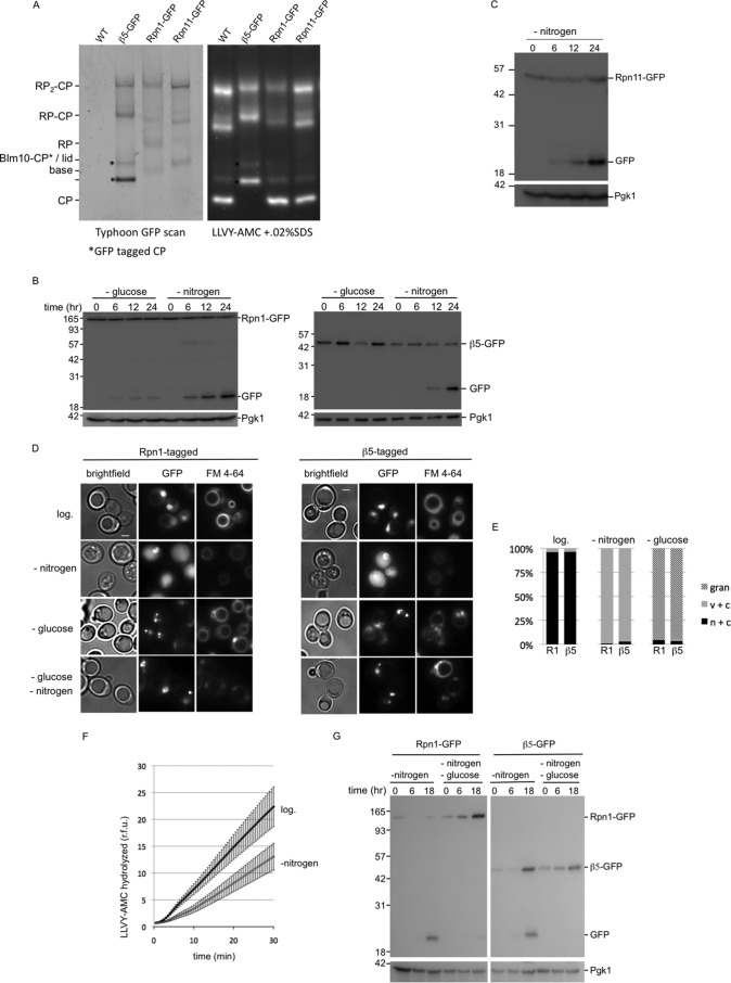
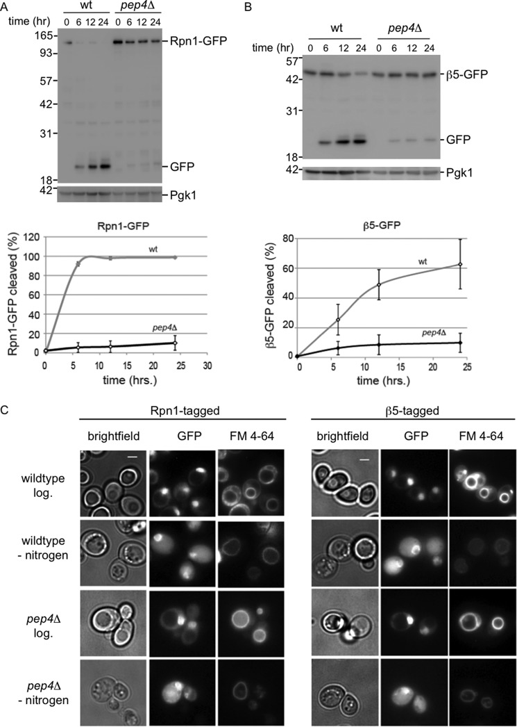
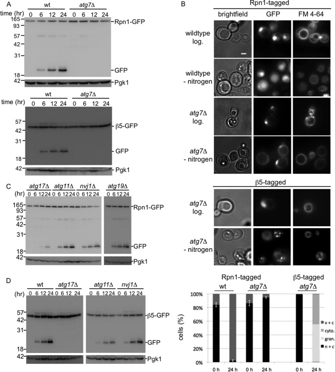
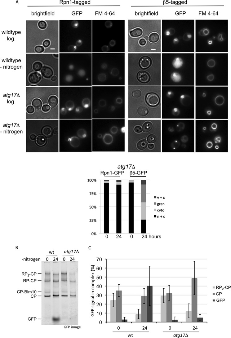
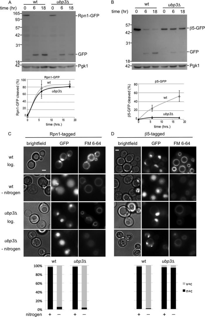
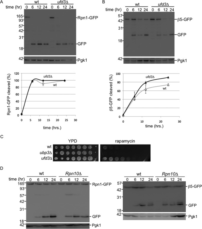
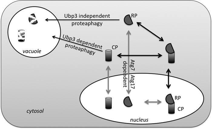
The proteasome is responsible for the degradation of many cellular proteins. If and how this abundant and normally stable complex is degraded by cells is largely unknown. Here we show that in yeast, upon nitrogen starvation, proteasomes are targeted for vacuolar degradation through autophagy. Using GFP-tagged proteasome subunits, we observed that autophagy of a core particle (CP) subunit depends on the deubiquitinating enzyme Ubp3, although a regulatory particle (RP) subunit does not. Furthermore, upon blocking of autophagy, RP remained largely nuclear, although CP largely localized to the cytosol as well as granular structures within the cytosol. In all, our data reveal a regulated process for the removal of proteasomes upon nitrogen starvation. This process involves CP and RP dissociation, nuclear export, and independent vacuolar targeting of CP and RP. Thus, in addition to the well characterized transcriptional up-regulation of genes encoding proteasome subunits, cells are also capable of down-regulating cellular levels of proteasomes through proteaphagy.

Keywords: autophagy; deubiquitylation (deubiquitination); nitrogen starvation; proteaphagy; proteasome; protein translocation; ribophagy; vacuole; yeast.


Figures

FIGURE 1.
FIGURE 1.
Nitrogen starvation targets proteasomes to the vacuole. A, strains were C-terminally tagged with GFP at the endogenous locus of the core particle subunit β5, the regulatory particle subunit Rpn1, or the regulatory particle subunit Rpn11. Cell lysates of indicated strains were resolved on native gel and scanned to determine incorporation of GFP-tagged subunits in proteasome complexes. Next, gels were stained with proteasome fluorogenic substrate LLVY-AMC, to visualize proteasome peptidase activity. * indicates CP with β5-GFP; GFP-tagged CP showed a migrational shift compared with untagged CP. The different subunits can be detected in assembled proteasomes and the appropriate subcomplexes (i.e. β5 in CP; Rpn1 in RP and base; and Rpn11 in RP and lid). B, strains expressing Rpn1-GFP or β5-GFP were grown logarithmically in YPD to A600 1.5, washed, and transferred to minimal media lacking either glucose or nitrogen. Cells were collected and lysed at indicated time points. Samples were resolved on SDS-PAGE and immunoblotted for GFP and the loading control Pgk1. C, strains expressing Rpn11-GFP were analyzed upon nitrogen starvation as described in B. D, strains expressing Rpn1-GFP or β5-GFP were analyzed by fluorescent microscopy for GFP localization prior to starvation or after incubation for 24 h in minimal media lacking glucose or nitrogen. Cells were stained with FM 4-64 to visualize vacuoles. Representative images are shown, and quantifications are shown in E. E, at least 100 cells were used to quantify the localization as displayed in the stacked bar graph as percentage. n + c, nuclear and cytosolic staining with stronger signals in nucleus; c + v, little nuclear staining and clear vacuolar localization; gran, presence of distinct granular localization sometimes combined with weak nuclear and cytosolic staining but no vacuolar staining. Three independent experiments showed similar amounts and localization results. F, cells were lysed before and after nitrogen starvation. Equal amounts of protein were assayed for hydrolysis of LLVY-AMC. Accumulation of fluorescent AMC over time is displayed as relative fluorescence units (r.f.u.). The average of six independent experiments are shown with S.E. Activity upon starvation is reduced to 58% and the difference was highly significant (paired Student's t test p = 0.01, n = 6). G, experiment as B only cells were starved in minimal media lacking nitrogen or nitrogen and glucose. Localization of the fluorescent signal in double-starved cells (see D) resembled that of glucose starvation, showing granules and little to no vacuolar localization.
FIGURE 2.
FIGURE 2.
Vacuolar hydrolases are responsible for cleavage of tagged proteasome subunits. A and B, wild type or pep4Δ strains with gene fusions encoding Rpn1-GFP (A) or β5-GFP (B) were starved for nitrogen in the presence of 1 mm PMSF and lysed. Samples were analyzed as in Fig. 1C. Three independent immunoblots were quantified and corrected for input (Pgk1 amounts), and percentage cleaved (displayed on y axis) was calculated as amount of Rpn1-GFP signal divided by the amount of Rpn1-GFP signal plus free GFP signal for each time point. C, cells were analyzed by fluorescent microscopy to visualize proteasome subunits (GFP) and vacuoles (FM 4-64) prior to starvation or after incubation for 24 h in minimal media lacking nitrogen.
FIGURE 3.
FIGURE 3.
Proteasomes are targeted to the vacuole by autophagy. A, wild type or atg7Δ strains with gene fusions encoding Rpn1-GFP or β5-GFP were starved for nitrogen and lysed. Samples were resolved on SDS-PAGE and immunoblotted for GFP and the loading control Pgk1. B, microscope images and quantification (n > 100) of cells as in A. n + c, nuclear and cytosolic staining with stronger signals in nucleus; c + v, little nuclear staining and clear vacuolar localization; cyto, clear cytosolic staining without nuclear enrichment and no vacuolar staining; gran, presence of distinct granular localization sometimes combined with weak nuclear and cytosolic staining but no vacuolar staining. Three independent experiments showed similar amounts and localization results. C and D, gene fusions encoding Rpn1-GFP (C) or β5-GFP (D) were introduced in strains deleted for indicated genes and analyzed as in A.
FIGURE 4.
FIGURE 4.
Different processing of proteasome CP and RP autophagy. A, microscope images and quantification (n > 100) of atg17Δ cells. n + c, nuclear and cytosolic staining with stronger signals in nucleus; c + v, little nuclear staining and clear vacuolar localization; cyto, clear cytosolic staining with little to no vacuolar or nuclear staining; gran, presence of distinct granular localization sometimes combined with weak nuclear and cytosolic staining but no vacuolar staining. Note that for Rpn1-GFP after nitrogen starvation in the atg17Δ cells, the nuclear to cytosol ratio becomes somewhat smaller, but it remains enriched in the nucleus and hence no difference in our scoring. B, wild type and atg17Δ cells expressing β5-GFP were lysed, and samples were resolved on native gel. Gels were scanned on Typhoon 9410 imager to identify the complexes in which β5-GFP was incorporated. C, quantification of native gels from B. For each condition, the percentage of β5-GFP in a particular proteasome complex was determined. Displayed is the average of three independent experiments. Error bars indicate S.E.
FIGURE 5.
FIGURE 5.
Proteasome core particles and regulatory particles depend differently on Ubp3 for vacuolar targeting. A and B, wild type or ubp3Δ strains with gene fusions encoding Rpn1-GFP (A) or β5-GFP (B) were analyzed for vacuolar targeting of RP and CP, respectively, upon nitrogen starvation. Samples were resolved on SDS-PAGE and immunoblotted for GFP and the loading control Pgk1. Lower panel shows quantifications as in Fig. 2. Shown are the averages from 3 to 5 experiments, and error bars indicate S.E. C and D, microscope images and quantification (n > 100) of cells from A and B before and after 24 h of starvation. c + v, little nuclear staining and clear vacuolar localization; n + c, nuclear and cytosolic staining with stronger signals in nucleus, except for β5-GFP ubp3Δ cells where there was clearly stronger signal in the cytosol. Irrespectively, all n + c all have little to no vacuolar staining.
FIGURE 6.
FIGURE 6.
Proteaphagy does not depend on Ufd3 or Rpn10. A and B, CP proteaphagy is distinct from 60S ribophagy as it does not depend on Ufd3. Strains expressing Rpn1-GFP (A) or β5-GFP (B), either in wild type or UFD3 deletion background, were starved for nitrogen and lysed. Samples were resolved on SDS-PAGE and immunoblotted for GFP and the loading control Pgk1 (top panel). Lower panel shows quantification of three independent experiments. Error bars indicate S.E. C, strains were plated in 4-fold serial dilution on indicated plates. D, nitrogen starvation induced proteaphagy does require Rpn10. Indicated strains expressing Rpn1-GFP (left) or β5-GFP (right) were analyzed as in A.
FIGURE 7.
FIGURE 7.
Proposed model for proteaphagy. During logarithmic growth ∼70% of yeast proteasomes are nuclear. Nitrogen starvation induces autophagy of proteasomes. However, similar to the observations for ribosomes, two subcomplexes (CP and RP) show a different requirement for components that target them to the vacuole as well as different fates upon disruption of autophagy. This indicates that CP and RP dissociate. It is currently not clear whether this dissociation occurs prior to or following nuclear export resulting in two possible models of nitrogen starvation-induced proteaphagy.

References

    1. Reggiori F., and Klionsky D. J. (2013) Autophagic processes in yeast: mechanism, machinery and regulation. Genetics 194, 341–361 - PMC - PubMed
    1. Suzuki K. (2013) Selective autophagy in budding yeast. Cell Death Differ. 20, 43–48 - PMC - PubMed
    1. Finley D. (2009) Recognition and processing of ubiquitin-protein conjugates by the proteasome. Annu. Rev. Biochem. 78, 477–513 - PMC - PubMed
    1. Inobe T., and Matouschek A. (2014) Paradigms of protein degradation by the proteasome. Curr. Opin. Struct. Biol. 24, 156–164 - PMC - PubMed
    1. Warner J. R. (1999) The economics of ribosome biosynthesis in yeast. Trends Biochem. Sci. 24, 437–440 - PubMed

Publication types

MeSH terms

Substances

LinkOut - more resources