. 2016;7(2):98-109.
doi: 10.1080/21505594.2015.1131381. Epub 2015 Dec 15.

Autophagy postpones apoptotic cell death in PRRSV infection through Bad-Beclin1 interaction

Affiliations

Autophagy postpones apoptotic cell death in PRRSV infection through Bad-Beclin1 interaction

Ao Zhou et al. Virulence. 2016.

Abstract

Autophagy and apoptosis play significant roles in PRRSV infection and replication. However, the interaction between these 2 processes in PRRSV replication is still far from been completely understood. In our studies, the exposure of MARC-145 cells to PRRSV confirmed the activation of autophagy and subsequent induction of apoptosis. The inhibition of autophagy by 3-methyladenine (3-MA) caused a significant increase in PRRSV-induced apoptosis, showing a potential connection between both mechanisms. Moreover, we observed an increase in Bad expression (a pro-apoptotic protein) and Beclin1 (an autophagy regulator) in virus-infected cells up to 36h. Co-immunoprecipitation assays showed the formation of Bad and Beclin1 complex in PRRSV infected cells. Accordingly, Bad co-localized with Beclin1 in MARC-145 infected cells. Knockdown of Beclin1 significantly decreased PRRSV replication and PRRSV-induced autophagy, while Bad silencing resulted in increased autophagy and enhanced viral replication. Furthermore, PRRSV infection phosphorylated Bad (Ser112) to promote cellular survival. These results demonstrate that autophagy can favor PRRSV replication by postponing apoptosis through the formation of a Bad-Beclin1 complex.

Keywords: Apoptosis; Autophagy; Bad; Beclin1; Porcine reproductive and respiratory syndrome virus.


Figures

Figure 1.
Figure 1.
PRRSV induced cell autophagy and apoptosis in MARC-145 cells. (A) LC3 punctate distribution in mock-infected and PRRSV infected cells. Cells were fixed at 24hpi and stained with antibodies against LC3. Cellular nuclei were counterstained by DAPI. (B) Western blot analysis of LC3-I (16kDa), LC3-II (18kDa), and β-actin (42 kDa) expression were performed in mock-infected (ctrl) and PRRSV-infected cells at 6, 12, 18, 24, 36 and 48 hpi. (C) and (D) Quantitative measurement of LC3-II/ LC3-I and Beclin 1 proteins in MARC-145 during PRRSV infection according to the result of western blot in (B). Amounts of proteins were normalized to β-actin that controlled for protein loading by using ImageJ software. All experiments were repeated three times. (E) Apoptosis induced by PRRSV was detected at 12, 24, 36 and 48 hpi by Hoechst 33258 staining in MARC-145 cells.
Figure 2.
Figure 2.
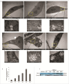
PRRSV-induced autophagy delays cell apoptosis. (A) Electron microscopy pictures were taken of MARC-145 cells uninfected or infected for 6h, 12h, 24h, 36h and 48h respectively. Autophagosome (square) and nucleus (N) are indicated. (B) The quantification of the number of autophagosome-like vesicles per cell profile in mock- and PRRSV cell at different time points was calculated with at least 50 cells. (C) Western blotting analysis of anti-apoptotic protein Bcl-xl at indicated time points.
Figure 3.
Figure 3.
Inhibition of autophagy increased PRRSV-induced apoptosis in MARC-145 cells. (A) and (B) MARC-145 cells were treated with DMSO, rapamycin and 3-MA followed by PRRSV infection, respectively, and then were trypsinized and harvested. The cells were incubated with AnnexinV APC and stained with 7-ADD for 15 minutes at room temperature. The percentage of apoptostic cells was analyzed using FACS can flow cytometer with CellQuest pro software. (C) Beclin1 silencing by RNAi in MARC-145 cells. Western blot was performed using anti-Beclin1 antibody (D) MARC-145 cells were transfected with Beclin1 siRNA prior to PRRSV infection. After 24 hpi, Cell lysates were subjected to Western blot analysis with specific antibody. The blot was re-probed with an antibody to actin for comparison of protein load in each lane.
Figure 4.
Figure 4.
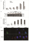
PRRSV infection regulated the expression of Bad and Beclin1 and intracellular distribution of Bad. (A) Total RNA was isolated from mock or PRRSV infected MARC-145 cells at a MOI of 1 at different time points and subjected to qPCR to analyze Bad and Beclin1 expression. β-actin was used as an internal control. (B) Cell lysates were analyzed for Beclin1 and Bad expression by Western blotting. The blot was reacted with anti- β-actin antibody for comparison of protein load. (C) The fold changes of protein are presented after normalizing with internal control and were arbitrarily set as 1. Data presented were representative of three independent experiments. (D) Fluorescence microscopy images showing immunofluorescence staining for Bad (green), PRRSV NSP2 (red) and DAPI (blue) in mock cells and PRRSV-infected MARC-145 cells.
Figure 5.
Figure 5.
(See previous page) Bad regulated PRRSV-induced autophagy in MARC-145 cells. (A) The knockdown efficiency of Beclin 1 protein was detected in untreated control cells (MOCK), control RNAi-treated cells (NC), and beclin 1-silenced cells (siBad-1 or siBad-2) 48h after transfection by protein gel blot analysis. The β-actin as control was used to confirm equal loading of protein. (B) The silencing of Bad reduced the cleavage of caspase3 while increasing the expressing of LC3ІІ. (C) Confocal fluorescence microscopy analysis of LC3 punctate and PRRSV replication in Bad and Beclin1 knockdown. The wild type cells (MOCK) and RNAi-transfected cells (siNC, siBad and siBeclin1) were infected with PRRSV at MOI of 1. At 36h post-infection, cells were fixed and permeabilized. LC3 was stained with rabbit anti-LC3 antibody (green) and PRRSV NSP2 was with stained mouse anti-NSP2 antibody (red). The nuclei were stained with DAPI (blue).
Figure 6.
Figure 6.
Bad interacts with Beclin1 in PRRSV infection. (A) MARC-145 cells were mock-infected or infected with PRRSV, and cell lysates extracted after 36hpi were immunoprecipitated with rabbit monoclonal Beclin1antibody. Interaction of Beclin1, Bax, Bad or Bcl-2 was detected by immunoblotting with specific antibody. (B) Differential co-localization of endogenous Bad with Beclin1 upon PRRSV infection. Mock-infected or infected MARC-145 cells were stained for endogenous Bad (red) and Beclin1 (green). The nuclear was stained with DAPI (blue). (C) and (D) Western blotting analysis of p-Bad and the optical density ratio of p-Bad/GAPDH was calculated. The data were presented as mean ± SD of three independent experiments (t-test,*p<0 .05).

References

    1. Thorburn A. Apoptosis and autophagy: regulatory connections between two supposedly different processes. Apoptosis 2008; 13:1-9; PMID:17990121; http://dx.doi.org/10.1007/s10495-007-0154-9 - DOI - PMC - PubMed
    1. Ding WX, Ni HM, Gao W, Hou YF, Melan MA, Chen X, Stolz DB, Shao ZM, Yin XM. Differential effects of endoplasmic reticulum stress induced autophagy on cell survival. J Biol Chem 2007; 282(7):4702-10; PMID:17135238; http://dx.doi.org/10.1074/jbc.M609267200 - DOI - PubMed
    1. Nikoletopoulou V, Markaki M, Palikaras K, Tavernarakis N. Crosstalk between apoptosis, necrosis and autophagy. BiochimBiophysActa 2013; 1833(12):3448-59 - PubMed
    1. Wang W, Zhou J, Shi J, Zhang Y, Liu S, Liu Y, Zheng D. Human T-cell leukemia virus type 1 Tax-deregulated autophagy pathway and c-FLIP expression contribute to resistance against death receptor-mediated apoptosis. J Virol 2014; 88(5):2786-98; PMID:24352466; http://dx.doi.org/10.1128/JVI.03025-13 - DOI - PMC - PubMed
    1. Gannagé M, Dormann D, Albrecht R, Dengjel J, Torossi T, Rämer PC, Lee M, Strowig T, Arrey F, Conenello G, et al. . Matrix protein 2 of influenza A virus blocks autophagosome fusion with lysosomes. Cell Host Microbe 2009; 6(4):367-80; http://dx.doi.org/10.1016/j.chom.2009.09.005 - DOI - PMC - PubMed

Publication types

MeSH terms

LinkOut - more resources