. 2015 Dec 16:5:18316.
doi: 10.1038/srep18316.

Sustained intraocular VEGF neutralization results in retinal neurodegeneration in the Ins2(Akita) diabetic mouse

Affiliations

Sustained intraocular VEGF neutralization results in retinal neurodegeneration in the Ins2(Akita) diabetic mouse

Jose R Hombrebueno et al. Sci Rep. .

Abstract

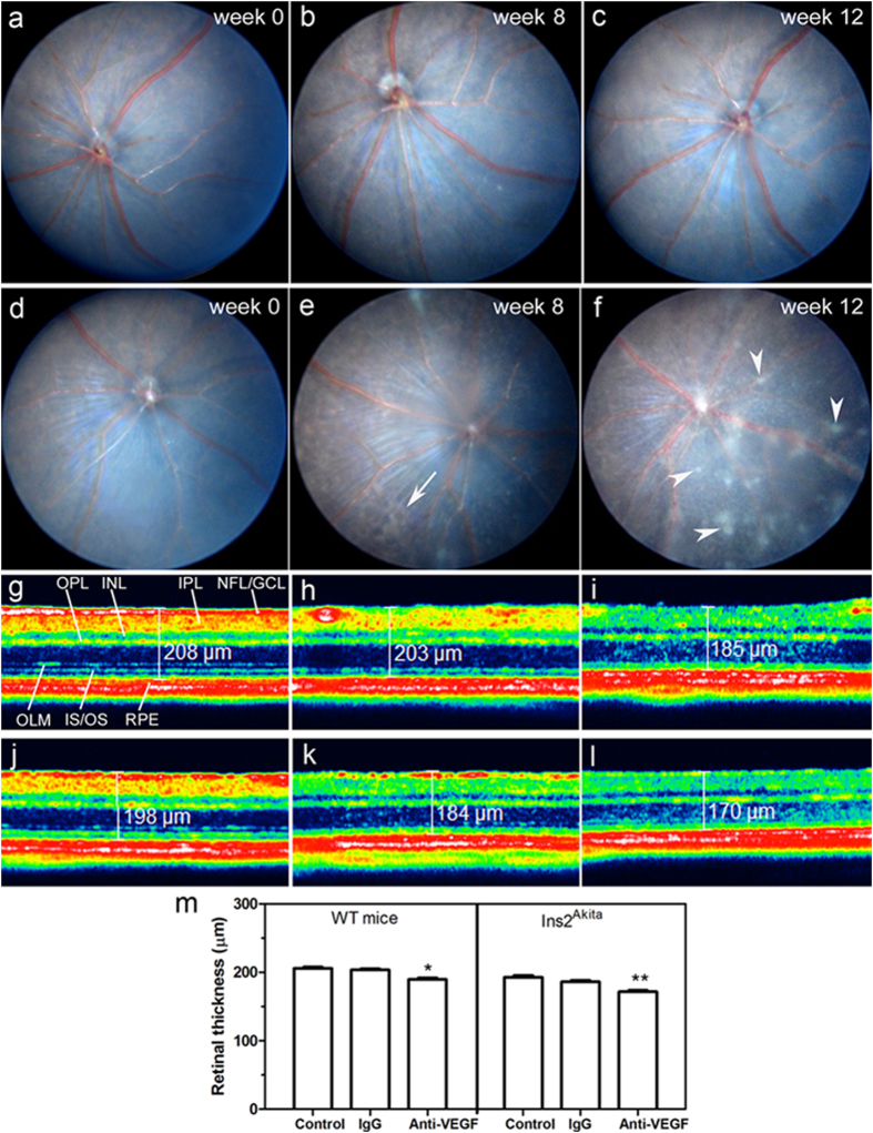
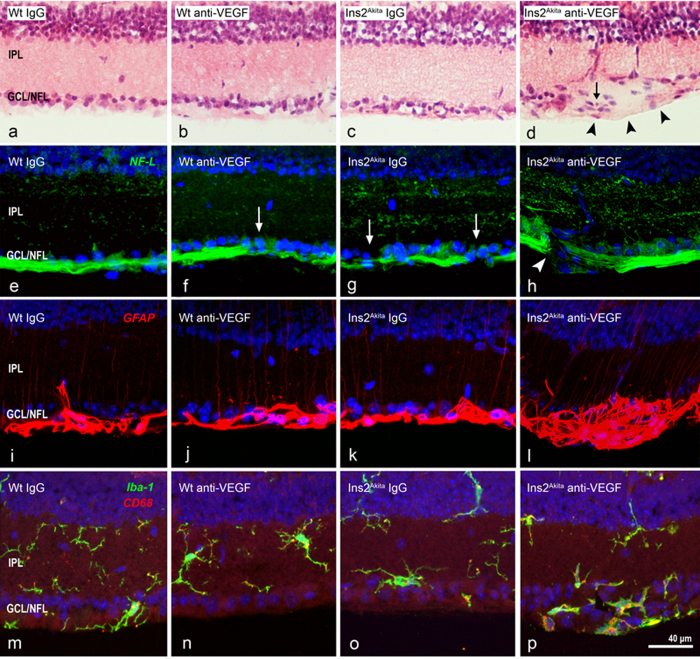
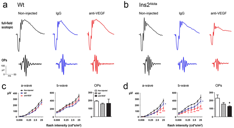
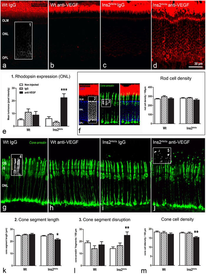
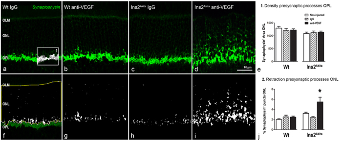
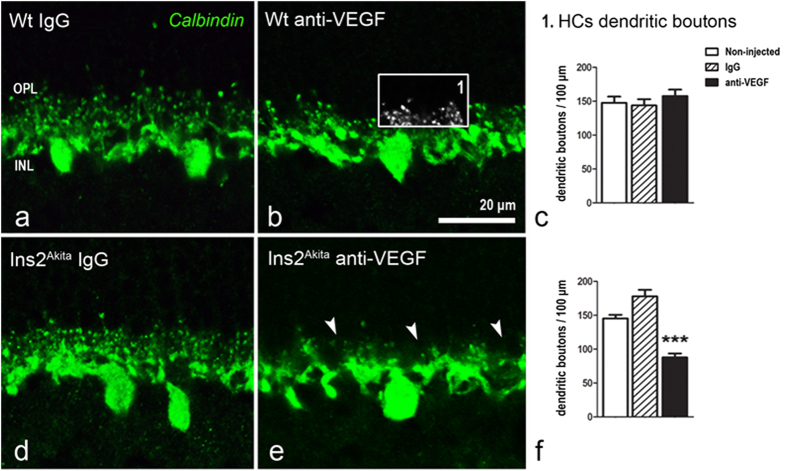
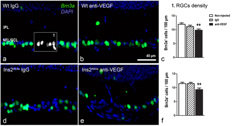
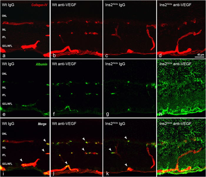
Current therapies that target vascular endothelial growth factor (VEGF) have become a mainstream therapy for the management of diabetic macular oedema. The treatment involves monthly repeated intravitreal injections of VEGF inhibitors. VEGF is an important growth factor for many retinal cells, including different types of neurons. In this study, we investigated the adverse effect of multiple intravitreal anti-VEGF injections (200 ng/μl/eye anti-mouse VEGF164, once every 2 weeks totalling 5-6 injections) to retinal neurons in Ins2(Akita) diabetic mice. Funduscopic examination revealed the development of cotton wool spot-like lesions in anti-VEGF treated Ins2(Akita) mice after 5 injections. Histological investigation showed focal swellings of retinal nerve fibres with neurofilament disruption. Furthermore, anti-VEGF-treated Ins2(Akita) mice exhibited impaired electroretinographic responses, characterized by reduced scotopic a- and b-wave and oscillatory potentials. Immunofluorescent staining revealed impairment of photoreceptors, disruptions of synaptic structures and loss of amacrine and retinal ganglion cells in anti-VEGF treated Ins2(Akita) mice. Anti-VEGF-treated WT mice also presented mild amacrine and ganglion cell death, but no overt abnormalities in photoreceptors and synaptic structures. At the vascular level, exacerbated albumin leakage was observed in anti-VEGF injected diabetic mice. Our results suggest that sustained intraocular VEGF neutralization induces retinal neurodegeneration and vascular damage in the diabetic eye.


Figures

Figure 1
Figure 1. Clinical examinations following intravitreal injections of anti-VEGF.
Fundus images from WT (a–c) or Ins2Akita (d–f) mice at baseline (week 0) (a,d), 8 weeks (b,e) or 12 weeks (c,f) after intravitreal injections of anti-VEGF. Arrows indicate brownish irregular shaped lesions; Arrowheads indicate cotton wool spot-like lesions. SD-OCT representative images from WT (g–i) or Ins2Akita (j–l) mouse retinas of non-injected controls (g,j), intravitreal IgG (h,k) or anti-VEGF (i,l) treated mice at 12 weeks post-injection. (m) Quantitative analysis of neuroretinal thickness in WT and Ins2Akita non-injected, IgG or anti-VEGF treated mice (n = 6 eyes per strain/condition). Results are presented as mean ± SEM. *P < 0.05, **P < 0.01 compared to non-injected controls of the same strain. One-way ANOVA. NFL/GCL, nerve fibre/ganglion cell layer; IPL, inner plexiform layer; INL, inner nuclear layer; OPL, outer plexiform layer; OLM, outer limiting membrane; IS/OS, photoreceptor inner/outer segments; RPE, retinal pigment epithelium.
Figure 2
Figure 2. Histopathology of cotton wool spot (CWS)-like lesions after anti-VEGF treatment.
Retinal sections from WT and Ins2Akita mice after 5 intravitreal injections of IgG or anti-VEGF were processed for H&E staining (a–d) or NF-L (e–h), GFAP (i–l) and Iba-1/CD68 (m–p) immunoreactivities. (d) CWS-lesion at the GCL/NFL (arrowheads) accompanied by atypical cell infiltration (arrow). (f,g) Slight disruption of NF-L+ axonal fibres (arrows) in anti-VEGF treated WT (f) or IgG treated Ins2Akita (g) mice. (h) Severe disruption of NF-L+ axonal fibres (arrowhead) in anti-VEGF treated Ins2Akita mice. (l,p) Focal up-regulation of GFAP and infiltration of Iba-1+CD68+ microglial cells in CWS-lesions. IPL, inner plexiform layer; GCL/NFL, ganglion cell/nerve fibre layer.
Figure 3
Figure 3. Abnormal electroretinogram (ERG) responses after anti-VEGF treatment.
Scotopic ERG responses in WT and Ins2Akita mice after 5 intravitreal injections of IgG or anti-VEGF. (a,b) Representative scotopic ERG responses from WT (a) and Ins2Akita (b) mice of different treatment groups. (c,d) The amplitude (μV) of a-wave, b-wave and oscillatory potentials in WT (c) and Ins2Akita (d) mice. (c,d) n = 5 mice per strain/condition. Results are presented as mean ± SEM. *P < 0.05, **P < 0.01, ***P < 0.001 compared to non-injected controls of the same strain. Two-way ANOVA.
Figure 4
Figure 4. Photoreceptor abnormalities after anti-VEGF treatment.
Retinal sections from WT and Ins2Akita mice after 5 intravitreal injections of IgG or anti-VEGF processed for rhodopsin or cone-arrestin immunostaining. (a–d) Confocal images of rhodopsin immunoreactivity of different treatment groups. (e,f) Mean luminance values of rhodopsin immunostaining at the ONL (box1) and rod photoreceptor cell density (difference between DAPI+ nuclei (box2) and cone somata (box3) at the ONL). (g–j) Confocal images of cone-arrestin immunoreactivity of different treatment groups. (k-m) Quantitative analysis of cone segment length (box4) (k), cone segment disruption (cone-arrestin+ free particles above cone outer segments (box5)) (l) and cone photoreceptor density (m) in different treatment groups. n ≥ 20 retinal images per strain/condition. Results are presented as mean ± SEM. *P < 0.05, **P < 0.01, ***P < 0.001 compared to non-injected controls of the same strain. One-way ANOVA. OS, outer segments; IS, inner segments; OLM, outer limiting membrane: ONL, outer nuclear layer; OPL, outer plexiform layer.
Figure 5
Figure 5. Pre-synaptic changes at the OPL after anti-VEGF treatment.
Retinal sections from WT and Ins2Akita mice after 5 intravitreal injections of IgG or anti-VEGF processed for synaptophysin immunoreactivity. (a–d) Confocal images of synaptophysin immunoreactivity from different treatment groups. (e) Quantitative analysis of synaptophysin+ area at the OPL (box1) and (fj) ONL (thresholded white pixels enclosed in the yellow area (f)) in different treatment groups. n ≥ 20 retinal images per strain/condition. Results are presented as mean ± SEM. *P < 0.05 compared to non-injected controls of the same strain. One-way ANOVA. OLM, outer limiting membrane: ONL, outer nuclear layer; OPL, outer plexiform layer.
Figure 6
Figure 6. Horizontal cell dendritic boutons after anti-VEGF treatment.
Retinal sections from WT (a–c) and Ins2Akita mice (d–f) after 5 intravitreal injections of IgG or anti-VEGF processed for calbindin immunoreactivity. (e) Loss of horizontal cell dendritic boutons (arrowheads) at the OPL in anti-VEGF treated Ins2Akita mice. The density of horizontal cell dendritic boutons (box1) in WT (c) and Ins2Akita (f) mice. n ≥ 20 retinal images per strain/condition. Results are presented as mean ± SEM. ***P < 0.001 compared to non-injected controls of the same strain. One-way ANOVA. OPL, outer plexiform layer; INL, inner nuclear layer.
Figure 7
Figure 7. Amacrine cell loss in WT and Ins2Akita anti-VEGF treated mice.
Retinal sections from Ins2Akita mice after 5 intravitreal injections of IgG or anti-VEGF processed for GABA (a,b) and GlyT1 (d,e) immunoreactivities. (c,f,i) The density of GABAergic (box1), glycinergic (box2) and total amacrine cells (GABAergic + glycinergic) at the INL in different treatment groups. n ≥ 20 retinal images per strain/condition. Results are presented as mean ± SEM. *P < 0.05, **P < 0.01, ***P < 0.001 compared to non-injected controls of the same strain. One-way ANOVA. INL, inner nuclear layer; IPL, inner nuclear layer.
Figure 8
Figure 8. Retinal ganglion cell loss in WT and Ins2Akita anti-VEGF treated mice.
Retinal sections from WT (a–c) and Ins2Akita mice (d–f) after 5 intravitreal injections of IgG or anti-VEGF processed for Brn3a immunoreactivity. (c,f) The density of Brn3a+ cells in different treatment groups. n ≥ 20 retinal images per strain/condition. Results are presented as mean ± SEM. **P < 0.01 compared to non-injected controls of the same strain. One-way ANOVA. IPL, inner nuclear layer; NFL/GCL, nerve fibre/ganglion cell layer.
Figure 9
Figure 9. Retinal vascular changes in WT and Ins2Akita anti-VEGF treated mice.
Retinal photomicrographs of collagen-IV (red) and albumin (green) immunoreactivities in WT (a,b,e,f,i-i) and Ins2Akita (c,d,g,h,k,l) after 5 intravitreal injections of IgG or anti-VEGF. (i–k) Albumin confined within retinal vessels (arrowheads). ONL, outer nuclear layer; INL, inner nuclear layer; IPL, inner nuclear layer; GCL/NFL, ganglion cell layer/nerve fibre layer.

References

    1. Klein R., Klein B. E., Moss S. E., Davis M. D. & DeMets D. L. The Wisconsin epidemiologic study of diabetic retinopathy. II. Prevalence and risk of diabetic retinopathy when age at diagnosis is less than 30 years. Arch Ophthalmol 102, 520–526 (1984). - PubMed
    1. Klein B. E. Overview of epidemiologic studies of diabetic retinopathy. Ophthalmic Epidemiol 14, 179–183 (2007). - PubMed
    1. Klein R., Klein B. E., Moss S. E., Davis M. D. & DeMets D. L. The Wisconsin epidemiologic study of diabetic retinopathy. III. Prevalence and risk of diabetic retinopathy when age at diagnosis is 30 or more years. Arch Ophthalmol 102, 527–532 (1984). - PubMed
    1. Diabetes C. et al. Modern-day clinical course of type 1 diabetes mellitus after 30 years’ duration: the diabetes control and complications trial/epidemiology of diabetes interventions and complications and Pittsburgh epidemiology of diabetes complications experience (1983-2005). Arch Intern Med 169, 1307–1316 (2009). - PMC - PubMed
    1. Jyothi S. & Sivaprasad S. Five-year visual outcome following laser photocoagulation of diabetic macular oedema. Eye (Lond) 25, 851–858; quiz 859 (2011). - PMC - PubMed

Publication types

MeSH terms

Substances